| Journal of Clinical and Analytical Medicine
1
Hegemann Hastalığı / Hegemann’s Disease
A Rare Cause of Elbow Pain: Hegemann’s Disease
Nadir Bir Dirsek Ağrısı Nedeni: Hegemann Hastalığı
DOI: 10.4328/JCAM.1061 Received:03.05.2012 Accepted: 27.05.2012 Printed: 01.07.2015 J Clin Anal Med 2015;6(4): 499-501 Corresponding Author: Yunus İmren, Kars Devlet Hastanesi Ortopedi ve Travmatoloji Kliniği, Örnek Mahallesi, Hastane Sokak, 36200 Kars, Türkiye.
GSM: +905423661038 E-Mail: [email protected]
Özet
Travma olmaksızın dirsek ağrısı çocuklarda ergenlik öncesi dönemde nadir gö-rülür. Bu dönemde kronik dirsek ağrısı, şişlik ve eklem hareket kaybı osteokond-ral lezyonlara bağlı olabilir. Osteokondritis dissekans, subkondosteokond-ral kemik ile eklem kıkırdağının subkondral kemikteki bir lezyona bağlı ayrışması olarak tanımlanır. Özellikle diz, ayak bileği ve dirsek eklemlerinde görülür. Dirsek osteokondrozunda birincil olarak kapitellum etkilenmekteyken, troklea osteonekrozu ile ilgili az sayı-da olgu bildirilmiştir. Bu yazısayı-da yakın zamansayı-da travma öyküsü olmayan, ergenlik öncesi dönemde bulunan, klinik ve radyolojik olarak tek taraflı troklea osteokond-ral lezyonu ile uyumlu olan bir olgu sunulmuştur. Hastada son 3 haftadır daha önce belirti vermeyen, günlük aktivitelerde ortaya çıkan sağ dirsek ağrısı mevcuttu. 2 hafta uzun kol atelde takip edilen hastada şikayetlerin devamı üzerine dirsek MR görüntülemesine ihtiyaç duyuldu. Tanı konduktan sonra cerrahi dışı tedavi ile ba-şarılı sonuç elde edildi. Çocuklarda rezidüel deformitenin tanınması ve uygun te-davisi için takipte dikkatli olunmalıdır.
Anahtar Kelimeler
Osteokondrit; Dirsek; Ağrı
Abstract
Non-traumatic elbow pain is rarely seen in children and pre-adolescents. Osteo-chondral lesions may be the source of chronic elbow pain, swelling, and loss of motion in children or adolescents. Osteochondritis dissecans (OCD) is described as a lesion of subchondral bone resulting in separation of the articular cartilage and subchondral bone. It is found primarily in the knee, ankle, and elbow joints. Since osteochondrosis of the elbow primarily involves capitellum, few papers in-volving osteonecrosis of the trochlea have been reported. This paper discusses a pre-adolescent boy with clinical and radiographic signs consistent with unilateral osteochondral lesion of the trochlea humeri, with no history of recent trauma. The patient had insidious onset of right elbow pain during daily activities for the last 3 weeks. After usage of long arm splint for 2 weeks, persistence of the symptoms necessitated MRI of the affected elbow. After the diagnosis, non-operative man-agement was achieved. Care should be taken for the affected children to recog-nize any residual deformity and to treat it properly at follow up.
Keywords
Osteochondritis; Elbow; Pain
Tuğrul Alıcı1, Yunus İmren2, Hakan Gündeş3 1Özel Avusturya Sen Jorj Hastanesi, Ortopedi ve Travmatoloji Kliniği, İstanbul, 2Kars Devlet Hastanesi, Ortopedi ve Travmatoloji Kliniği, Kars, 3İstanbul Medipol Üniversitesi Ortopedi ve Travmatoloji Ana Bilim Dalı, İstanbul, Türkiye
Bu olgu sunumu 31 Ekim-5 Kasım 2011 tarihleri arasında Antalya’da düzenlenen 22. Ulusal Ortopedi ve Travmatoloji Kongresi’nde poster bildirisidir.
| Journal of Clinical and Analytical Medicine Hegemann Hastalığı / Hegemann’s Disease
2
Introduction
Non-traumatic elbow pain is rarely seen in children and pre-adolescents. A differential diagnosis of elbow pain should in-clude ulnar neuritis, osteochondritis dissecans, triceps tendini-tis, instability and epicondylitis [1]. Osteochondral lesions may be the source of chronic elbow pain, swelling, and loss of motion in children or adolescents. Virtually dominant arm is involved. Osteochondritis is defined as an inflammation of bone and car-tilage. Osteochondritis dissecans (OCD) is described as a lesion of subchondral bone resulting in separation of the articular car-tilage and subchondral bone. The term “osteochondritis disse-cans” was given to this condition by Franz Konig in 1888 (1). He described a case of knee pain that appeared to suggest a sub-chondral inflammatory process dissecting a fragment of carti-lage from the femoral condyle. OCD involves localized avascular necrosis of subchondral bone and subsequent loss of structural support for the adjacent articular cartilage. It is found primarily in the knee, ankle, and elbow joints [2]. Osteochondrosis of the elbow primarily involves capitellum, which was first described by Panner in 1927. Few papers involving osteonecrosis of the trochlea have been reported. Uhrmacher firstly described this condition in 1933, and then Hegemann presented three cases in 1951. Conservative treatment is usually reserved for early lesions with intact overlying cartilage and nearly half of the lesions tend to heal treated non-operatively. Strengthening exercises may be helpful after relief of symptoms. Surgery is indicated for patients who do not respond to conservative treat-ment, or those who have mechanical symptoms with unstable fragments and loose bodies in the elbow joint. This paper dis-cusses a pre-adolescent boy with clinical and radiographic signs consistent with unilateral osteochondral lesion of the trochlea humeri, with no history of recent trauma.
Case Report
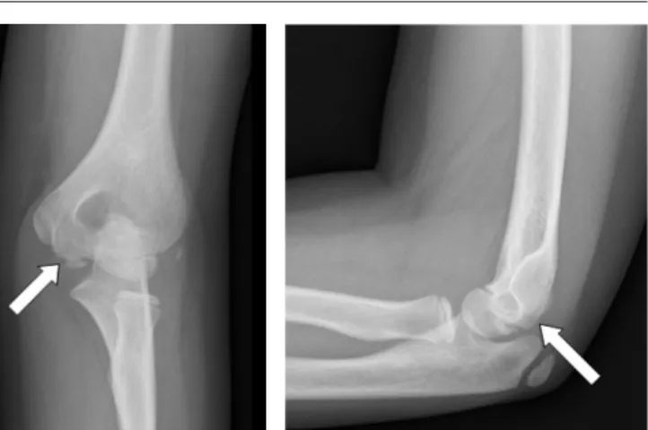
A right-handed 12-year-old boy had insidious onset of right el-bow pain during daily activities for the last 3 weeks. No history of recent trauma, vigorous activity, and locking episode of the elbow joint were noted. There was no history of corticosteroid use and bone disease in his family. Physical examination re-vealed mild swelling and palpation induced tenderness on the medial epicondyle and 25° loss of active elbow extension. The carrying angle was 5° on the right and 13° on the left. There was no evidence of medial or lateral instability. Mental examination and laboratory data were within normal limits. Radiographs of the right elbow showed a slight radiolucent defect in the troch-lear notch with well-defined margins (Fig.1). After usage of long arm splint for 2 weeks, persistence of the symptoms necessi-tated MRI of the affected elbow. The diagnosis of aseptic necro-sis of the humeral trochlea was made by characteristic signal patterns of the magnetic resonance imaging. Avascular necro-sis was traditionally seen as a geographic area that exhibited low signal on both T1- and T2-weighted images (Fig.2). Fluid collection was also noted in the radiohumeral joint. After the diagnosis, a long arm splint was applied for further 2 weeks. 2 months after removal of the splint, motion pain of the elbow joint disappeared and a full range of movement was achieved at 6 months after his first visit.
Discussion
Osteochondritis dissecans (OCD) is a relatively common condi-tion, seen predominantly in adolescents and young adults, often in males. It is recently considered as a form of osteochondral fracture caused by chronic repetitive injury. OCD is a pain syn-drome primarily affecting the knee, secondarily the ankle, and occasionally the elbow involving the humerus. The overall inci-dence of OCD has been reported between 15-29/100000 with a significant number of lesions being asymptomatic, and male to female ratio is 2/1. Increasing incidence of OCD is found in other joints such as the ankle, elbow, shoulder, and hip (1,2). It’s well-known that the ossification center of the trochlea develops between 8 and 13 years of age in boys and fuses with the me-taphysis of the humerus between 13 and 16 years of age [2]. Beyer et al. [3] reviewed the literature for aseptic osteonecrosis of the trochlea and reported that this condition was primarily seen in older children and pre-adolescents. The main symptoms Figure 2. Right elbow axial T1 MR image showing avascular necrosis of the trochlea (A). Right elbow axial T2 MR image showing patchy avascular necrosis of the trochlea (B). Right el-bow coronal T1 MR image showing the affected part of troch-lea (C).Right elbow coronal T2 MR image showing the trochtroch-lear avascular necrosis (D). Sagittal T2-weighted MR image of the elbow delineating the lesion of the trochlea and effusion in the joint space (E).
Figure 1. AP and lateral views of right elbow showing a slight radiolucent defect in the trochlear notch (A, B).
A
C
D
E
B
| Journal of Clinical and Analytical Medicine
500
| Journal of Clinical and Analytical Medicine Hegemann Hastalığı / Hegemann’s Disease
3
were either swelling of the elbow or a limited range of move-ment without any history of recent trauma. Physical examina-tion usually reveals the tenderness around the elbow joint, and the loss of elbow extension. Our patient’s complaint was only the pain and restriction of elbow motion, so the carrying angle was affected. Many theories have been proposed for the eti-ology of osteochondritis dissecans such as ischemia, trauma, and genetic predisposition. However, none of them has been accepted universally. The ischemic theory is based primarily on the histopathologic characteristics of the lesion and also the vascular anatomy of the distal part of the humerus. Haraldsson [4] has shown that the vascular supply of the capitellum was limited with end arteries entering only the posterior portion of the distal humerus in people aged from 5 to 19 years. On the other hand, Yamaguchi et al. [5] indicated that the medial and lateral aspects of the trochlea were supplied by separate vas-cular arcades and there was a watershed area in the central part, therefore capitellum was mostly involved. Trauma has also been suggested as a cause of osteochondritis dissecans. His-tory of frequent repetitive overuse of the elbow is common in people who have osteochondritis dissecans [6]. Some patients may have genetic predisposition, as suggested by the rate of osteochondritis dissecans occurring bilaterally and in multiple locations, although hereditary influences are probably slight. Kenniston JA et al. [6] presented a case report of fraternal twins with OCD lesions in their non-dominant arms without a known history of repetitive injury to the elbow. Conservative treatment was preferred for our patient since he had early lesion with no mechanical symptoms and the overlying cartilage of the elbow was intact. After initial subchondral rarefaction, condensation occured, and contour of humeral trochlea was preserved. He was closely observed for degenerative sequela which could be seen in nearly half of patients despite non-operative treatment. The entity of osteochondritis dissecans remains controversial, primarily due to debate over its etiology. The natural history is poorly understood and long-term sequelae include degenerative arthritis. Treatment of osteochondritis dissecans of the elbow is based on the radiographic stability of the fragment. Regardless of the etiology of osteochondral injury, the role of imaging is to provide information regarding the integrity of the overlying articular cartilage, the viability of the separated fragment, and the presence of associated intra-articular bodies [7]. The integ-rity of the articular surface and the stability of the lesion can be carefully evaluated with MRI and arthroscopy. MRI also provides superior soft tissue imaging and allows visualization of marrow and vasculature changes in bone. The most common indications for elbow arthroscopy include removal of loose bodies, syno-vectomy, debridement and/or excision of osteophytes, capsular release, and the assessment and treatment of osteochondritis dissecans [8]. Intact lesions generally respond to non-operative management, including rest, splinting, and NSAIDs. Although osteochondrosis of the humeral trochlea has similarities with Perthes disease, fortunately Hegemann’s disease is a relatively more benign condition due to non-weight bearing property of the upper extremity. Non-operative management is good for lesions in skeletally immature patients given the potential for healing associated with normal subsequent function and radio-graphs. Care should be taken for the affected children to
recog-nize any residual deformity and to treat it properly at follow up.
Competing interests
The authors declare that they have no competing interests.
References
1. Patel N, Weiner SD. Osteochondritis dissecans involving the trochlea: report of two patients (three elbows) and review of the literature. J Pediatr Orthop 2002;22(1):48-51.
2. Ito K, Ogino T, Aoki M, Wada T, Ishii S. Growth disturbance in aseptic osteone-crosis of the humeral trochlea (Hegemann’s Disease): a case report with develop-mental distal radioulnar joint incongruency. J Pediatr Orthop 2004;24(2):201-4. 3. Beyer WF, Heppt P, Glückert K, Willauschus W. Aseptic necrosis of the humeral trochlea (Hegemann’s Disease). Arch Orthop Trauma Surg 1990;110(1):45-8. 4. Le TB, Mont MA, Jones LC, LaPorte DM, Hungerford DS. Atraumatic osteonecro-sis of the adult elbow. Clin Orthop Relat Res 2000;(373):141-5.
5. Yamaguchi K, Sweet FA, Bindra R, Morrey BF, Gelberman RH. The extraosse-ous and intraosseextraosse-ous arterial anatomy of the adult elbow. J Bone Joint Surg Am 1997;79(11):1653-62.
6. Kenniston JA, Beredjiklian PK, Bozentka DJ. Osteochondritis dissecans of the capitellum in fraternal twins: case report. J Hand Surg Am 2008;33(8):1380-3. 7. Steinbach LS, Fritz RC, Tirman PF, Uffman M. Magnetic Resonance Imaging of the Elbow. Eur J Radiol 1997;25(3):223-41.
8. Dodson CC, Nho SJ, Williams RJ 3rd, Altchek DW. Elbow arthroscopy. J Am Acad Orthop Surg 2008;16(10):574-85.
How to cite this article:
Alıcı T, İmren Y, Gündeş H. A Rare Cause of Elbow Pain: Hegemann’s Disease. J Clin Anal Med 2015;6(4): 499-501.
Journal of Clinical and Analytical Medicine | 501