Perfusion 2015, Vol. 30(3) 239 –242 © The Author(s) 2014 Reprints and permissions: sagepub.co.uk/journalsPermissions.nav DOI: 10.1177/0267659114540025 prf.sagepub.com
Introduction
The number of clinical applications of extracorporeal membrane oxygenation (ECMO) has progressively increased, with a total of over 50,000 patients as of January 2013, more than half of whom are neonates with an overall mortality rate of over 50%.1 Obviously, such interventions are related to long hospitalization periods with high costs. Although the poly-methylpentene (PMP) oxygenators have significant advantages in ECMO implementation, their usage may be limited in some situations, which may be related to economic con-straints or legal issues regarding to cessation of the unprecedented PMP oxygenators in the market.1 Shipment of the Quadrox iD, (Maquet Cardiopulmonary AG, Hirrlingen, Germany), which had been the only available PMP oxygenator for ECMO implementation in the U.S., had been blocked by FDA for several weeks for safety concerns, which led the ECMO Centers to find a rapid solution, such as using a hollow-fiber oxygenator in the ECMO setup.
In this report, we aimed to share our initial clinical experience about the mandatory usage of a hollow-fiber membrane oxygenator, due to limited funds, in the
ECMO circuit, which provides a cost-effective solution for the treatment of patients during the neonatal period.
Patients and Methods
A retrospective analysis of the cardiac surgery database was performed in order to identify patients for whom the ECMO intervention was performed between the years 2012 and 2013. The patient characteristics, indications for the ECMO usage, perioperative course and details about the system setup were obtained for this analysis. The
Cost-effective usage of membrane
oxygenators in extracorporeal
membrane oxygenation in infants
A Özyüksel, C Ersoy, A Akçevin, H Türkoğlu, AE Çiçek,
A Kahraman, B Kayhan and E Cantürk
Abstract
Although the poly-methylpentene (PMP) oxygenators have significant advantages in ECMO implementation, their usage may be limited in some situations, which may be related to economic constraints. In this report, we aimed to emphasize our cost-effective usage of a membrane oxygenator at the ECMO setup. We implemented ECMO with eight Capiox®
FX05 or Baby RX05 hollow-fiber membrane oxygenators in five neonatal patients. The average ECMO duration was 121 hours (ranging from 41 to 272 hours). Following the termination of the ECMO, the system was broken down into its components for macroscopic analysis. Neither gross blood clots nor plasma leakage were observed in any of the components. The integration of a centrifugal pump and a separate hollow-fiber oxygenator may provide a cost-effective ECMO implementation setup with no adverse effects which may be an encouraging alternative for the low cost usage of ECMO in neonates.
Keywords
extracorporeal membrane oxygenation; oxygenator; congenital heart defect; infant; centrifugal pump
Department of Cardiovascular Surgery, Medipol University, Istanbul, Turkey
Corresponding author:
Arda Özyüksel, MD Medipol University TEM Otoyolu Göztepe Çıkışı No: 1, Bağcılar İstanbul Turkey. Email: [email protected] 540024PRF0010.1177/0267659114540025PerfusionÖzyüksel et al. research-article2014 Original paper by guest on March 17, 2015 prf.sagepub.com Downloaded from
240 Perfusion 30(3)
ECMO system consisted of a centrifugal pump, RotaFlow® (Maquet Cardiopulmonary AG, Hirrlingen, Germany) and a hollow-fiber oxygenator, Capiox® FX05 or Capiox® Baby RX05 (Terumo Corporation, Tokyo, Japan) (Figure 1). After the initial de-airing with the multiple electrolyte solution Isolyte-S®, (Eczacıbaşı-Baxter, Istanbul, Turkey), the ECMO system was primed with fresh frozen plasma (FFP) and erythrocyte suspension (ES). Neither heparin nor any other additive was used at the prime solution. The total time needed for the priming of the system was 3 minutes. Mediastinal access was preferred for the venous drainage and arterial return. The venous drainage was performed by the DLP® Single Stage Venous Cannula (Medtronic Inc., Minneapolis, MN) with the cannula sizes between 12-20 French. The arterial return cannula, DLP® Pediatric One Piece Arterial Cannula (Medtronic Inc., Minneapolis, MN) at the sizes between 8-12 French was inserted into the ascending aorta.
Results
A total of 5 patients (2 males, 3 females) had ECMO implementation. The average age was 7 months (ranging from 8 days to 12 months), the average weight was 6.240 grams (ranging from 2.400 grams to 10 kg) and the aver-age body surface area was 0.31 m2 (ranging from 0.17 m2 to 0.45 m2). The operative characteristics of the patients are summarized in Table 1. The primary indication for
ECMO implementation was post-pericardiotomy low car-diac output status and the associated respiratory compli-cations. All implementations were performed with sternotomy, followed by cannulation using the venoarte-rial route; the venous cannula at the right atrium and the arterial cannula at the ascending aorta. All of the ECMO implementations were at the early postoperative period except for patient no.5, who could not be weaned from cardiopulmonary bypass (CPB) and, therefore, the ECMO implementation had to start in the operating room. Two of the ECMO implementations (patients no.1 and 2) were initiated following cardiopulmonary arrest. All the patients received moderate doses of inotropic infusions and peritoneal dialysis. Initial ECMO settings were aimed to bypass >75% of the cardiac output, with maintenance of adequate oxygenation, mean arterial pressure and acid-base status. All the ECMO support was conducted under normothermia. The patients who had cardiac arrest before ECMO initiation were passively cooled down to a body temperature of 34ºC. Activated clotting time (ACT) count and arterial blood gas analysis were performed at two hour intervals unless more frequent studies were needed. The goal was to achieve an ACT value between 150 and 200 seconds by means of continuous intravenous heparin administration. Platelet, ES and FFP transfusions were held on a routine basis in order to provide a hematocrit level of 35% and platelet count of 100,000/mm3. The mechanical ventilation support was set at 6-8 breaths/min,
Figure 1. Schematic presentation of the extracorporeal membrane oxygenation system with separate membrane oxygenator.
by guest on March 17, 2015
prf.sagepub.com
Özyüksel et al. 241
positive end expiratory pressure (PEEP) at 5-8 cmH2O and FiO2 at 0.50-0.60, respectively. The average ECMO duration was 121 hours (ranging from 41 to 272 hours).
A total number of eight oxygenators (3 Capiox® Baby RX05, 5 Capiox® FX05) were used in five patients. The criteria to replace the membrane oxygenator during ECMO implementation was a drop in partial blood oxy-gen pressure below 70mmHg in arterial gas analysis. The membrane oxygenator was replaced at the 92nd hour, 173rd hour (after 81 hours of usage) and 272nd hour (after 99 hours of usage) on patient no.1 and at the 90th hour and 166th hour (after 76 hours of usage) on patient no.2 during the ECMO period, where the final replacements indicate ECMO termination. The analysis of the ECMO implementation is summarized in Table 2. Following the termination of ECMO, the system was broken down into its components for macroscopic anal-ysis. Neither gross blood clots nor plasma leakage were observed in any of the ECMO systems. Three patients could not be weaned from the ECMO intervention. The reasons for mortality in patients no.1, 2 and 3 were necrotizing fulminant pneumonia, intracranial hemor-rhage with disseminated intravascular coagulation and sepsis with multiorgan failure, respectively. Patients no.4 and no.5 were successfully weaned and discharged without any complication at the 48th and 20th days after the termination of ECMO, respectively.
Discussion
Many clinical indications leading to cardiac and/or respiratory complications have been successfully treated with ECMO implementation today. The usage of ECMO
represents a wide spectrum of patients, more than 13,000 of whom have been treated with survival to dis-charge rates of 40%, 49% and 39% for neonates, pediat-ric and adults, respectively.2 As well as in primary cardiac etiologies, initiation of a rapid ECMO therapy before the circulatory collapse comes into consideration and is an important strategy in such patients.3,4 Since the primary purpose of the hollow-fiber oxygenator design is to be used in a CPB setup, the data about its clinical usage in ECMO systems is limited.5 In our patients, we observed that it provides a cost-effective alternative without adverse effects when it has been integrated in the ECMO system. The low prime volume and wide blood flow range enables the safe clinical usage in patients especially under the weight of 10 kg.
Plasma leakage, which seems to be a major problem when a hollow-fiber oxygenator is used in a prolonged implementation, was not encountered in our patients, but could have necessitate the earlier replacement of the oxy-genator than expected. We also did not encounter any significant hemolysis over the physiological range of the newborns. Routine blood parameters (direct-indirect bilirubin, free plasma hemoglobin) for hemolysis were continuously monitored in order to determine an early finding of hemolysis at a level more than the physiologi-cal jaundice of the newborn. Oxygenator types, mean venous inlet pressure and mean pump speed are related with the risk of hemolysis in ECMO.6
We would like to clarify one important point about our setup. The cost of one Capiox® series oxygenator is about $500 (USD) whereas a typical ECMO oxygenator is about $6000 (USD) in our country. We had to use a cheaper CPB oxygenator instead of a routine use of PMP
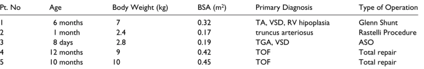
Table 1. The operative characteristics of the patients.
Pt. No Age Body Weight (kg) BSA (m2) Primary Diagnosis Type of Operation
1 6 months 7 0.32 TA, VSD, RV hipoplasia Glenn Shunt
2 1 month 2.4 0.17 truncus arteriosus Rastelli Procedure
3 8 days 2.8 0.19 TGA, VSD ASO
4 12 months 9 0.42 TOF Total repair
5 10 months 10 0.45 TOF Total repair
TA: Tricuspid atresia; VSD: Ventricular septal defect; RV: Right ventricle; TGA: Transposition of the great arteries; TOF: Tetralogy of Fallot; ASO: Arterial switch operation.
Table 2. The analysis of the ECMO duration.
Pt. No ECMO
implementation time
ECMO
on CPR Aortic Cannula (Fr) Flow (Lt/min/m2)
ECMO duration (hours)
Mortality
1 po. 5. day Yes 12 768 272 +
2 po. 6. day Yes 8 408 66 +
3 po. 3. day No 8 455 41 +
4 po.12. hour No 12 1008 63 –
5 early after CPB No 12 1080 166 –
PO: Postoperative; CPB: Cardiopulmonary bypass; CPR: Cardiopulmonary resuscitation.
by guest on March 17, 2015
prf.sagepub.com
242 Perfusion 30(3)
ECMO oxygenator because of limited funds and reim-bursements. We do not recommend replacing the rou-tine use of CPB oxygenators with PMP ECMO oxygenators. When funds are limited, then we recom-mend the use of hollow-fiber CPB oxygenators instead of PMP ones in neonatal ECMO patients, in whom even multiple replacements are more cost effective than using PMP ECMO oxygenators.
Although this report includes a limited number of patients, we wanted to share our experience with this setup. The safe period of the membrane oxygenators is obviously shorter than the spiral coil silicone mem-branes and PMP oxygenators which were originally invented for prolonged usages.7 We do not find it neces-sary to conduct a clinical trial in order to compare PMP ECMO oxygenators with hollow-fiber CPB oxygenat-ors, since our only thought was to create a solution in patients in this limited number of our patients where the funds were limited.
Conclusion
A centrifugal pump and a separate hollow-fiber oxy-genator may provide a cost-effective ECMO setup without plasma leakage, hemolysis and thromboem-bolic complications. This simple and low cost setup may encourage the usage of ECMO with extended indi-cations in the neonatal period, in particular, when the funds are limited.
Author Note
Presented at the 7th Symposium on Pediatric Extracorporeal Life Support Systems, September 28th, 2013; Istanbul, Turkey
Declaration of Conflicting Interest
The authors declare that there is no conflict of interest.
Funding
This research received no specific grant from any funding agency in the public, commercial, or not-for-profit sectors.
References
1. Undar A, Wang S, Palanzo D. Impact of polymethylpen-tene oxygenators on outcomes of all extracorporeal life support patients in the United States. Artif Organs 2013; 37: 1080–1081.
2. Extracorporeal Life Support Organization (ELSO). Extracorporeal Life Support Registry Report International Summary. Ann Arbor, MI: ELSO; 2013.
3. Yerebakan C, Ozyuksel A, Yildirim O et al. Extracorporeal membrane oxygenation for the treatment of acute res-piratory failure due to resres-piratory syncytial virus after congenital heart surgery. Wien Med Wochenschr 2013; 429–431.
4. Bryner BS, West BT, Hirschi RB et al. Congenital dia-phragmatic hernia requiring extracorporeal membrane oxygenation: dose timing of repair matter? J Pediatr Surg 2009; 44; 1165–1171.
5. Talor J, Yee S, Rider A et al. Comparison of perfusion quality in hollow-fiber membrane oxygenators for neo-natal extracorporeal life support. Artif Organs 2010; 34: E110–116. doi: 10.1111/j.1525–1594.2009.00971.x. 6. Lou S, Maclaren G, Best D et al. Hemolysis in
pediat-ric patients receiving centrifugal-pump extracorporeal membrane oxygenation: prevalence, risk factors, and outcomes. Crit Care Med. 2014; 42: 1213–1220.
7. Yu K, Long C, Hei F et al. Clinical evaluation of two dif-ferent extracorporeal membrane oxygenation systems: a single center report. Artif Organs 2011; 35: 733–737.
by guest on March 17, 2015
prf.sagepub.com
Downloaded from
View publication stats View publication stats