Low Vitamin D Deficiency Associated With Thyroid Disease
Among Type 2 Diabetic Mellitus Patients
Abdulbari Benera, b, c, g, Yasar Ozdenkayad, Abdulla O.A.A. Al-Hamaqe, Cem Cahit Barisikf, Mustafa Ozturkc
Abstract
Background: The aim of this study was to investigate the relation-ship between vitamin D deficiency and thyroid diseases among type 2 diabetes mellitus (T2DM) patients.
Methods: This was a cohort case and control study, 546 T2DM pa-tients and 546 control study participants were enrolled, aged between 25 and 65 years. The subjects were also investigated for fasting blood glucose levels (FBG), post prandial glucose (PPG,) glycosylated he-moglobin (HbA1c), thyroid stimulating hormone (TSH), T3, T4, and presence of other comorbid conditions. Thyroid fine needle aspiration biopsy was suggested to patients whose thyroid nodules were greater than 1.00 cm.
Results: There were significant differences between T2DM patients and control subjects regarding BMI (kg/m2), physical activity, ciga-rette smoking, sheesha smoking, family history of diabetes, hyperten-sion and family history of thyroid nodules. The clinical biochemistry values among T2DM for vitamin D, calcium, magnesium, potassium, phosphorous, fasting blood glucose, cholesterol, HbA1c, HLDL, LDL, triglyceride, systolic blood pressure (SBP) and diastolic blood pressure (DBP) were lower than control subjects, but higher in creati-nine, albumin, TSH, T3, and T4 which appeared statistically signifi-cant differences (P < 0.001). Also, the study revealed statistically sig-nificant differences between subjects vitamin D deficiency and with
thyroid nodules for calcium, magnesium, phosphorous, HbA1c, high density lipoprotein (HDL), SBP and DBP, TSH, T3, and T4 among T2DM patients and control subjects (P < 0.001). Multivariable step-wise logistic regression analysis showed that TSH, HbA1c, vitamin D deficiency, SBP (mm Hg), BMI, family history of DM, serum calcium level and family history of thyroid were considered at higher risk as predictors of thyroid among T2DM patients.
Conclusions: This study suggests that obesity, HbA1c, the environ-ment, and genetic susceptibility among T2DM, may increase the risk of thyroid disease and cancer. Although evidence has shown that thy-roid cancer incidence has been rising more rapidly over time than the occurrence of cancers of other sites, due to an increase of obesity, dia-betes and lack of physical activity, this study lacks of direct evidence supporting this conclusion.
Keywords: Vitamin D deficiency; Thyroid disease; TSH; HbA1C; T2DM; Prevention
Introduction
Type 2 diabetes mellitus (T2DM) and thyroid dysfunction are the main threats in developed and developing countries and impairs the health and economic status [1, 2]. T2DM increases the risk of coronary heart disease [3, 4] and thyroid dysfunc-tion [5-9] in the long-term. T2DM and thyroid dysfuncdysfunc-tion are the primary reasons for mortality and morbidity in most high-income and developing countries [5-9].
Thyroid disorders and diabetes are the two most widespread endocrinological medical conditions seen in general clinical medical practice [4]. Achieving good glycemic control prevents cardiovascular risk associated with diabetes [1-5]. However, sev-eral studies have shown a higher prevalence of thyroid dysfunc-tion occurring among T2DM patients, and vice versa [7-12].
Positive correlations between vitamin D deficiency and thyroid dysfunction among T2DM patients have been reported by several authors [11-15]. The impact of vitamin D deficiency on thyroid diseases which are highly correlated as the two most common endocrinological medical conditions was reported in clinical practice [13-15]. We designed the present study to test the hypothesis that lower vitamin D levels may be related to the occurrence of thyroid disease. This study is a cross-sec-tional case-control analysis in which we examined the levels
Manuscript submitted June 7, 2018, accepted June 18, 2018
aDepartment of Biostatistics and Medical Informatics, Cerrahpasa Faculty of
Medicine, Istanbul University, Istanbul, Turkey
bDepartment of Evidence for Population Health Unit, School of Epidemiology
and Health Sciences, The university of Manchester, Manchester, UK
cDepartment. of Endocrinology, Medipol International School of Medicine,
Istanbul Medipol University, Istanbul, Turkey
dQatar Diabetic Associations and Qatar National Research Foundation, Doha,
Qatar
eDepartment of Surgery, Medipol School of Medicine, Istanbul Medipol
Uni-versity, Istanbul, Turkey
fDepartment of Radiology and Pathology, Medipol School of Medicine,
Istan-bul Medipol University, IstanIstan-bul, Turkey
gCorresponding Author: Abdulbari Bener, Department of Biostatistics and
Medical Informatics, Cerrahpaşa Faculty of Medicine, Istanbul University and Istanbul Medipol University, International School of Medicine, 34098 Cerrah-pasa, Istanbul, Turkey. Email: abdulbari.bener@istanbul.edu.tr
of 25(OH)D in patients with thyroid among both T2DM and in controls. The aim of this study was to explore the association between vitamin D deficiency and thyroid dysfunction among T2DM patients.
Materials and Methods
The design of this study was a case and control. The study in-volved participants between the ages of 25 and 65 who visited the diabetes, endocrinology, thyroid surgery and outpatient clinics in the Mega Medipol International School of Medi-cine Hospital and Medipol Hospital from March 2016 to May 2018. This case and control study was based on 546 type 2 diabetic and 546 control subjects. IRB ethical approval for the present study was taken from the Medipol International School of Medicine, Istanbul Medipol University, and patients gave informed written consent before starting.
Laboratory measurements
Case patients were considered to have DM if they had a his-tory of DM and were currently taking any oral medications for diabetes. According to the World Health Organization (WHO) and the International Diabetes Federation (IDF) [1, 2], the criterion for diagnosing DM is when fasting venous blood glucose (FBG) concentration is equal to or higher than 7.0 mmol/L and/or postprandial blood glucose (PPG) concentra-tion is higher than 11.1 mmol/L [1, 3]. A subject was included in the control group (subjects without diabetes) if FBG was less than 7.0 mmol/L (126 mg/dL) and glycosylated hemo-globin (HbA1c) less than 48 mmol/mol (6.5%) and no pre-scribed diabetic medications were reported.
These subjects were also investigated for FBG, serum cho-lesterol, serum triglycerides, high density lipoprotein (HDL), low density lipoprotein (LDL), PPG, HbA1c, very low density lipoprotein (VLDL), blood urea, and serum creatinine.
Blood collection and serum measurements of vitamin D
The collected venous blood sample, and serum were separated and stored at -70 °C until examination. Serum 25-hydroxyvita-min D (25OHD), a vita25-hydroxyvita-min D metabolite, was measured using a commercially obtainable kit (DiaSorin Corporate Headquarter, Saluggia, Italy). The treated samples were then assayed using the competitive binding radioimmunoassay (RIA) technique. Subjects were classified into two categories as : 1) Vitamin D deficiency, 25(OH)D < 20 ng/mL; 2) Vitamin sufficiency, 25(OH)D > 20 ng/mL on the basis of previous recommenda-tions by Bener et al [3] and Mazokopakis and Kotsiris [14] and Holick et al [15].
Thyroid evaluation
A thyroid nodule is a lump that occurs from the abnormal
growth of thyroid cells, located at the base of the neck and pro-duces the hormones thyroxine and triiodothyronine [16-19]. Thyroid functioning was assessed by measuring thyroid stimu-lating hormone (TSH), free-T3 (FT3), and free-T4 (FT4), us-ing immunochemoluminescent assays by an automated ana-lyzer [20], (Immulite 2000; Diagnostic Products, Los Angeles, CA, USA). A 10-MHz linear probe (Logiq 5 Pro, GE Medical Systems, WI, USA) was used by same physician (CCB) for thyroid ultrasonography. TSH was measured by immunoassay [17]. The measurement of FT3 and FT4 are the most clinically relevant for the evaluation of thyroid disorders, with total thy-roid hormones being affected by variations in binding protein concentrations. It is essential that free thyroid hormone meas-urement accurately assess hormone concentration even in the presence of significant variation Thyroid fine needle aspiration biopsy was suggested to patients whose thyroid nodules were greater than 1.00 cm.
Questionnaire , physical examination and measurements
This study included several parameters such as socio-demo-graphic characteristics, lifestyle habits and biochemical test results. Patients were categorized as physically active if they walked or cycled for more than 30 min per day.
The data was analyzed using the Statistical Package for the Social Sciences (IBM SPSS Statistics for Windows, Ver-sion 22.0.). The significance of differences between mean val-ues of two continuous variables was determined by Student’s t-test for normally distributed data and by Mann-Whitney test for not normal distribution data. Chi-square and Fisher’s ex-act tests performed for significance differences between two or more categorical groups. Multivariate logistic regression analysis was performed to predict the risk factors for the pres-ence of thyroid nodules. The cut-off value for determining sig-nificance was chosen as 0.05.
Results
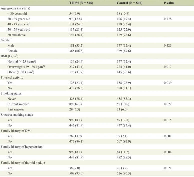
Table 1 presents socio-demographic and clinical characteris-tics of studied T2DM patients and control subjects. There were statistically significant differences between T2DM patients and control subjects regarding BMI, physical activity, cigarette smoking, sheesha smoking, family history of diabetes, hyper-tension and family history thyroid nodules.
Table 2 shows the clinical biochemistry baseline values among T2DM and control subjects. The clinical biochemistry values among T2DM for vitamin D (P < 0.001), calcium (P < 0.001), magnesium (P < 0.001), potassium (P = 0.012), phos-phorous (P < 0.001), fasting blood glucose (P < 0.001), choles-terol (P < 0.001), HbA1c (P < 0.001), HLDL (P < 0.001), LDL (P = 0.021), albumin (P < 0.001), triglyceride (P = 0.005), SBP (mm Hg) (P < 0.001) and diastolic blood pressure (DBP) (P < 0.001), TSH (P < 0.001), T3 (P < 0.001), and T4 (P < 0.001) were lower than control subjects and statistically significant differences (P < 0.001).
vi-tamin D deficiency and sufficiency with and without thyroid nodules among T2DM and control subject. There were statisti-cally significant differences between subjects vitamin D defi-ciency and suffidefi-ciency with and those without thyroid nodules for vitamin D (P < 0.001), calcium (P < 0.001), magnesium (P < 0.001), phosphorous (P < 0.001), HbA1c (P < 0.001), HDL (P < 0.001), SBP (P = 0.006) and, DBP (P = 0.005), TSH (P < 0.001), T3 (P < 0.001), and T4 (P < 0.001) among T2DM. Sim-ilarly, there were statistically significant differences between subjects vitamin D deficiency and sufficiency among control subjects for vitamin D (P < 0.001), calcium (P = 0.002), mag-nesium (P < 0.001), HbA1c (P < 0.001), FBG (P < 0.001), HDL (P < 0.001), bilirubin (P = 0.003) and, DBP (P = 0.027),
TSH (P < 0.001), T3 (P < 0.001), and T4 (P = 0.002).
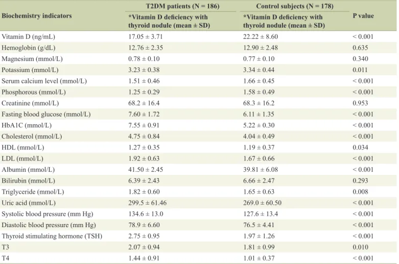
Table 4 presents clinical biochemistry baseline based on vitamin D deficiency level and with thyroid nodules among T2DM and control subject. There were statistically significant differences between both groups for vitamin D (P < 0.001), potassium (P = 0.011), calcium (P < 0.001), phosphorous (P < 0.001), fasting blood glucose (P < 0.001), HbA1c (P < 0.001), HDL (P = 0.034), LDL (P < 0.001), albumin (P < 0.001), tri-glyceride (P = 0.008), uric acid (P < 0.001), SBP (P = 0.006) and DBP (P < 0.001), TSH (P < 0.001), T3 (P = 0.010), and T4 (P < 0.001).
Table 5 gives multivariable stepwise logistic regression analysis of independent predictors for the presence of
thy-Table 1. Socio-Demographic and Clinical Characteristics of Studied T2DM Patients and Control Subjects (N = 1,092) T2DM (N = 546) Control (N = 546) P value Age groups (in years)
< 30 years old 54 (9.9) 58 (10.8) 30 - 39 years old 97 (17.8) 106 (19.4) 0.778 40 - 49 years old 134 (24.5) 128 (23.4) 50 - 59 years old 117 (21.4) 125 (22.9) 60 and above 144 (26.4) 129 (23.6) Gender Male 181 (33.2) 177 (32.4) 0.423 Female 365 (66.8) 369 (67.6) BMI (kg/m2) Normal (< 25 kg/m2) 136 (24.9) 177 (32.4) Overweight (29 - 30 kg/m2) 237 (43.4) 224 (41.0) 0.017 Obese (> 30 kg/m2) 173 (31.7) 145 (26.6) Physical activity Yes 128 (23.4) 158 (28.9) 0.039 No 418 (76.6) 388 (71.1) Smoking status Never 428 (78.4) 455 (83.3) Current smoker 89 (16.3) 58 (10.6) 0.022 Past smoker 29 (5.3) 33 (6.0)
Sheesha smoking status
Yes 99 (18.1) 69 (12.8) 0.015
No 447 (81.9) 477 (87.4)
Family history of DM
Yes 76 (13.9) 39 (7.1) 0.001
No 473 (86.1) 507 (92.9)
Family history of hypertension
Yes 99 (18.1) 64 (11.7) 0.004
No 447 (81.9) 482 (88.3)
Family history of thyroid nodule
Yes 38 (7.0) 20 (3.7) 0.021
roid nodules. TSH mIU/L (OR: 4.41, 95% CI: 3.22 - 5.61, P < 0.001), HbA1c (OR: 3.67, 95% CI: 2.84 - 4.92, P < 0.001), vi-tamin D deficiency (OR: 3.34 95% CI: 1.60 - 5.50, P < 0.001), SBP (mm Hg) (OR: 2.70, 95% CI: 2.39 - 3.10, P < 0.001), family history of T2DM (OR: 2.41, 95% CI: 2.13 - 3.87, P < 0.001), BMI (OR: 2.23, 95% CI: 1.94 - 3.06, P = 0.005), serum calcium level (mmol/L) (P = 0.008), and family history of thy-roid (P = 0.010) were considered at higher risk as predictors of thyroid among T2DM patients.
Discussion
There are very few studies which determine the relationship between vitamin D deficiency and thyroid diseases among T2DM with thyroid diseases patients and controls subject as a worldwide. Our study has revealed a higher prevalence vita-min D deficiency with thyroid diseases in T2DM patients com-pared to control subjects. It has been recognized that vitamin D deficiency is a global major public health problem worldwide. In the current study, we conducted a matched case-control study to explore the relationship between vitamin D and thy-roid disease among T2DM patients. Vitamin D deficiency has been correlated with thyroid antibody levels in both adult [18, 19] and children populations [20]. This is consistent with
cur-rent study outcome.
Also, vitamin D deficiency and T2DM are usually recog-nized as a complication and risk for thyroid disease. There-fore, effective controls of vitamin D and T2DM are essential to reduce the occurrence of thyroid diseases in the middle age group and may affect the quality of life. Further, the current subjects with T2DM also had a higher prevalence and larger thyroid nodules size which is consistent with a previous re-ported a positive correlation between insulin resistance and thyroid nodule size [4-10]. The present study demonstrated very low mean vitamin D level deficiency with thyroid nod-ule compared to vitamin D sufficiency without thyroid nodnod-ule. This is confirmative with the previous reported studies [11, 12, 18, 19].
In fact, T2DM and thyroid diseases [5-9] are highly cor-related as the two commonest endocrinological medical con-ditions reported and linked with the vitamin D deficiency in general clinical practice [10-15]. Further, several studies re-ported that thyroid volume is related to a variety of risk fac-tors such as iodine deficiency and supply, BMI, age, gender, smoking status, genetic factors, impaired fasting glucose and diabetes mellitus [3, 19-21]. Additionally, a study has investi-gated the role of environmental and lifestyle factors [3]. It is worth to note that the possible role of vitamin D insufficiency/ deficiency can be considered in the pathogenesis of both DM
Table 2. Clinical Biochemistry Baseline Value Among T2DM and Control Subjects (N = 1,092)
Variables T2DM (N = 546, mean ± SD) Control (N = 546, mean ± SD) P value
Vitamin D (ng/mL) 17.24 ± 3.71 23.67 ± 5.46 0.001 Hemoglobin (g/dL) 12.53 ± 2.45 13.10 ± 2.52 0.276 Magnesium (mmol/L) 0.76 ± 0.09 0.90 ± 0.10 < 0.001 Potassium (mmol/L) 3.39 ± 0.46 3.77 ± 0.53 < 0.001 Calcium (mmol/L) 1.63 ± 0.56 1.75 ± 0.68 0.001 Phosphorous (mmol/L) 1.46 ± 0.62 1.59 ± 0.52 0.038 Creatinine (mmol/L) 68.67 ± 16.17 67.33 ± 16.84 0.101
Fasting blood glucose (mmol/L) 7.49 ± 1.89 6.50 ± 1.98 < 0.001
HbA1C (mmol/L) 7.32 ± 0.97 5.29 ± 0.62 < 0.001 Cholesterol (mmol/L) 4.69 ± 0.71 4.03 ± 0.50 < 0.001 HDL (mmol/L) 0.96 ± 0.21 1.14 ± 0.34 0.911 LDL (mmol/L) 1.63 ± 0.64 1.54 ± 0.61 0.002 Albumin (mmol/L) 41.48 ± 5.57 39.81 ± 5.54 < 0.001 Bilirubin (mmol/L) 6.98 ± 2.43 6.99 ± 2.54 0.015 Triglyceride (mmol/L) 1.71 ± 0.69 1.59 ± 0.68 < 0.001
Uric acid (mmol/L) 278.1 ± 64.57 266.9 ± 61.7 < 0.001
Systolic blood pressure (mm Hg) 130.7 ± 12.9 126.8 ± 12.2 0.009
Diastolic blood pressure (mm Hg) 77.6 ± 6.8 75.9 ± 4.86 0.002
Thyroid stimulating hormone (TSH) 2.58 ± 1.06 2.31 ± 1.19 < 0.001
T3 1.89 ± 1.06 1.57 ± 0.93 < 0.001
T4 1.19 ± 0.70 0.97 ± 0.35 < 0.001
TSH (reference range: 0.35 - 4.0 mIU/L), free triiodothyronine (FT3) (reference range: 1.71 - 4.71 pg/mL), and free thyroxine (FT4) (reference range: 0.8 - 1.9 ng/dL).
and thyroid disease. However, vitamin D deficiency could be also secondary to these diseases. Oral anti-diabetic medica-tions as well as therapeutic dietary restriction could affect vi-tamin D levels in patients with diabetes. In addition, thyroid dysfunction could also modify vitamin D intake, absorption or metabolism.
In a few recent studies, thyroid cancer risk factors includ-ing iodine deficiency, environmental, genetic, family history of DM and hypertension and lifestyle factors [19-22]. Since the complex interplay between vitamin D and thyroid autoimmun-ity in the context of T2DM has been investigated more recently [22], this confirmative with the present study hypothesis could be substantiated on the basis of our data results. Thyroid can-cer incidence has been raising very rapidly over time than the occurrence of cancers of other sites, due to increase of obesity, diabetes and lack of physical activity leading to increase risk of thyroid cancer [23-25]. Our results were similar to previous studies particularly that vitamin D deficiency was associated with an increased risk of thyroid disease among T2DM [19, 22, 26].
Furthermore, increased thyroid cancer risk in diabetics
might be related to several factors including, abnormal HbA1C and metabolic syndrome, triglyceride levels, obesity, dietary, and lifestyle [7]. Obese subjects were at 10 times more risk of developing diabetes [23], and obesity was associated with an increased risk of thyroid cancer [25]. Our sample demonstrat-ed significant prevalence of thyroid diseases among T2DM compared to controls. Finally, this study confirms previous re-ported studies [18, 19] to determine the correlations between vitamin D deficiency and thyroid diseases in diabetic patients in Turkish population and also indicates T2DM patients whose prevalence is very poorly described in earlier studies [11, 18].
Limitations and strengths of study
The study has several strengths, but also some weaknesses. The present study has several limitations. Firstly, the sample might be partially biased which may not be ideal for a matched case and control study. This is due to the possibility of selec-tion bias as subjects were selected from patients who visited a tertiary hospital. Another limitation pertains to the
unavail-Table 3. Clinical Biochemistry Baseline Based on Vitamin D Deficiency and Sufficiency With and Without Thyroid Nodules Among T2DM and Control Subject (N = 1,092)
Biochemistry indicators
T2DM patients(N = 546) Control subjects(N = 546) *Vitamin D
defi-ciency with thy-roid nodule (N = 186, mean ± SD) **Vitamin D suf-ficiency without thyroid nodule (N = 360, mean ± SD) P value *Vitamin D defi-ciency with thy-roid nodule (N = 178, mean ± SD) **Vitamin D suf-ficiency without thyroid nodule (N = 368, mean ± SD) P value Vitamin D (ng/mL) 17.05 ± 3.71 21.76 ± 4.96 0.001 22.22 ± 8.60 25.43 ± 9.07 < 0.001 Hemoglobin (g/dL) 12.76 ± 2.35 13.09 ± 2.48 0.248 12.90 ± 2.48 13.10 ± 2.56 0.275 Magnesium (mmol/L) 0.78 ± 0.10 0.92 ± 0.11 < 0.001 0.77 ± 0.10 0.89 ± 0.13 < 0.001 Potassium (mmol/L) 3.23 ± 0.38 3.47 ± 0.49 0.001 3.34 ± 0.44 3.58 ± 0.60 < 0.001 Serum calcium level (mmol/L) 1.51 ± 0.46 1.70 ± 0.60 < 0.001 1.66 ± 0.45 1.84 ± 0.84 0.002 Phosphorous (mmol/L) 1.25 ± 0.29 1.58 ± 0.71 0.001 1.58 ± 0.49 1.60 ± 0.99 0.983
Creatinine(mmol/L) 68.2 ± 16.4 68.1 ± 16.7 0.642 68.3 ± 16.2 67.2 ± 17.5 0.492
Fasting blood glucose(mmol/L) 7.60 ± 1.72 7.43 ± 1.96 0.351 6.11 ± 1.35 6.88 ± 2.37 < 0.001
HbA1C (mmol/L) 7.55 ± 0.91 7.19 ± 0.97 0.001 5.22 ± 0.30 5.14 ± 0.32 0.002 Cholesterol (mmol/L) 4.75 ± 0.84 4.65 ± 0.64 0.117 4.04 ± 0.49 4.00 ± 0.51 0.847 HDL (mmol/L) 1.27 ± 0.35 1.45 ± 0.25 0.001 1.19 ± 0.37 1.08 ± 0.30 < 0.001 LDL (mmol/L) 1.92 ± 0.63 1.54 ± 0.65 0.045 1.67 ± 0.66 1.41 ± 0.60 < 0.001 Albumin (mmol/L) 41.50 ± 2.45 40.75 ± 3.92 0.386 39.81 ± 6.08 40.75 ± 3.92 0.470 Bilirubin (mmol/L) 6.39 ± 2.43 7.28 ± 2.38 0.253 6.66 ± 2.47 7.31 ± 2.58 0.003 Triglyceride (mmol/L) 1.82 ± 0.60 1.50 ± 0.72 0.036 1.65 ± 0.63 1.154 ± 0.71 0.083 Uric acid (mmol/L) 299.5 ± 61.46 280.1 ± 64.85 < 0.001 269.0 ± 60.50 264.9 ± 62.90 0.444 Systolic blood pressure (mm Hg) 134.6 ± 13.0 131.31 ± 13.6 0.006 127.6 ± 13.4 126.2 ± 130.76 0.139 Diastolic blood pressure (mm Hg) 78.9 ± 6.60 77.2 ± 6. 7 < 0.001 76.5 ± 4.41 75.5 ± 5.21 0.027 Thyroid stimulating hormone (TSH) 2.75 ± 0.95 2.40 ± 1.05 0.005 1.97 ± 1.26 2.65 ± 1.02 < 0.001
T3 2.07 ± 0.94 1.81 ± 1.10 < 0.001 1.81 ± 0.99 1.33 ± 0.81 < 0.001
T4 1.44 ± 0.91 1.07 ± 0.52 < 0.001 1.01 ± 0.37 0.92 ± 0.32 0.002
ability of data on the frequency of thyroid examinations in our study population. Lastly, there was no cytological or histologi-cal results for each nodule. The greatest strengths of this study are the very large sample of participants and the large data-set of risk factor variables such as TSH, T3, T4, body weight, physical activity, smoking cigarette, diet, BMI, family history of diabetes, hypertension and thyroid. Furthermore, the dis-crimination between the T2DM subjects and the control group
was based on fasting blood glucose and HbA1c measurements, which secured a clear distinction between both groups.
Conclusions
This study suggests that obesity, HbA1c, the environment, and genetic susceptibility among T2DM may increase the risk of
Table 4. Clinical Biochemistry Baseline Based on Vitamin D Deficiency With Thyroid Nodules Among T2DM and Compared to Control Subject (N = 364)
Biochemistry indicators *Vitamin D deficiency with T2DM patients (N = 186) Control subjects (N = 178) P value thyroid nodule (mean ± SD) *Vitamin D deficiency with thyroid nodule (mean ± SD)
Vitamin D (ng/mL) 17.05 ± 3.71 22.22 ± 8.60 < 0.001
Hemoglobin (g/dL) 12.76 ± 2.35 12.90 ± 2.48 0.635
Magnesium (mmol/L) 0.78 ± 0.10 0.77 ± 0.10 0.340
Potassium (mmol/L) 3.23 ± 0.38 3.34 ± 0.44 0.011
Serum calcium level (mmol/L) 1.51 ± 0.46 1.66 ± 0.45 < 0.001
Phosphorous (mmol/L) 1.25 ± 0.29 1.58 ± 0.49 < 0.001
Creatinine (mmol/L) 68.2 ± 16.4 68.3 ± 16.2 0.953
Fasting blood glucose (mmol/L) 7.60 ± 1.72 6.11 ± 1.35 < 0.001
HbA1C (mmol/L) 7.55 ± 0.91 5.22 ± 0.30 < 0.001 Cholesterol (mmol/L) 4.75 ± 0.84 4.04 ± 0.49 < 0.001 HDL (mmol/L) 1.27 ± 0.35 1.19 ± 0.37 0.034 LDL (mmol/L) 1.92 ± 0.63 1.67 ± 0.66 < 0.001 Albumin (mmol/L) 41.50 ± 2.45 39.81 ± 6.08 < 0.001 Bilirubin (mmol/L) 6.39 ± 2.43 6.66 ± 2.47 0.293 Triglyceride (mmol/L) 1.82 ± 0.60 1.65 ± 0.63 0.008
Uric acid (mmol/L) 299.5 ± 61.46 269.0 ± 60.50 < 0.001
Systolic blood pressure (mm Hg) 134.6 ± 13.0 127.6 ± 13.4 < 0.001
Diastolic blood pressure (mm Hg) 78.9 ± 6.60 76.5 ± 4.41 < 0.001
Thyroid stimulating hormone (TSH) 2.75 ± 0.95 1.97 ± 1.26 < 0.001
T3 2.07 ± 0.94 1.81 ± 0.99 0.010
T4 1.44 ± 0.91 1.01 ± 0.37 < 0.001
*Vitamin D deficiency: 25(OH)D level < 20 ng/mL.
Table 5. Multivariate Logistic Regression Analysis for Predictors Presence of Thyroid Disorder Among T2DM Patients (N = 1,092) Independent variables Adjusted odds ratio 95% Confidence interval P value
TSH, mIU/L 4.41 3.22 - 5.61 < 0.001
HbA1C (mmol/L) 3.67 2.84 - 4.92 < 0.001
Vitamin D deficiency 3.34 1.60 - 5.50 < 0.001
Systolic blood pressure, mm Hg 2.7 2.39 - 3.10 < 0.001
Family history of T2DM 2.41 2.13 - 3.87 < 0.001
BMI ( kg/m2) 2.23 1.94 - 3.06 0.005
Serum calcium level (mmol/L) 1.98 1.66 - 2.84 0.008
thyroid disease and cancer. There is a strong positive associa-tions between the vitamin D deficiency and thyroid diseases population among T2DM patients. This study reveals that an increase in thyroid diseases might be caused by increases in metabolic syndrome, vitamin D deficiency, HbA1C, diabetes and obesity. Although evidence has shown that thyroid cancer incidence has been rising more rapidly over time than the oc-currence of cancers of other sites due to an increase of obesity, diabetes and lack of physical activity, this study lacks of direct evidence supporting this conclusion.
Acknowledgments
This work was generously supported and funded by the Qatar Diabetes Association, Qatar Foundation. The authors would like to thank the Istanbul Medipol University for their support and ethical approval (Research Protocol and IRB# 10840098-604.01.01-E.8421).
Author Contributions
AB, MO and YO designed and supervised the study and were involved in data collection, statistical analysis and the writing of the paper. AOAA, CCB were involved in study designed, interpretation of data and writing of the manuscript. All au-thors approved the final version.
Conflict of Interest
No conflict of interest was declared by the authors.
Financial Support
The authors declared that this study has received no financial support.
References
1. Alberti KG, Eckel RH, Grundy SM, Zimmet PZ, Clee-man JI, Donato KA, Fruchart JC, et al. Harmonizing the metabolic syndrome: a joint interim statement of the In-ternational Diabetes Federation Task Force on Epidemi-ology and Prevention; National Heart, Lung, and Blood Institute; American Heart Association; World Heart Fed-eration; International Atherosclerosis Society; and Inter-national Association for the Study of Obesity. Circula-tion. 2009;120(16):1640-1645.
2. Bener A, Zirie M, Musallam M, Khader YS, Al-Hamaq AO. Prevalence of metabolic syndrome according to Adult Treatment Panel III and International Diabetes Fed-eration criteria: a population-based study. Metab Syndr Relat Disord. 2009;7(3):221-229.
3. Bener A, Al-Hamaq AO, Kurtulus EM, Abdullatef WK,
Zirie M. The role of vitamin D, obesity and physical exer-cise in regulation of glycemia in Type 2 Diabetes Mellitus patients. Diabetes Metab Syndr. 2016;10(4):198-204. 4. Al-Wazzan HT, Daban AH, Askar RA. El-Shazly MK.
Prevalence and associated factors of thyroid dysfunc-tion among type 2 diabetic patients in Kuwait. Alex Bull. 2010;46(2):141-148.
5. Papazafiropoulou A, Sotiropoulos A, Kokolaki A, Kard-ara M, Stamataki P, Pappas S. Prevalence of thyroid dys-function among greek type 2 diabetic patients attending an outpatient clinic. J Clin Med Res. 2010;2(2):75-78. 6. Akbar DH, Ahmed MM, Al-Mughales J. Thyroid
dys-function and thyroid autoimmunity in Saudi type 2 dia-betics. Acta Diabetol. 2006;43(1):14-18.
7. Bener A, Ozdenkaya Y, Barişik CC, Ozturk M. The im-pact of metabolic syndrome on increased risk of thyroid nodules and size. Health Service Res Man Epidemiology. 2018;5:1-6.
8. Sarfo-Kantanka O, Sarfo FS, Ansah EO, Yorke E, Ak-palu J, Nkum BC, Eghan B. Frequency and determi-nants of thyroid autoimmunity in Ghanaian type 2 dia-betes patients: a case-control study. BMC Endocr Disord. 2017;17(1):2.
9. Centeno Maxzud M, Gomez Rasjido L, Fregenal M, Ari-as Calafiore F, Cordoba Lanus M, D'Urso M, Luciardi H. [Prevalence of thyroid dysfunction in patients with type 2 diabetes mellitus]. Medicina (B Aires). 2016;76(6):355-358.
10. Van Belle TL, Gysemans C, Mathieu C. Vitamin D and diabetes: the odd couple. Trends Endocrinol Metab. 2013;24(11):561-568.
11. Bozkurt NC, Karbek B, Ucan B, Sahin M, Cakal E, Oz-bek M, Delibasi T. The association between severity of vitamin D deficiency and Hashimoto's thyroiditis. Endocr Pract. 2013;19(3):479-484.
12. Kim D. Low vitamin D status is associated with hypo-thyroid Hashimoto's hypo-thyroiditis. Hormones (Athens). 2016;15(3):385-393.
13. Kivity S, Agmon-Levin N, Zisappl M, Shapira Y, Nagy EV, Danko K, Szekanecz Z, et al. Vitamin D and autoim-mune thyroid diseases. Cell Mol Immunol. 2011;8(3):243-247.
14. Mazokopakis EE, Kotsiris DA. Hashimoto's autoimmune thyroiditis and vitamin D deficiency. Current aspects. Hell J Nucl Med. 2014;17(1):37-40.
15. Holick MF. Vitamin D deficiency. N Engl J Med. 2007;357(3):266-281.
16. Brunn J, Block U, Ruf G, Bos I, Kunze WP, Scriba PC. [Volumetric analysis of thyroid lobes by real-time ul-trasound (author's transl)]. Dtsch Med Wochenschr. 1981;106(41):1338-1340.
17. Gharib H, Tuttle RM, Baskin HJ, Fish LH, Singer PA, McDermott MT. Subclinical thyroid dysfunction: a joint statement on management from the American Association of Clinical Endocrinologists, the American Thyroid As-sociation, and the Endocrine Society. J Clin Endocrinol Metab. 2005;90(1):581-585; discussion 586-587.
18. Camurdan OM, Doger E, Bideci A, Celik N, Cinaz P. Vitamin D status in children with Hashimoto thyroiditis.
J Pediatr Endocrinol Metab. 2012;25(5-6):467-470. 19. Shin DY, Kim KJ, Kim D, Hwang S, Lee EJ. Low
se-rum vitamin D is associated with anti-thyroid peroxi-dase antibody in autoimmune thyroiditis. Yonsei Med J. 2014;55(2):476-481.
20. Welsh KJ, Soldin SJ. Diagnosis of endocrine disease: How reliable are free thyroid and total T3 hormone as-says? Eur J Endocrinol. 2016;175(6):R255-R263. 21. Palma CC, Pavesi M, Nogueira VG, Clemente EL,
Vas-concellos Mde F, Pereira LCJ, Pacheco FF, et al. Preva-lence of thyroid dysfunction in patients with diabetes mellitus. Diabetol Metab Syndr. 2013;5(1):58.
22. Toulis K, Tsekmekidou X, Potolidis E, Didangelos T, Gotzamani-Psarrakou A, Zebekakis P, Daniilidis M, et al. Thyroid autoimmunity in the context of type 2 diabetes mellitus: implications for Vitamin D. Int J Endocrinol. 2015;2015:710363.
23. Aschebrook-Kilfoy B, Sabra MM, Brenner A, Moore SC, Ron E, Schatzkin A, Hollenbeck A, et al. Diabetes and thyroid cancer risk in the National Institutes of Health-AARP Diet and Health Study. Thyroid. 2011;21(9):957-963.
24. Schmid D, Behrens G, Jochem C, Keimling M, Leitz-mann M. Physical activity, diabetes, and risk of thyroid cancer: a systematic review and meta-analysis. Eur J Epi-demiol. 2013;28(12):945-958.
25. Leitzmann MF, Brenner A, Moore SC, Koebnick C, Park Y, Hollenbeck A, Schatzkin A, et al. Prospective study of body mass index, physical activity and thyroid cancer. Int J Cancer. 2010;126(12):2947-2956.
26. D'Aurizio F, Villalta D, Metus P, Doretto P, Tozzo-li R. Is vitamin D a player or not in the pathophysiol-ogy of autoimmune thyroid diseases? Autoimmun Rev. 2015;14(5):363-369.

