Radyoloji / Radiology ARAŞTIRMA YAZISI / ORIGINAL ARTICLE
500
https://doi.org/10.31067/0.2019.182 ACU Sağlık Bil Derg 2019; 10(3):500-503
1Baskent University Faculty of
Medicine, Department of Radiology, Izmir, Turkey
2Baskent University Faculty
of Medicine, Department of Ophthalmology, Izmir, Turkey
3Baskent University Faculty of
Medicine, Department of Radiology, Ankara, Turkey
4Selcuk University, Department of
Biostatistics, Konya, Turkey
Elçin Aydın, Assis. Prof. Hasan Yerli, Prof. Sinan Emre, Prof.
Işıl Kurultay Ersan, MD, Specialiast Muhteşem Ağıldere, Prof. Mustafa Agah Tekindal, Assoc. Prof.
Eye Elasticity in Patients With
Primary Open Angle Glaucoma
Elçin Aydın1 , Hasan Yerli1 , Sinan Emre2 , Işıl Kurultay Ersan2 ,
Muhteşem Ağıldere3 , Mustafa Agah Tekindal4
ABSTRACT
Purpose: The present study was performed to assess elasticity changes in the ocular and periocular structures of patients with primary open angle glaucoma (POAG) via sonoelastography.
Patients and Methods: Forty-three patients (19 male, 24 female; ranging from 45 to 80, mean age, 63.20 ± 2.31 years) diagnosed with POAG were prospectively analyzed. This patient group was compared with a control group of 28 individuals (17 male, 11 female; ranging from 40 to 85, mean age, 63.50 ± 1.89 years) who had no eye diseases apart from refractive errors. Following an ophthalmologic assessment and B-mode sonography, all participants underwent semiquantitative sonoelastographic analysis. The elasticity of the retrobulbar fat tissue, optic nerve, lens, and retina–choroid–sclera complex (RCSC) was compared between the two groups via sonoelastography. Student’s t test and the Mann–Whitney U test were used for statistical assessment.
Results: There was no significant differences in the degree of elasticity of the ocular and periocular structures between patients with and without POAG.
Conclusion: No difference was detected between elasticity levels of ocular and periocular structures in between patients with and without POAG.
Keywords: Glaucoma, elastography, ultrasonography
PRİMER AÇIK AÇILI GLOKOMLU HASTALARDA GÖZ ELASTİSİTESİ ÖZET
Amaç: Bu çalışmada primer açık açılı glokom (PAAG) hastalarının oküler ve perioküler yapılarının elastisite deği-şikliklerinin sonoelastografi ile değerlendirilmesi amaçlandı.
Hastalar ve Yöntem: PAAG’li 43 hasta (19 erkek, 24 kadın; 45-80 arası, ortalama yaş, 63.20 ± 2.31 yıl) prospektif olarak incelendi. Bu hasta grubu refraksiyon kusuru dışında göz hastalığı olmayan 28 birey (17 erkek, 11 kadın, 40-85 yaş arası, ortalama yaş, 63.50 ± 1.89 yıl) kontrol grubu ile karşılaştırıldı. Oftalmolojik değerlendirme ve B-mod ultrasonografi sonrasında, tüm katılımcılara semikuantatif sonoelastografik analiz yapıldı. Retrobulber yağ dokusu, optik sinir, lens ve retina-koroid-sklera kompleksinin (RKSK) elastikliği iki grup arasında sonoelastog-rafi ile karşılaştırıldı. Student t-test ve Mann–Whitney U testi istatistik değerlendirme için kullanıldı.
Bulgular: PAAG’li hastalar ve kontrol grubu arasında ölçülen perioküler ve oküler parametrelerde ortalama strain oranı belirgin farklılık göstermemektedir.
Sonuç: Elastisite oranları perioküler ve oküler yapılarda PAAG ve PAAG olmayan hastalar arasında farklılık göster-memektedir.
Anahtar sözcükler: Glokom, elastografi, ultrasonografi
Correspondence: Assis. Prof. Elçin Aydın
Baskent University Faculty of Medicine, Department of Radiology, Izmir, Turkey Phone: +90 532 641 91 64 E-mail: [email protected]
Received : December 14, 2017 Revised : January 14, 2018 Accepted : January 15, 2018
Aydın E et al.
501
ACU Sağlık Bil Derg 2019; 10(3):500-503
G
laucoma is a multifactorial chronic opticneuro-pathy characterized by a progressive loss in retinal ganglion cells and axons. In glaucoma patients, thinning of the retinal nerve fiber and changing in the optic nerve endings are observed. Ganglion cell damage predominantly takes place in the axons at the lamina crib-rosa level (1-4). Changes in the optic disk are characteri-zed by glaucoma(or characteristics of glaucoma), such as increased cup/disk ratio, bleeding or bisected disc and it cannot be easily differentiated by the naked eye (5,7). Sonoelastography is an advanced technique that measures elasticity of a tissue using sound waves processed by a soft-ware program. This technique has proved to contribute to the successful differentiation of malign(malignant) and benign lesions in numerous studies on breast, thyroid and prostate tissues, as malign(malignant) tissues have a hard-er texture (8,9). Thhard-ere is a limited numbhard-er of studies in the field of ophthalmology. It is suggested in the literature that there may be changes in the elasticity of the ocular and periocular structures in diabetic patients following panreti-nal photocoagulation and in ocular structures in highly my-opic patients (10,11). In a study on a group of patients who have lost their visions due to glaucoma, the retrobulber fat tissue strain value was significantly higher than the respec-tive value for the optic nerve (12). In our study, we analyzed the contribution of sonoelastography in foreseeing the prognosis of the disease and patientfollow-up by identi-fying the potential changes in the elasticity of retina-cho-roid-sclera complex (RCSC), optic nerve (ON), retrobulber fat tissue (RF) and lens (L) in primary open angle glaucoma (POAG) patients without visual impairments.
Materials and methods
PatientsForty three POAG patients (19 males, 24 females; mean age 63.2, ranging from 45 to 80) treated in the ophthalmology department of Baskent University hospital were included in the study. Twenty eight patients (11 females, 17 males and mean age 63.5, ranging from 40 to 85) who were found to have no additional ophthalmologic or systemic disease other than refractive errors (<-6 D myopia or <+3 D hyper-opia) in the ophthalmologic examination were selected for the control group. The project was approved by Baskent University Ethics Committee (Project number: KA15/359) and informed consents were received from all patients.
B-mode Sonography and Sonoelastography
B-mode sonography and sonoelastography were per-formed in a single (right) eye with standard ophthalmologic
assessment. Sonoelastography measurements were read by an experienced observer. The participants were ex-amined in the supine position. All images were obtained via sonography equipment (Arietta V70, Hitachi-Aloka Medical, Japan) with 7-13 MHz high-resolution probe. Following B-mode sonography, strain elastography meth-od was used for the assessment of RF, ON, L and RCSC elasticity. The US probe was placed in contact with the eyelid, and rhythmic, small compressions were applied manually by the operator. Sonoelastography was dis-played via a color-coded image in a selected rectangular area superimposed over the B-mode US image obtained simultaneously. A compression was performed by creat-ing sinusoidal waves to identify the response of the tis-sue to the compression where the whole site is observed in color. Strain ratios of two tissues observed of similar depth were measured in percentage. Elasticity ratios of eye structures against each other were measured by using the region of interest (ROI). The elasticity image was dis-played with color mapping for each pixel according to the degree of strain with the ROI. On the color scale, red color represented the softest component, while green and blue color represented the intermediate and hardest compo-nents, respectively (Figure 1 and 2). Measurements were recorded in a transverse section through the equator of the eyeball. The ON was identified and used as a landmark for the imaging of ocular and orbital structures, including the RF and posterior RCSC. The L was used as a landmark for comparing the elasticity of the anterior RCSC. It was used for ratio analysis, including anterior RCSC in the son-oelastography screen. Strain elastography ratios for both groups were measured: ON/RF, optic nerve to RCSC on the right of the optic nerve (ON/RCSC1), optic nerve to RCSC
Figure 1. Transverse B scan image (a) and respective grayscale ultrasound elastographic image (b) of eye. Optic nerve (ON), retrobulbar fat tissue (RF), choroid-sclera complex to the right of the optic nerve (RCSC1), retina-choroid-sclera complex (RCSC2) to the left of the optic nerve.
Eye Elasticity in Patients with Glaucoma
502 ACU Sağlık Bil Derg 2019; 10(3):500-503 on the left of the optic nerve (ON/RCSC2), and ratios of
RCSCs against each other on both sides of the ON (RCSC1/ RCSC2), L/anterior RCSC (L/RCSC3). The semi-quantitative values obtained as a result of the procedure were com-pared to the control group of similar age group.
Statistical analysis
For discrete and continuous variables, descriptive statis-tics (mean, standard deviation, median, minimum value, maximum value, and percentile) were given. In addition, the homogeneity of the variances, which is one of the prerequisites of parametric tests, was checked through Levene’s test. The assumption of normality was tested via the Shapiro-Wilk test. To compare the differences be-tween the two groups, the Student’s t test was used when the parametric test prerequisites were fulfilled, and the Mann Whitney–U test was used when such prerequisites
were not fulfilled. The data were evaluated via SPPS 20 (IBM Corp. Released 2011. IBM SPSS Statistics for Windows, Version 20.0. Armonk, NY: IBM Corp.). p<0.05 and p<0.01 were taken as significance levels.
Results
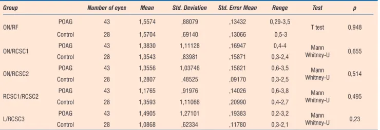
The tests applied to both groups and their results are presented in Table 1. In glaucoma patients, mean strain elastography ratios for ON/RF, ON/RCSC1, ON/RCSC2, RCSC1/RCSC2, L/RCSC3 were measured as 1,55 ±0,88 (0,29-3,5), 1,38±1,11 (0,4-4), 1,35±1,03 (0,6-3,5), 1,17± 0,91 (0,6-3,8), and 1,49±1,27 (0,2-3,2) respectively. Same ratios were measured in the control group as 1,57±0,69 (0,5-3), 1,35±0,83 (0,3-2,4), 1,28±0,48 (0,3-2,5), 1,35±1,11 (0,4-2,7), and 1,08±0,62 (0,3-2,1) respectively. These results did not reveal a statistically significant difference in strain elas-tography ratios for ON/RF, ON/RCSC1, ON/RCSC2, RCSC1/ RCSC2, L/RCSC3 between POAG patients and the control group (p>0.05).
Discussion
In our study, we used the strain ratio elastography meth-od to foresee the certain potential elasticity changes in the ocular and periocular structures in the early phases of POAG. Strain ratio elastography is a semi-quantitative method that reveals the hardness of a tissue by compar-ing the difference between the compliances of target area A and target area B. In strain elastography, a single tissue is chosen as the reference point. The difference between strain values of tissues that are assumed to be diseased is calculated according to the reference tissue (8-13).
Figure 2. Transverse B scan image (a) and respective grayscale ultrasound elastographic image (b) of eye. Anterior retina-choroid-sclera complex (RCSC3): C, lens (L).
Table 1. The mean strain elastıgraphy ratios
Group Number of eyes Mean Std. Deviation Std. Error Mean Range Test p
ON/RF POAG 43 1,5574 ,88079 ,13432 0,29-3,5 T test 0,948
Control 28 1,5704 ,69140 ,13066 0,5-3
ON/RCSC1 POAG 43 1,3830 1,11128 ,16947 0,4-4 Mann
Whitney-U 0,655
Control 28 1,3543 ,83981 ,15871 0,3-2,4
ON/RCSC2 POAG 43 1,3556 1,03746 ,15821 0,6-3,5 Whitney-UMann 0,514
Control 28 1,2807 ,48525 ,09170 0,3-2,5
RCSC1/RCSC2 POAG 43 1,1765 ,91976 ,14026 0,6-3,8 Whitney-UMann 0,495
Control 28 1,3593 1,11066 ,20990 0,4-2,7
L/RCSC3 POAG 43 1,4905 1,27101 ,19383 0,2-3,2 Whitney-UMann 0,23
Control 28 1,0868 ,62334 ,11780 0,3-2,1
Optic nerve/retrobulbar fat tissue (ON/RF), optic nerve/choroid-sclera complex (ON/RCSC1) to the right of the optic nerve, retina- optic nerve/ retina-choroid sclera complex (ON/RCSC2) to the left of the optic nerve, ratio of retina-choroid-sclera complexes (RCSC1/RCSC2) on both sides of the optic nerve, lens/ anterior retina-choroid-sclera complex (L/RCSC3)
Aydın E et al.
503
ACU Sağlık Bil Derg 2019; 10(3):500-503 Pekel et al.’s study (10) on diabetic patients with
retinopa-thy using strain ratio elastography method suggests that panretinal photocoagulation decreases RCSC elasticity. Another sonoelastography study by Pekel et al. on high myopia patients, high myopia was found to be related with more elastic posterior vitreous. The elasticity of the posteri-or RCSC increases as the axial length is increased (11). Among studies using the strain ratio elastography meth-od, the study by Vural et al. on 14 patients have found that RF and ON strain ratios were different in the patient group with sight impairment due to glaucoma compared to the control group (12). In their study, Detorakis et al. have as-sessed 5 patients who have gone blind in a single eye for various reasons (2 optic neuropathy, 2 absolute glaucoma, 1 retinal detachment). In this study, the posterior vitreous displayed a low level of elasticity while the anterior vitre-ous displayed an intermediate level of elasticity, whereas the medial and lateral rectus muscle elasticity was high-er in primary position compared to the adduction or ab-duction positions (13). Meanwhile in our study, a larger patient population under glaucoma treatment without a loss of vision due to glaucoma has been compared with a control group of similar age group with no other chronic References
1. Quigley HA, Addicks EM, Green WR. Optic nerve damage in human glaucoma III. Quantitative correlation of nerve fiber loss and visual field defect in glaucoma, ischemic neuropathy, papilledema, and toxic neuropathy. Arch Ophthalmol 1982;100:135–46. [CrossRef] 2. Quigley HA, Dunkelberger GR, Green WR. Retinal ganglion cell
atrophy correlated with automated perimetry in human eyes with glaucoma. Am J Ophthalmol 1989;107:453–64. [CrossRef]
3. Sommer A, Katz J, Quigley HA, Miller NR, Robin AL, Richter RC, Witt KA. Clinically detectable nerve fiber atrophy precedes the onset of glaucomatous field loss. Arch Ophthalmol 1991;109:77–83. [CrossRef]
4. Kerrigan-Baumrind LA, Quigley HA, Pease ME, Kerrigan DF, Mitchell RS. Number of ganglion cells in glaucoma eyes compared with threshold visual field tests in the same persons. Invest Ophthalmol Vis Sci 2000;41:741–8.
5. Hoh ST. Evaluating the optic nerve and retinal nerve fibre layer: the roles of Heidelberg retina tomography, scanning laser polarimetry and optical coherence tomography. Ann Acad Med Singapore 2007;36:194–202.
6. Mackenzie PJ, Mikelberg FS. Evaluating optic nerve damage: pearls and pitfalls. Open Ophthalmol J 2009;3:54–8. [CrossRef]
disease apart from refractive errors. Measurements were taken in ON, RF, RCSC and L areas of the eye. Our results contradict with findings of the above mentioned studies, as we did not detect a significant difference between elas-ticity measurements of ocular and periocular structures. In our opinion, the most likely reason for this result is that sonoelastographic assessment based on current technol-ogy fails to detect changes in elasticity in the different ar-eas of a small organ like the eye and the spatial resolution may not be sufficient. Another reason could be that there is no difference in elasticity in early phases of the disease. Further studies will contribute to a better understanding of our findings in this field.
Our study has had its limitations, the first one was the lack of interobserver and intraobserver variability analysis and the second one was the use of semi-quantitative mea-surement as opposed to the quantitative.
Conclusion
In the present study, no difference was detected between elasticity levels of ocular and periocular structures in POAG patients.
7. Hawker MJ, Vernon SA, Tattersall CL, Dua HS. Detecting glaucoma with RADAAR. the Bridlington Eye Assessment Project. Br J Ophthalmol 2006;90:744–8. [CrossRef]
8. Itoh A, Ueno E, Tohno E, Kamma H, Takahashi H, Shiina T. Breast Disease: Clinical application of US elastography for diagnosis. Radiology 2006;239:341–50. [CrossRef]
9. Yerli H, Yilmaz T, Oztop I. Clinical importance of diastolic sonoelastographic scoring in the management of thyroid nodules. AJNR Am J Neuroradiol 2013;34:E27–30. [CrossRef]
10. Pekel G, Ağladıoğlu K, Acer S, Yağcı R, Kaşıkçı A. Evaluation of ocular and periocular elasticity after panretinal photocoagulation: An ultrasonic elastography study. Curr Eye Res 2015;40:332–7. [CrossRef]
11. Pekel G, Ağladıoğlu K, Acer S, Bozkurt K, Çetin EN, Yağcı R. Evaluation of ocular elasticity in high myopia. Optom Vis Sci 2015;92:573–8. [CrossRef]
12. Vural M, Acar D, Toprak U, Alp MN, Köz ÖG, Sayin B, Abat GK. The evaluation of the retrobulbar orbital fat tissue and optic nerve with strain ratio elastography. Med Ultrason 2015;17:45–8. [CrossRef] 13. Detorakis ET, Drakonaki EE, Tsilimbaris MK, Pallikaris IG, Giarmenitis
S. Real-time ultrasound elastographic imaging of ocular and periocular tissues: A feasibility study. Ophthalmic Surg Lasers Imaging 2010;41:135–41. [CrossRef]