The role of ultrasonographic findings to predict molecular subtype,
histologic grade, and hormone receptor status of breast cancer
Filiz Çelebi
Kezban Nur Pilancı
Çetin Ordu
Filiz Ağacayak
Gül Alço
Serkan İlgün
Dauren Sarsenov
Zeynep Erdoğan
Vahit Özmen
B
reast cancer is the most common malignant tumor and the major cause of death from cancer among women worldwide. Breast cancer is also a heterogeneous and com-plex disease with different morphologic, biologic, and molecular characteristics (1). Although histopathologic characteristics of tumors have been used to determine prognosis and treatment of breast cancer, they do not provide sufficient information due to tumor heterogeneity. For this reason, several distinct molecular subtypes of breast cancer have been defined based on gene expression patterns (2). The St. Gallen International Expert Consensus determined a new biologic classification system based on the expression of tu-mor markers: estrogen receptor (ER), progesterone receptor (PR), human epidermal growth factor 2–neu (HER2), and more recently, Ki-67, which are evaluated routinely because of their utility in guiding clinical care. The classification system categorizes invasive breast car-cinomas into five molecular subtypes: luminal A, luminal B (HER 2-), luminal B (HER 2+), HER 2, and triple-negative (1–5).Molecular subtyping of breast cancer is a common practice for individualized cancer management, to understand prognosis of disease and avoid overtreatment. Radiologic im-aging has an important role in diagnosis, stim-aging, treatment, and follow-up of patients with breast cancer, and it may also help to predict molecular subtypes of patients with breast cancer for guiding treatment (4, 5). It is important for breast radiologists to understand the differences of these molecular subtypes.
Many studies have already determined the imaging features of breast cancer and a few studies focused on the association between ultrasonography (US) findings, different histo-logic grades, and hormone receptor status. However, the relationship between US features From the Departments of Radiology (F.Ç. elbuken.
[email protected], F.A.), Radiation Oncology (G.A.), and General Surgery (S.İ., D.S., V.Ö.), Florence Nightingale Hospital, İstanbul, Turkey; the Departments of Oncology (K.N.P., Ç.O.) and Physical Therapy and Rehabilitation (Z.E.), İstanbul Bilim University, İstanbul, Turkey.
Received 29 November 2014; revision requested 13 January 2015; final revision received 19 May 2015; accepted 27 May 2015.
Published online 10 September 2015. DOI 10.5152/dir.2015.14515
Diagn Interv Radiol 2015; 21: 448–453 © Turkish Society of Radiology 2015
BREAST IMAGING
ORIGINAL ARTICLE
PURPOSE
The correlation between imaging findings and pathologic characteristics of tumors may provide information for diagnosis and treatment of cancer. The aim of this study is to determine whether ultrasound features of breast cancer are associated with molecular subtype, histologic grade, and hormone receptor status, as well as assess the predictive value of these features.
METHODS
A total of 201 consecutive invasive breast cancer patients were reviewed from the database accord-ing to the Breast Imagaccord-ing and Reportaccord-ing Data System (BI-RADS). Tumor margins were classified as circumscribed and noncircumscribed. Noncircumscribed group was divided into indistinct, spiculat-ed, angular, and microlobulated. The posterior acoustic features were divided into four categories: shadowing, enhancement, no change, and mixed pattern.
RESULTS
Tumors with posterior shadowing were more likely to be of nontriple negative subtype (odds ratio [OR], 7.42; 95% CI, 2.10–24.99; P = 0.002), low histologic grade (grade 1 or 2 vs. grade 3: OR, 2.42; 95% CI, 1.34–4.35; P = 0.003) and having at least one positive receptor (OR, 3.36; 95% CI, 1.55–7.26; P = 0.002). Tumors with circumscribed margins were more often triple-negative subtype (OR, 6.72; 95% CI, 2.56–17.65; P < 0.001), high grade (grade 3 vs. grade 1 or 2: OR, 5.42; 95% CI, 2.66–11.00; P < 0.001) and hormone receptor negative (OR, 4.87; 95% CI, 2.37–9.99; P < 0.001).
CONCLUSION
Sonographic features are strongly associated with molecular subtype, histologic grade, and hormone receptor status of the tumor. These findings may separate triple-negative breast cancer from other molecular subtypes.
US to predict molecular subtypes, histologic grade, and hormone receptor status of breast cancer •
449
and molecular subtypes is not clear yet. Theprediction of triple-negative molecular sub-type by US may be important for diagnosis, prognosis, treatment, and understanding of the biologic behavior. It may also predict treatment efficacy of breast cancer. The pur-pose of our study was to investigate wheth-er US features (e.g., tumor margins and posterior acoustic features) of breast cancer are associated with molecular subtypes, histologic grade, and hormone receptor status, as well as assess the predictive value of these features.
Methods
Patients
The US features of 220 consecutive pri-mary invasive breast cancer patients, who were treated and followed up at our breast cancer center between November 2011 and August 2013, were retrospectively eval-uated using electronic database. Of 220 patients, 19 patients with treatment of neo-adjuvant chemotherapy, prior cancer histo-ry, pregnancy, and bilateral and recurrent breast cancer were excluded. Thirty-three of 201 invasive breast cancers (16.4%) were multifocal; in these cases, the largest lesion was evaluated for the purpose of this study. All patients had histologically proven breast cancer and molecular subtypes from surgi-cal specimens. The study was approved by the institutional review board.
Ultrasonography
US scans were performed with a 13–5 MHz linear transducer (Acuson Antares, Sie-mens Medical) and evaluated by two breast radiologists who had at least five-year
expe-rience on breast imaging. Both radiologists were blinded to the histopathology results. One radiologist assessed the US images of each tumor from the PACS and the soft copy images; the second radiologist was consulted if a case was unclear. All US ex-ams were performed by radiologists, and multiple images were recorded.
US findings of margins and posterior acous-tic features were retrospectively analyzed based on the criteria of the breast imaging re-porting and data system (BI-RADS) (6, 7).
Posterior acoustic features were divided into four categories: shadowing, enhance-ment, mixed pattern, and no change. Tumor margins were categorized as circumscribed and noncircumscribed. Noncircumscribed category was divided into subgroups as indistinct, spiculated, angular, and microl-obulated. These US findings were then cor-related with molecular subtype, histologic grade, and hormone receptor status. Non-circumscribed and Non-circumscribed groups were compared with each other.
Histologic analysis
Histologic grading was based on the modified Scarff-Bloom-Richardson system (8) and classified as: grade 1 (well-differen-tiated), grade 2 (moderately differentiated) and grade 3 (poorly differentiated). For the purpose of the study, grade 1 and 2 were considered as low grade, whereas grade 3 was considered as high grade. Differentia-tion between ductal and lobular carcino-mas was made using E-cadherin stains in cases of equivocal histologic appearance. Immunohistochemistry
The expression status of the ER, PR, HER2, and Ki-67 antigen was assessed by an immunohistochemical analysis with an-tibodies. Breast cancers were classified as hormone receptor-positive and negative and grouped into five molecular subtypes. Cells that had receptors for one of the es-trogen or progesterone hormones, or both of them (ER+ and/or PR+), were considered “hormone receptor-positive.” Molecular subtypes were defined as follows: Luminal A: ER+, PR+, HER2-, and low Ki-67 index; luminal B (HER2-): ER+, PR+ or PR-, HER2-, and high Ki-67 index; luminal B (HER2+): ER+, PR+, HER2+; HER2: ER-, PR-, HER2+; and triple-negative: ER-, PR-, HER2-. In our study, the Ki-67 index was scored as high when 14% or more of the tumor cells were immunohistostained in accordance with the St. Gallen International Expert
Consen-sus guidelines (1). Immunohistochemistry results were taken from the reports and re-corded.
Statistical analysis
All analyses were performed with the use of statistical software (SPSS, version 17.0; SPSS), with P < 0.05 and 95% CI indicating a significant difference. Logistic regression analyses were used to determine the asso-ciation between tumor molecular subtype, grade, hormone receptor status, and US features. Distribution of demographic, ra-diologic, and pathologic findings according to molecular classification were evaluated using Pearson’s chi-square test and Fish-er Freeman Halton test with Monte Carlo procedure. Multivariate logistic regression analyses were used to evaluate the associ-ation between posterior acoustic changes and tumor margins.
Results
Our study population’s tumor character-istics and US imaging features are shown in Table 1. The mean age at diagnosis was 50±23 years (range, 23–83 years) and the ultrasonographic mean tumor size was 22±14.1 mm. Histologic grade was 1 or 2 in 73 patients (36.3%) and grade 3 in 128 patients (63.7%). Among 201 patients with invasive breast cancer, molecular subtype was luminal A in 58 (28.9%), luminal B in 99 (49.3%), HER2 in 18 (9.0%), and triple-neg-ative in 26 (12.9%). Tumors with hormone receptor-positive status (n=154) were more likely to have noncircumscribed margins (n=108, 70.1%) and tumors with hormone receptor-negative status (n=47) were more likely to have circumscribed margins (n=32, 68.1%) (P < 0.001).
Tumors with posterior shadowing were more likely to be of nontriple-negative subtype (odds ratio [OR], 7.42; 95% CI, 2.10–24.99; P = 0.002), low histologic grade (grade 1 or 2 vs. grade 3: OR, 2.42; 95% CI, 1.34–4.35; P = 0.003), and having at least one positive hormone receptor (OR, 3.36; 95% CI, 1.55–7.26; P = 0.002) (Fig. 1).
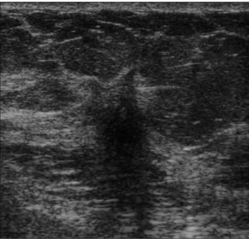
Tumors with circumscribed margins were more often triple-negative subtype (OR, 6.72; 95% CI, 2.56–17.65; P < 0.001), high grade (grade 3 vs. grade 1 or 2: OR, 5.42; 95% CI, 2.66–11.00; P < 0.001) and hormone receptor-negative (OR, 4.87; 95% CI, 2.37– 9.99; P < 0.001) (Fig. 2). Tumors with poste-rior acoustic enhancement were commonly high grade (OR, 1.65, 95% CI, 0.92–2.97) Main points
•
Radiologic imaging has an important role in diagnosis, staging, treatment, and follow-up of patients with breast cancer, and it may also help to predict molecular subtypes of patients with breast cancer for guiding treatment.•
The prediction of triple-negative molecular subtype by ultrasonography (US) may be important for diagnosis and management.•
Breast US performed by experienced radiologists may also help to predict hormone receptor status.•
Sonographic features such as margins and posterior acoustic features were found to be significantly associated with molecular subtype, histologic grade, and hormone receptor status.though there was no statistical significance (P =0.095). There was, however, a strong as-sociation between posterior enhancement and hormone receptor negativity (OR, 2.46; 95% CI, 1.26–4.8; P = 0.009) and triple nega-tivity (OR, 3.98; 95% CI, 1.59–9.95; P = 0.003). Posterior tumor enhancement and circum-scribed margins were together significantly associated with high grade, and they were different from tumors with only posterior acoustic enhancement. This situation in-creases the likelihood of hormone receptor negativity having a triple-negative status. Together, posterior shadowing and noncir-cumscribed margins were significantly cor-related with low grade, hormone receptor positivity and nontriple-negative subtype (Table 2, Fig. 3).
In our study most patients had luminal A and luminal B subtypes (78%) and only 13% had triple-negative breast cancer. Luminal A (n=41, 70%) and luminal B (n=68, 68.7%) subtypes were more often associated with noncircumscribed margins. Triple-negative breast cancers often had circumscribed margins (n=20, 76.9%), with only 23% of them having noncircumscribed margins (P < 0.001) (Table 1).
Discussion
Our study showed that sonographic fea-tures such as posterior acoustic feafea-tures and margins were significantly associat-ed with molecular subtype and histologic grade. Luminal A and luminal B subtypes were more often associated with noncir-cumscribed margins and triple-negative breast cancers commonly had circum-scribed margins.
Breast cancer is a heterogeneous disease with different histopathologic and biolog-ic features. Hence, suitable classifbiolog-ication is needed for appropriate individual manage-ment (9–11). Currently due to the inade-quate prognostic power and predictive ac-curacy of existing classifications, a modified classification according to molecular char-acteristics of breast cancer was defined by the 13th St. Gallen Breast Cancer Conference to categorize breast cancers into molecular subtypes (1). Despite the lack of a complete overlap among molecular classes and their immunohistochemical status, the St. Gallen Breast Cancer Conference accepted immu-nohistochemistry as a basic methodology to identify breast cancer subtypes. This new molecular classification may overcome the limitations of previous schemes (9). Most of
Figure 1. A 55-year-old woman with hormone receptor-positive (luminal B subtype) invasive ductal
carcinoma (low grade). US image shows noncircumscribed hypoechoic mass with indistinct margins and posterior acoustic shadowing in the right upper breast.
Figure 2. A 44-year-old woman with triple-negative invasive ductal carcinoma (high grade). US image
the patients in our study had luminal A and B molecular subtypes (78%) and only 13% had triple-negative breast cancer.
The use of breast US exam has become an effective method to differentiate benign from malignant lesions, especially in young women with dense breast tissue (12). Al-though many previous studies have focused on the sonographic appearance of breast cancer according to grade, the same is not true for the association between tumor sub-types (3, 8). We have very limited imaging data with regards to the determination of the sonographic features of molecular subtypes. As compared with hormone receptor-neg-ative status, tumors with hormone recep-tor-positive status were more likely to have noncircumscribed margins. In our study, tu-mors with posterior shadowing and noncir-cumscribed margins were more likely to be of nontriple-negative subtype.
Previously, the majority of malignant breast tumors were expected to have
pos-terior acoustic shadowing at US and have poorly defined spiculated margins (13, 14). However, it is now widely known that many tumors may have variable posterior acoustic features; new studies have shown that well-defined margins and posterior enhancement are more likely to represent higher grade tumors and negative receptor status (13, 15, 16). Our results are consistent with these findings and there is a strong statistically significant correlation between these parameters.
Lacroix et al. (20) determined that grade 1 tumors (low grade) and grade 2 tumors (intermediate grade) show stromal reac-tion, which results in spicules and perile-sional hyperechogenic halo, while grade 3 (high grade) tumors do not develop stromal reaction and have a round shape. Conversely, Lamb et al. (15) found that higher grade tumors are significantly more likely than lower grade ones to show in-distinct margins and posterior acoustic
enhancement. Rotstein and Neerhut (11) mentioned that grade 3 invasive ductal cancers show the classic feature of acous-tic shadowing. These studies did not use BI-RADS lexicon to categorize sonographic features. All these studies showed different results and there was no concordance be-tween them.
The presence of receptors is important for good prognosis and indicates hormone sensitivity. Shin et al. (16) showed that masses with circumscribed margins and posterior acoustic enhancement are asso-ciated with high grade and negative hor-mone status. In our study, similar to these results, tumors with circumscribed margins and posterior enhancement were more likely to be triple-negative, high grade, and hormone-receptor negative. Receptor pos-itivity is associated with an irregular shape and spiculated margins, and it is significant-ly more frequent than triple-negative can-cers (11). Our results are consistent with this Table 1. Distribution of demographic, radiologic, and pathologic findings according to molecular classification
Independent variables, n (%)
All cases Lum A Lum B Her2 TNBC
Dependent variables n=201 n=58 n=99 n=18 n=26 P a Age (years) 0.038 ≤50 102 (50.7) 23 (39.7) 52 (52.5) 8 (44.4) 19 (73.1) >50 99 (49.3) 35 (60.3) 47 (47.5) 10 (55.6) 7 (26.9) Menopausal status 0.189 Premenopausal 96 (47.8) 24 (41.4) 48 (48.5) 7 (38.9) 17 (65.4) Postmenopausal 105 (52.2) 34 (58.6) 51 (51.5) 11 (61.1) 9 (34.6)
Sonographic tumor size (mm) 0.014
<20 106 (52.7) 39 (67.2) 51 (51.5) 5 (27.8) 11 (42.3) ≥20 95 (47.3) 19 (32.8) 48 (48.5) 13 (72.2) 15 (57.7)
Margins <0.001
Circumscribed 78 (38.8) 17 (29.3) 31 (31.3) 10 (55.6) 20 (76.9) Noncircumscribed 123 (61.2) 41 (70.7) 68 (68.7) 8 (44.4) 6 (23.1)
Posterior acoustic features 0.041
Shadowing 88 (43.8) 29 (50.0) 49 (49.5) 7 (38.9) 3 (11.5) Mixed 23 (11.4) 4 (6.9) 12 (12.1) 3 (16.7) 4 (15.4) Enhancement 27 (13.4) 7 (12.1) 10 (10.1) 2 (11.1) 8 (30.8) No change 63 (31.3) 18 (31.0) 28 (28.3) 6 (33.3) 11 (42.3) Tumor grade <0.001 1 or 2 73 (36.3) 45 (77.6) 25 (25.3) 2 (11.1) 1 (3.8) 3 128 (63.7) 13 (22.4) 74 (74.7) 16 (88.9) 25 (96.2)
Lum A, luminal A breast cancer; Lum B, luminal B breast cancer; Her2, human epidermal growth factor 2–neu; TNBC, triple-negative breast cancer. aPearson’s chi-square test.
finding and a strong relationship was ob-served between noncircumscribed margins and hormone receptor positivity.
Blaichman et al. (3) determined that tri-ple-negative cancers were almost always high grade at diagnosis and showed pos-terior enhancement more commonly. Ad-ditionally, the presence of shadowing was strongly associated with low grade (about 92% were low grade in their study). Kojima
et al. (17) mentioned that triple-negative breast cancers were more likely to be lob-ulated in shape and having circumscribed margins and less likely to show posterior at-tenuating. Our results confirm the findings of these studies. Triple-negative breast can-cers were likely to be histologically inter-mediate and high-grade tumors. This trend was the same for patients with ER-/PR-/ HER2+ breast cancers, compared with
pa-tients with ER+/PR-/HER2- breast cancers. Although a triple-negative breast cancer may have similar sonographic features to a benign lesion, sonographic imaging recog-nition can assist in both pretreatment plan-ning and understanding biologic behavior of this entity (18).
The posterior enhancement is seen more often in hormone receptor-negative HER2+ cancers (50%) than hormone receptor-pos-itive HER2- cancers (29%) (16). Triple-neg-ative tumors present with round, oval, and lobular shapes having an indistinct or microlobulated contour (19, 20). Posterior enhancement is determined in 35.5% to 49% of triple-negative cancers and 50% of hormone receptor-negative HER2+ cancers (16, 18). In our study, we did not investigate the enhancement for all subtypes separate-ly; however, posterior acoustic enhance-ment was more common in the triple-nega-tive subtype than in the other subtypes.
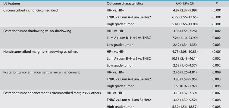
Histopathologic evaluation is an essen-tial requirement for breast cancer diagnosis and treatment. Advanced pathologic pro-cedures including ER, PR, and HER2/neu sta-tus are also needed as prognostic and pre-dictive factors. The reliability of hormone receptor status is dependent on tissue han-dling and processing. These steps can lead to false-negative results if quality control is not sufficient. The issues related to patholo-Table 2. Comparison of US features, hormone receptor status, histologic grade, and molecular subtypes
US features Outcome characteristics OR (95% CI) P
Circumscribed vs. noncircumscribed HR- vs. HR+ 4.87 (2.37–9.99) <0.001 TNBC vs. Lum A+Lum B+Her2 6.72 (2.56–17.65) <0.001 High grade tumor 5.41 (2.66–11.00) <0.001 Posterior tumor shadowing vs. no shadowing HR+ vs. HR - 3.36 (1.55–7.26) 0.002
Lum A+Lum B+Her2 vs. TNBC 7.24 (2.10–24.99) 0.002 Low grade tumor 2.42 (1.34–4.35) 0.003 Noncircumscribed margins+shadowing vs. others HR+ vs. HR- 4.75 (2.08–10.83) <0.001
Lum A+Lum B+Her2 vs. TNBC 10.58 (2.43–46.14) 0.002 Low grade tumor 2.53 (1.40–4.57) 0.002 Posterior tumor enhancement vs. no enhancement HR- vs. HR+ 2.46 (1.26–4.81) 0.009 TNBC vs. Lum A+Lum B+Her2 3.98 (1.59–9.95) 0.003 High grade tumor 1.65 (0.92–2.97) 0.095 Posterior tumor enhancement +circumscribed margins vs. others HR- vs. HR+ 3.18 (1.37–7.39) 0.007 TNBC vs. Lum A+Lum B+Her2 3.65 (1.39–9.52) 0.008 High grade tumor 5.39 (1.56–18.57) 0.008
US, ultrasonography; OR, odds ratio; CI, confidence interval; HR, hormone receptor; TNBC, triple-negative breast cancer; Lum A, luminal A breast cancer; Lum B, luminal B breast cancer; Her2, human epidermal growth factor 2–neu.
Logistic regression, P < 0.05.
Figure 3. Comparison of US features, hormone receptor (HR) status, histologic grade, and molecular
gy services in low-middle income countries include limited financial resources, limited equipment, as well as inadequate numbers of expert pathologists and technologists (21, 22). Due to these negative factors, hormone receptors are not routinely deter-mined. Our findings suggest that breast US performed by experienced radiologists may help predict the hormone receptor status and molecular subtypes of tumors. In coun-tries without resources for receptor testing, US features may be used to make the deci-sion for hormone therapy.
Our study has many limitations such as its retrospective design, small sample size, and lack of some sonographic features deter-mined in BI-RADS US lexicon. Furthermore, our results cannot completely reflect actu-al clinicactu-al conditions, because the anactu-alyses were carried out on selected images rather than images that were acquired during re-al-time scanning. Despite these limitations, our study provides a new additional insight into US features of breast cancer lesions.
In conclusion, sonographic features such as margins and posterior acoustic features were found to be significantly associated with molecular subtype, histologic grade, and hormone receptor status. Being able to predict the molecular subtype, especial-ly the triple-negative subtype by US might also have an important role for earlier man-agement and treatment. Further work with larger populations and prospective nature are necessary to determine the full poten-tial of US in the evaluation of the molecular subtypes of malignant breast lesions. Conflict of interest disclosure
The authors declared no conflicts of interest.
References
1. Yaganawa M, Ikemot K, Kawauchi S, et al. Lumi-nal A and LumiLumi-nal B (HER2 negative) subtypes of breast cancer consist of a mixture of tumors with different genotype. BMC Res Notes 2012; 5:376. [CrossRef]
2. Irshad A, Leddy R, Pisano E, et al. Assessing the role of ultrasound in predicting the biological behavior of breast cancer. AJR Am J Roentge-nol 2013; 200:284–290. [CrossRef]
3. Blaichman J, Marcus JC, Alsaadi T, et al. Sono-graphic appearance of ductal carcinoma of the breast according to histologic grade. AJR Am J Roentgenol 2012; 199:W402–W408. [CrossRef]
4. Reis-Filho JS, Simpson PT, Gale T, et al. Molec-ular evolution of breast cancer. J Pathol 2005; 205:248–254. [CrossRef]
5. Bosch A, Eroles P, Zaragoza R, et al. Triple nega-tive breast cancer: Molecular features, patho-genesis, treatment and current lines of research. Cancer Treat Rev 2010; 36:206–215. [CrossRef]
6. Levy L, Suissa M, Chiche JF, Teman G, Martin B. BIRADS ultrasonography. Eur J Radiol 2007; 61:202–211. [CrossRef]
7. Costantini M, Belli P, Lombardi R, et al. Charac-terization of solid breast masses use of the so-nographic breast imaging reporting and data system lexicon. J Ultrasound Med 2006; 25:649. 8. Bloom HJG, Richarson WW. Histologic grading
and prognosis in breast cancer: A study of 1709 cases of which 359 have been followed for 15 years. Br J Cancer 1957; 11:353–377.
9. Viale G. The current state of breast cancer classi-fication. Ann Oncol 2012; 23(Suppl 10):207–210. 10. Elsawaf Z, Sinn HS, Rom J, et al. Biological sub-types of triple negative breast cancer are asso-ciated with distinct morphological changes and clinical behavior. The Breast 2013; 22:986–992.
[CrossRef]
11. Rotstein AH, Neerhut PK. Ultrasound character-istics of histologically proven grade 3 invasive ductal breast carcinoma. Australas Radiol 2005; 49:476–479. [CrossRef]
12. Skaane P, Engedal K. Analysis of sonographic features in the differentiation of fibroadeno-ma and invasive ductal carcinofibroadeno-ma. AJR Am J Roentgenol 1998; 170:109–114. [CrossRef]
13. Aho M, Irshad A, Ackerman SJ. Correlation of sono-graphic features of invasive ductal carcinoma with age, tumor grade, and hormone-receptor status. J Clin Ultrasound 2013; 41:10–17. [CrossRef]
14. Weinstein SP, Conant EF, Mies C, et al. Posterior acoustic shadowing in benign breast lesions: sonographic-pathologic correlation. J Ultra-sound Med 2004; 23:73–83.
15. Lamb PM, Perry NM, Vinnicombe SJ, et al. Cor-relation between ultrasound characteristics, mammographic findings and histological grade in patients with invasive ductal carcino-ma of the breast. Clin Radiol 2000; 55:40–44.
[CrossRef]
16. Shin HJ, Kim HH, Huh MO, et al. Correlation between mammographic and sonographic findings and prognostic factors in patients with node-negative invasive breast cancer. Br J Radiol 2011; 84:19–30. [CrossRef]
17. Kojima Y, Tsunoda H. Mammography and ultra-sound features of triple negative breast cancer. Breast Cancer 2011; 18:146–151. [CrossRef]
18. Ko ES, Lee AH, Kim H, et al. Triple-negative breast cancer: correlation between imaging and pathological findings. Eur Radiol 2010; 20:1111–1117. [CrossRef]
19. Wang Y, Ikeda DM, Narasimhan B, et al. Estro-gen receptor-negative invasive breast cancer: imaging features of tumors with and without human epidermal growth factor receptor type 2 overexpression. Radiology 2008; 246:367– 375. [CrossRef]
20. Boisserie-Lacroix M, Mac Grogan G, Debled M, et al. Radiological features of triple negative breast cancers (about 73 cases). Diagn Interv Imaging 2012; 93:196–203. [CrossRef]
21. El Saghir NS, Adebamowo CA, Anderson BO, et al. Breast cancer management in low re-source countries (LRCs): consensus statement from the Breast Health Global Initiative. Breast 2011;20 (Suppl 2):S3–11. [CrossRef]
22. Shyyan R, Masood S, Badwe RA, et al. Breast can-cer in limited-resource countries: diagnosis and pathology. Breast J 2006; 12 (Suppl 1):S27–37.