World Glaucoma Association
The Global Glaucoma Network
www.worldglaucoma.org
ABSTRACT BOOK
GLAUCOMA
CONGRESS
JUNE 28–JULY 1 2017
HELSINKI
ABSTRACT BOOK
ABSTRACT BOOK
WGA Executive Office, c/o Schipluidenlaan 4, 1062 HE Amsterdam, The Netherlands Tel: +31 20 679 3411 E-mail: [email protected]
Published by Kugler Publications, P.O. Box 20538, 1001 NM Amsterdam, The Netherlands, on behalf of the World Glaucoma Association.
Typesetting: 3bergen, www.3bergen.com
© 2017. World Glaucoma Association.
No part of this publication may be reproduced, stored in a retrieval system, or transmitted in any form by any means, electronic, mechanical, photocopying or otherwise, without the prior consent of the copyright owners.
Disclaimer
The Hippocrates Glaucoma Foundation, based upon an agreement with the World Glaucoma Association, organizes the World Glaucoma Congress with the aim of providing education and scientific discourse in the field of glaucoma. The Hippocrates Glaucoma Foundation accepts no responsibility for any products, presen-tations, opinions, statements or positions expressed by speakers at the congress. Inclusion of material in the scientific program does not constitute an endorsement by The Hippocrates Glaucoma Foundation.
Download the most recent version at:
III 7th World Glaucoma Congress 2017 - Abstract Book
Grand Rounds
1
Film Festival
10
Rapid Fire
41
Poster Abstracts
65
• Epidemiology, Quality of Life and Health Economics
65
• Genetics, Genomics and Biomarkers
168
• Medical Treatment and Non-Incisional Surgery
196
• Structural and Functional Testing
300
• IOP Physiology and Pathophysiology
490
• Laboratory Sciences
536
• Other
584
• Surgery and Wound Healing
688
Index
930
This is a searchable PDF.
CYCLODIALYSIS CLEFT TREATMENT: HOW TO PRESERVE CONJUNCTIVAL
FILTRATION?
Carlos A. Arciniegas-Perasso
47-year-old male, diagnosed of hypertensive uveitis OD in 2014 (presumed Fuchs’ cyclitis).
Two acute episodes in the last 2 years. Controlled with medication until October 2016, when intraocular pressure (IOP) was 27 mmHg with maximal tolerated medical treatment. Visual acuity (VA) was 0.63 and no cellularity was observed in the anterior chamber.
MMC-enhanced ab-interno AC-subconjunctival shunting was performed (XEN). Successful positioning was achieved in the second attempt, a bleb was formed.
A very low IOP with a shallow anterior chamber (AC) was observed since the early postop. Indentation gonioscopy revealed a cyclodialysis cleft next to the implant (image 1). After 2 weeks of conservative measures IOP was still low and the VA worsened; an OCT disclosed macular folds (image 2A).
GR FF RF P I 2 7th World Glaucoma Congress 2017 - Abstract Book
Gonioscopically-assisted cyclocryopexy was done to close the cleft. IOP and VA improved just for one week. AC was partially refilled with viscoelastic. IOP spikes were detected afterwards, requiring AC tap and resum-ing glaucoma treatment. VA improved and glaucoma medication was gradually withdrawn, one dose of 5-FU (5 mg) was subconjunctivally administered.
Ten weeks after the initial glaucoma procedure, VA is 0.75, IOP is 10 mmHg with a low diffuse wide bleb and the cleft does not seem to open with gonioscopic indentation (image 3). Macular OCT returned to normal (image 2B). Cycloplegics and steroids are being tapered. See IOP, VA, meds chronologic graph (image 4).
GR FF RF P I 3 7th World Glaucoma Congress 2017 - Abstract Book
SUDDEN VISUAL SCOTOMA IN A WOMAN WITH EXFOLIATION GLAUCOMA
Luis Silva
Clinical Glaucoma Fellow, New York Eye and Ear Infirmary of Mount Sinai. 310 E 14th Street. Suite 304. South
building. New York NY, 10003. USA. Mentor: Robert Ritch, MD
We present the case of a 74 year-old woman with exfoliation glaucoma OS, status post selective laser trabec-uloplasty (SLT) x 2 OS, trabeculectomy with mitomycin C OS (December 2009), and phacoemulsification with PC IOL insertion OS (April, 2010). She used atorvastatin 20 mg for hyperlipidemia and aspirin 80 mg.
On July 2015, she was seen by an ophthalmologist for a regular follow-up. IOP was 24 mmHg OD and 12 mmHg OS. Cataract extraction was recommended OD, and she requested a second opinion.
She was seen by another ophthalmologist October 8th, 2015. IOP was 36 mmHg OD and 14 mmHg OS. The
patient mentioned that she received several drops to dilate the pupils, and dorzolamide/timolol OD, with which IOP decreased. After pupillary dilation, she complained of a sudden central scotoma OD. She was seen the following day. She was told that she wait a few days for recovery. Four days after the episode, she was reevaluated. The retina was reported as normal, and had posterior vitreous detachment OD. They ordered macular OCT and 10-2 visual fields.
On October 16th, 2015, the patient came to us for a second opinion. On examination, best corrected visual
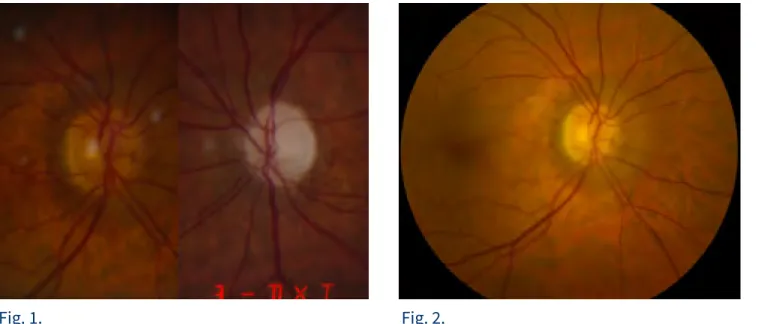
acuity was 20/25 OD and 20/30 OS. Slit lamp examination revealed a cystic avascular bleb OS, clear corneas, deep and quiet anterior chambers OU, iridodonesis OS, 1+ nuclear sclerosis with exfoliation material OD, PCIOL and mild posterior capsular opacity OS. Intraocular pressure was 21 mmHg OD and 14 mmHg OS, using dorzolamide/timolol 0.50% q.d. OD. Dark-room gonioscopy revealed grade IV angles OU. Stereoscopic optic nerve head exam revealed a 0.3 cup-to-disc ratio OD and a pale optic nerve with 0.9 vertically and 0.7 horizontally cup-to-disc ratio OS. There was no disc hemorrhage OU (Figures 1 and 2).
Fig. 1. Fig. 2.
A 10-2 SITA Standard visual field test had a temporal defect OD and decreased general sensitivity OS (Figure 3). A macular OCT revealed a hyperrefringence of the inner nuclear layer nasal to the fovea OD (Figure 4). A cardiovascular workup, including carotid and vertebral duplex Doppler study ruled out the presence of an embolic source for a retinal ischemic event. However, minor carotid ulceration cannot be totally ruled out, and a transesophageal ultrasound would be more precise. ESR, PCR, basic metabolic and lipid panel were within normal limit.
We concluded that the patient had exfoliation glaucoma OS, exfoliation syndrome with ocular hyperten-sion OD, and paracentral acute middle maculopathy (PAMM) OD. The use of several drops of topical phen-ylephrine to dilate the pupils may have been the triggering factor to this clinical presentation. PAMM has been described after the use of systemic pressor agents or vasoconstrictors, such as sympathomimetics
GR FF RF P I 4 7th World Glaucoma Congress 2017 - Abstract Book
To the best of our knowledge, this is the first reported case of PAMM after pupillary dilation in a patient with exfoliation syndrome/OHT and exfoliation glaucoma.
Fig. 3.
Fig. 4.
References:
1. Sarraf D et al. A new variant of acute macular neuroretinopathy associated with retinal capillary ischemia. JAMA Ophthalmol 2013; 131(10):1275-87.
2. Bhavsar K et al. Acute macular neuroretinopathy: A comprehendsive review of the literature. Surv Ophthalmol 2016; 1-28.
3. Rahimy E et al. Paracentral acute middle maculopathy: what we knew then and what we know now. Retina 2015; 35(10):1921-30.
Special thanks to Robert Ritch, MD, Jeffrey Odel MD, Reinaldo Garcia, MD and Donald Hood, MD for their valuable contribution. GR FF RF P I 5 7th World Glaucoma Congress 2017 - Abstract Book
INTERNAL CAROTID ARTERY ANEURYSM SIMULATING NORMAL TENSION
GLAUCOMA
Mário Pincelli Netto
Department of Ophthalmology, Federal University of São Paulo, São Paulo, Brazil; Glaucoma Unit, Ver Mais Oftalmologia, São Paulo, Brazil.
A 48-year-old afro-American female patient, presented with low vision acuity in her left eye and frontal oppressive headache for 2 years. She denied any personal, ophthalmological and family medical history. On her first examination, she had intraocular pressure = 12 mmHg in both eyes (AU) without any topical or systemic medication and a daily tension curve demonstrating a variation between 13 and 16 mmHg in the right eye (OD) and between 12 and 15 in the left eye (OS). The anterior biomicroscopy and pupillary reflex were normal in AU. The gonioscopy showed 360o open angle until scleral spur in AU. The pachymetry exam was 542μm in OD and 535μm in OS. On fundoscopy she had a cup disc ratio of 0.5 in OD (Figure 1); and 0.7, with a lower temporal hoyt, in the OS (Figure 2).
The visual field test was normal in OD (Figure 3), but a temporal superior arched loss of sensitivity, crossing the vertical mid line, was seen in the OS (Figure 4). As the functional defect was compatible with the struc-tural defect, and presented normal intraocular pressure measurements, (although she did not have epide-miologic findings for normal tension glaucoma), topical bimatoprost 0.03% in AU was initiated for normal tension glaucoma treatment.
GR FF RF P I 6 7th World Glaucoma Congress 2017 - Abstract Book
ophthalmologic exams compared to that performed at her first visit. A nuclear magnetic resonance was requested that showed an internal carotid aneurysm of about 3.5 mm x 6.5 mm, near the emergence of the ophthalmic artery, compressing the optic nerve (Figures 5, 6). The patient was referred to a neurosurgeon for an endovascular aneurysm occlusion.
After 3 months of surgery, she reported improvement of the headache symptoms, but lost diffuse sensitivity in the visual field test. This case alerts us to the importance of remembering that many neurological defects are still underdiagnosed, and that not only does glaucoma generate suspicious cuppings on the optic disc. The differentiation between glaucomatous and non-glaucomatous cuppings is still a challenge, and predic-tive factors such as visual acuity worse than 20/40, visual field defects respecting the vertical midline, age lower than 50 years and disproportionate pallor compared to the disc cupping, should be considered.
GR FF RF P I 7 7th World Glaucoma Congress 2017 - Abstract Book
AN INTERESTING CASE OF ANGLE CLOSURE GLAUCOMA
Ayyappa Reddy M1, Gowri J Murthy2, Jyothi Kattige3
1Glaucoma fellow, Prabha Eye Clinic and Research Centre, Bangalore; 2Chief, Glaucoma Services, Prabha
Eye Clinic and Research Centre, Bangalore; 3Consultant, Glaucoma Services, Prabha Eye Clinic and Research
Centre, Bangalore.
Case Summary: A 65 year old anesthetist was referred to us for management of uncontrolled intraocular pressure. His brothers were under treatment for angle closure glaucoma. One year ago he had under-gone cataract extraction with IOL implantation in the right eye. Subsequently in the right eye, he devel-oped central retinal vein occlusion with neovascular glaucoma for which he was treated with injection Bevacizumab and trabeculectomy. However the visual recovery was poor in the right eye with non resolv-ing choroidal detachment. He had been on topical antiglaucoma medications for the left eye and recently the intraocular pressure were not getting controlled adequately and he complained of gradual decrease in vision.
At presentation, in the left eye his best corrected visual acuity was 6/60; anterior segment examination showed nuclear sclerosis grade 3, with shallow anterior chamber, the intraocular pressure was 32 mmHg with no angle structures visible on gonioscopy. A diagnosis of angle closure glaucoma was made and patient underwent yag laser iridotomy. Post iridotomy, the intraocular pressures remained uncontrolled and on dilation increased to 44 mmHg with hyperemic optic disc. After initial management with intravenous manni-tol and systemic acetazolamide he underwent a planned phacoemulsification with trabeculectomy under LA. Postoperatively his visual acuity was CF 0.5 m; IOP decreased to 12 mmHg with well functioning trabe-culectomy bleb but had a shallow choroidal effusion. The effusion did not resolve with a course of systemic steroids. Further investigations were done for non resolving choroidal detachment including blood investi-gations to rule out uveitic entities and a Bscan ultrasound. All the investiinvesti-gations were in the normal range; on the Bscan increased scleral thickness of 3.1 mm was noted. Axial length of the eye was 22.8 mm. Now with a diagnosis of idiopathic uveal effusion syndrome patient underwent scleral window dissection with drainage of the choroidal fluid which subsequently resolved.
Following this success in the left eye, on patient request, a similar procedure for the right eye choroidal detachment was performed under very guarded visual prognosis. Post operatively the choroidal detach-ment resolved with no significant visual improvedetach-ment.
The left eye had a BCVA of 6/24, n8 with mild inflammation in the anterior segment which responded to topical dexamethasone and iop was well controlled in the range of 10-15 mmHg with brinzolamide eye drops, fundus showed pale disc with resolved choroidal detachment.
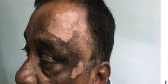
Six months post the left eye surgery, on routine review, hypopigmented facial lesions were noted. Dilated retinal examination also showed depigmented fundus picture with inflammatory yellowish nodules in the mid periphery. A diagnosis of ?sympathetic ophthalmia/ VKH syndrome was considered.
Fig. 1. Depigmented skin lesions
GR FF RF P I 8 7th World Glaucoma Congress 2017 - Abstract Book
Discussion: This is the first reported case of sympathetic ophthalmia following vortex vein decompres-sion. The case could be illustrative of a rare complication of scleral window dissection with exposure of the choroid, namely sympathetic ophthalmia. The points in favor of this are, the finding of Uveal Effusion Syndrome supported by the increased scleral thickness, and subsequent resolution of the choroidal detach-ment by scleral window dissection. This could have triggered an autoimmune response, leading to the typical picture of sympathetic ophthalmia.
The other possible differential diagnosis is VKH syndrome. The two entities are essentially similar, with the only difference being an antecedent history of trauma/ surgery in sympathetic ophthalmia. It could be that our patient might have had secondary angle closure on initial presentation due to ciliochoroidal effusion which was not evident due to the cataract (a UBM was not done pre operatively), which worsened following the surgery, and later developed into the full blown picture that we see now. The points against this are the complete resolution of the choroidal detachment following scleral window dissection, and the unrespon-siveness to systemic steroids.
The case illustrates two rare associations with angle closure glaucoma, and the successful management of the same.
Fig. 2+3. Sunset glow appearance of the fundus with Dalen Fuchs Nodules
GR FF RF P I 9 7th World Glaucoma Congress 2017 - Abstract Book
XEN MEGA-BLEB: SURGICAL MANAGEMENT
Ejaz Ansari*1, Richard Imonike1
1Maidstone & Tunbridge Wells NHS trust, Eye Ear and Mouth Unit, Maidstone, United Kingdom
We present a rare complication of XEN implant surgery. A 64 year old man underwent routine XEN implant + MMC+ Phaco + IOL. 4 weeks later he presented with dysaesthesia. There was a very extensive, raised drain-age bleb. Treatment included needling and also excision of the inferior bleb. The eye settled well with only a superiorly located bleb, IOP of 14 mmHg and much less discomfort.
11 7th World Glaucoma Congress 2017 - Abstract Book
GR
FF
RF
P
CO2 LASER: A NEW TOOL FOR NON PENETRATING GLAUCOMA SURGERY
Antoine Bastelica*1
1Ophthalmology, Atrium Vision Clinique Pasteur, Toulouse, France
We described our technique of using CO2 Laser technology for performing non-penetrating glaucoma
surgery. Laser assisted surgery enables good and reproducible technique for unroofing the Schlemm’s canal and exposing the trabeculo-descemetic membrane.
12 7th World Glaucoma Congress 2017 - Abstract Book
GR
FF
RF
P
CHILDHOOD GLAUCOMA…OUT OF A DESPERATE SITUATION
Natalia Volkova*1, Tatiana Iureva1
1Glaucoma, Irkutsk Branch of S. Fyodorov Eye Microsurgery Federal State Institution, Irkutsk, Russian
Federation
To present surgical tricks and tips of implantation of Ahmed valve system in childhood glaucoma. Original method of remote suturing of drainage body to sclera is presented. Initial ophthalmological status deter-mines peculiarities of implantation and position of tube. This technology allows achieving stable hypoten-sive effect, minimizing complications and determining it as a surgery of choice.
13 7th World Glaucoma Congress 2017 - Abstract Book
GR
FF
RF
P
BATTLE OF THE BULGES: A TALE OF TWO BLEBS
Cyril Jose*1, Murali Ariga1, Malarchelvi Palani1, M Nivean1
1Ophthalmology Glaucoma, M.N. Eye Hospital, Chennai, India
The first patient presented with foreign body sensation and cosmetic blemish with a history of trabeculec-tomy with mitrabeculec-tomycin C. The overhanging bleb was treated by bleb excision and resuturing. The second part of the video shows encysted bleb which was initially treated by needling and 5-FU. The encysted bleb was then treated by creating a 2*2 window followed by suturing the conjuctiva and tenon.
14 7th World Glaucoma Congress 2017 - Abstract Book
GR
FF
RF
P
MIGS PROCEDURES IN COMBINATION WITH PHACOEMULSIFICATION:
PATIENTS BENEFITS
Elena Tomilova*1
1Saint Petersburg Brunch, Ii Surgery Department, S. Fyodorov Eye Microsurgery Federal State Institution, Saint
Petersburg, Russian Federation
This video demonstrates two MIGS techniques: Endoscopic Cyclophotocoagulation and Selective
Trabeculotomy ab interno. Hence we would demonstrate how does it work, surgical technique, the tips and tricks to perform safe and effective surgery in different case scenarios and clinical results.
15 7th World Glaucoma Congress 2017 - Abstract Book
GR
FF
RF
P
CO2 LASER-ASSISTED SCLERECTOMY SURGERY (CLASS)
Zhu Li Yap*1, Shamira Perera1
1Singapore National Eye Centre, Singapore, Singapore
CLASS was developed as an alternative to manual deep Sclerectomy. It utilizes the IOPtiMate laser system, which is targeted towards ablating dry tissues and is highly absorbent by fluid, thus preventing the laser from penetrating into the anterior chamber. In our video, we demonstrate the steps of the surgery, discuss practical aspects of the procedure & explain post operative patient management.
16 7th World Glaucoma Congress 2017 - Abstract Book
GR
FF
RF
P
MANAGEMENT OF TUBE EXPOSURE
Gowri J Murthy*1, Jyoti Kattige1
1Glaucoma, Prabha Eye Clinic and Research Centre, Bangalore, India
Exposure of the Glaucoma drainage device tube is a late complication, which results in increased risk of endophthalmitis. The video demonstrates various surgical modalities of management of tube exposure, including, scleral patch grafts, conjunctival autografts, Amniotic membrane grafts, and posterior tube relo-cation, for the management of tube exposure.
17 7th World Glaucoma Congress 2017 - Abstract Book
GR
FF
RF
P
BRIDGING THE GAP
George Puthuran*1, S R Krishnadas1
1Glaucoma, Aravind Eye Care System, Madurai, India
TVT Study provides evidence that role of tube shunts have expanded beyond surgical management of refractory glaucomas. This video demonstrates how a majority of steps involved in aqueous shunt surgery could be perfected at wetlab. Intense wetlab training sessions under supervision of a trainer serves to maxi-mize learning and will ensure a smooth and safe transition to the operating room.
18 7th World Glaucoma Congress 2017 - Abstract Book
GR
FF
RF
P
BANDS AND PLATES
George Puthuran*1, Shashikant Shetty2, Naresh Babu2
1Glaucoma, 2Aravind Eye Care System, Madurai, India
Implantation of a glaucoma drainage device over or behind a pre existing encircling band is a successful management option for refractory glaucoma in patients who have undergone a previous scleral buckling procedure. In this video the authors wish to highlight certain operative techniques relevant to those eyes requiring aqueous shunts that have previously undergone retinal surgery.
19 7th World Glaucoma Congress 2017 - Abstract Book
GR
FF
RF
P
THE JOY OF INFERONASAL QUADRANT
George Puthuran*1, S R Krishnadas1
1Glaucoma, Aravind Eye Care System, Madurai, India
Superotemporal quadrant is preferred for placement of an aqueous drainage device. This video demon-strates the viability of inferonasal GDD implantation in the presence of pre existing superior conjunctival scarring. Available virgin conjunctiva, absence of oblique muscle complex and a cosmetically appealing result makes the inferonasal quadrant a real joy to work with for the glaucoma surgeon.
20 7th World Glaucoma Congress 2017 - Abstract Book
GR
FF
RF
P
SURGICAL MANAGEMENT OF HYPOTONY SYNDROME AFTER FILTERING
GLAUCOMA SURGERY
Dmitry Ivanov*1, Oleg Shilovskikh1, Ekaterina Ivanova1
1IRTC Eye Microsurgery Ekaterinburg Center, Ekaterinburg, Russian Federation
The film shows surgical management of severe hypotony after filtering surgery. A 2x2 mm fragment of Tenon’s capsule is formed to block the fistula. Filling of the fistula is performed ab interno in pseudophakic eyes, ab externo in phakic eyes. Ab interno trabeculotomy is performed. In most cases, this solves severe hypotony problem ; 16 of 18 operated eyes did not require additional surgery.
21 7th World Glaucoma Congress 2017 - Abstract Book
GR
FF
RF
P
SURGICAL MANAGEMENT OF POST TRABECULECTOMY SCLERAL MELTING
Suresh Kumar*1, Sahil Thakur1, Tanvi Soni1, Madhu Sharma1
1Ophthalmology, Government Medical College and Hospital, Chandigarh, India
Scleral melting is a devastating complication of MMC augmented trabeculectomies. In our case of a patient 2 weeks post trabeculectomy with flat anterior chamber and visible scleral melt under the bleb we used a combination of conjunctival advancement flap and sandwich amniotic membrane to close the leak. There were no patch related post-operative complications in the follow up period.
22 7th World Glaucoma Congress 2017 - Abstract Book
GR
FF
RF
P
PIGMENT DISPERSION SYNDROME SECONDARY TO IRIS-FIXATED PHAKIC
INTRAOCULAR LENS TO CORRECT MYOPIA
Ana Maria Vasquez*1, Yolanda Pazmiño2, Ana Roldan1
1Hospital Metropolitano, Instituto De Oftalmologia Y Glaucoma Vasquez, 2Hospital Metropolitano, Intituto De
Oftalmologia Y Glaucoma Vasquez, Quito, Ecuador
A 19 year-old patient with best corrected visual acuity of 20/40 in each eye, developed a postoperative pigment dispersion syndrome in both eyes after Artisan anterior iris-fixated intraocular lens implantation for correction of high myopia. The medically uncontrolled intraocular pressure and unresponsive SLT treat-ment necessitated implantation of Ahmed glaucoma valve in both eyes.
23 7th World Glaucoma Congress 2017 - Abstract Book
GR
FF
RF
P
ASTIGMATISM RELATED VISUAL LOSS IN THIN CYSTIC OVERHANGING BLEB
Talvir Sidhu*1,1, Saurabh Verma1, Tanuj Dada1
1Dr Rajendra Prasad Centre for Ophthalmic Sciences, All India Institute of Medical Sciences, New Delhi, India
A 37 year old JOAG patient, operated trabeculectomy, with thin cystic overhanging bleb in left eye had 19D atsigmatism on pentacam. Patient underwent excision of the overhanging part of bleb in left eye. Post-op bleb leak was noted for which entire bleb excision with conjunctival advancement with 10-0 MFN sutures get vertical steepening. Post-op astigmatism was 1.1D with improved vision.
24 7th World Glaucoma Congress 2017 - Abstract Book
GR
FF
RF
P
RAT MODEL OF GLAUCOMA FILTERING SURGERY
Surinder Pandav*1, Faisal Thattaruthody1, Madhuri Akella1, Natasha Gautam Seth1, Alka Khera1,
Simar Rajan Singh1, Nirbhai Singh1
1Advanced Eye Center, Postgraduate Institute of Medical Education & Research, Chandigarh, India
Rats are frequently used for medical research because they are easy to procure, cheap and their immuno-logical profile is well known. However, because of the small size, their use in glaucoma surgical research is difficult. This video describes step by step procedure to create a successful and reproducible glaucoma surgical model in rat eyes.
25 7th World Glaucoma Congress 2017 - Abstract Book
GR
FF
RF
P
SAFETY AND EFFICACY OF SIMULTANEOUS PENETRATING KERATOPLASTY
AND AHMED GLAUCOMA VALVE IMPLANT
Sonia Parreira*1, Sandra Barros1, Nadine Marques1, Ines Machado1
1Hospital Garcia de Orta, Almada, Portugal
This video demonstrates a combined surgery of penetrating keratoplasty and Ahmed glaucoma valve
implantation in a patient with history of failed trabeculectomy and bullous keratopathy. The valve tube was covered by an autologous partial thickness corneal graft. In the follow-up the corneal graft is transparent with no signs of rejection and implant has proven effective in controlling IOP.
26 7th World Glaucoma Congress 2017 - Abstract Book
GR
FF
RF
P
WATCH OUT FOR A PHACO-NIGHTMARE IN A GLAUCOMA EYE WITH THIN AND
AVASCULAR BLEB!
Sushma Tejwani*1, K Bhujang Shetty2
1Glaucoma, 2Narayana Nethralaya, Bangalore, India
This video demonstrates the bleb rupture while performing cataract surgery in patient with thin avascular bleb. Our worst fear came true when the bleb gave way, however a successful cataract surgery with closure of the bleb using amniotic membrane graft at the same time was performed. Thus with anticipation, early identification and timely management, a potentially blinding disaster was averted.
27 7th World Glaucoma Congress 2017 - Abstract Book
GR
FF
RF
P
SYNTHETIC TRAINING MODEL WITH ANGLE AND SCHLEMM’S CANAL FOR
GATT PROCEDURE
Michael Banitt*1
1University of Washington, University of Washington, Seattle, United States
This is a synthetic training model with a flexible base to simulate natural eye motions and allow for tilting of the head/eye. The angle can be seen with a standard direct gonioprism and has a patent Schlemm’s canal that can be canulated ab interno (or externo) and allow for simulation of gonioscopy assisted transluminal trabeculotomy (GATT). The video demonstrates GATT with prolene suture.
28 7th World Glaucoma Congress 2017 - Abstract Book
GR
FF
RF
P
RESECTION OF CILIARY BODY TUMOR PRESENTING AS NEOVASCULAR
GLAUCOMA
Daniela Alvarez Ascencio*1, Armando Castillejos-Chevez1, Cecilio Velasco-Barona 2
1Glaucoma, 2Anterior Segment, Asociacion Para Evitar la Ceguera en Mexico, Mexico City, Mexico
Ultrasound Biomicroscopy shows 2.30x4.04 mmx2.03 mm ciliary body tumor. Technique: temporal conjunc-tival incision, 8 mm scleral flap extending from mVII to mXI. 7 mm full thickness incision in the anterior limbus, incision of the scleral flap from limbus to equator. Block resection of the iris and ciliary body from mVII to mXI. After anterior vitrectomy, sclera and conjunctiva were closed.
29 7th World Glaucoma Congress 2017 - Abstract Book
GR
FF
RF
P
MANAGEMENT OF TUBE EROSION COMPLICATIONS
SriRamani Gollakota*1, Chandra Sekhar Garudadri1, Sirisha Senthil1
1Glaucoma, L.V.Prasad Eye Institute, Hyderabad, India
Glaucoma Drainage Devices (GDDs) play a significant role in the treatment of refractory glaucoma. Although safe and effective, longterm tube and plate related complications can be sight threatening. Postoperative tube and implant plate related complications, their early identification and appropriate management strate-gies are presented in this video
30 7th World Glaucoma Congress 2017 - Abstract Book
GR
FF
RF
P
HOOKING THE SINKING IOL JUST IN THE NICK OF TIME!!
Sushma Tejwani1, Vijna Kamath B*1, K Bhujang Shetty2
1Glaucoma, 2Chairman, Narayana Nethralaya, Bangalore, India
This video demonstrates the management of a case of secondary glaucoma where the lens was absorbed and the anterior and posterior capsules were fused together forming a dense plaque. Glaucoma and cata-ract surgery was planned. During the IOL placement in sulcus, inferior zonular instability was noticed as the IOL started sinking down. IOL was hooked out in time and fixed to the iris with sutures.
31 7th World Glaucoma Congress 2017 - Abstract Book
GR
FF
RF
P
AQUEOUS DRAINAGE IMPLANT IN AN EYE WITH SECONDARY GLAUCOMA
POST VITREO RETINAL SURGERY IS NOT EASY!
Sriramani Gollakota*1, Chandra Sekhar Garudadri1, Sirisha Senthil1
1Glaucoma, L.V.Prasad Eye Institute, Hyderabad, India
Implantation of Glaucoma Drainage device (GDD) in glaucoma following Vitreo-retinal (VR) surgery provides more predictable outcomes but is technically more challenging due to the presence of a retinal implant and common occurrence of obstruction due to fibrous capping of the distal tube ostium. This video shows various techniques of implantation of GDD, difficulties faced and their outcome in such eyes
32 7th World Glaucoma Congress 2017 - Abstract Book
GR
FF
RF
P
TO THE EDGE OF DARKNESS AND BACK!
Sumit Chowdhury*1, Suchanda Sar1, Subhendu Boral2, Shyamashree Sil1, Arijit Mitra1
1Glaucoma, 2Retina, Disha Eye Hospital, Kolkata, India
Lady presented with early corneal decompensation with IOL haptic touching endothelium & increased IOP. IOL redialling attempted. Sudden, unexpected complication suprachoroidal haemorrhage (SCH) occurred. Urgent Vitreo-Retinal intervention-SCH drained. An Inferior RD occurred during SCH drainage. PPV done. PFCL injected. Laser done. Silicone oil injected. Patient recovered good vision and a disaster avoided.
33 7th World Glaucoma Congress 2017 - Abstract Book
GR
FF
RF
P
TRABECULECTOMY IN MICROSPHEROPHAKIA
Sriramani Gollakota*1, Chandra Sekhar Garudadri1, Sirisha Senthil1
1Glaucoma, L.V.Prasad Eye Institute, Hyderabad, India
Trabeculectomy in eyes with Microsphaerophakia is a challenge. Careful pre-operative assessment and meticulous surgical technique would help prevent complications of shallow anterior chamber, trauma to lens and vitreous loss which are reported in such eyes. We demonstrate surgical steps, tips and techniques that are necessary to avoid intra and post-operative complications
34 7th World Glaucoma Congress 2017 - Abstract Book
GR
FF
RF
P
MANAGING LATE COMPLICATIONS OF TRABECULECTOMY
Neha Midha*1, Nasiq Hasan1, Tanuj Dada1
1Dr. Rajendra Prasad Centre for Ophthalmic Sciences, All india Institute of Ophthalmic Sciences, Delhi, India
After trabeculectomy with mitomycin-C, a patient presented with thin-walled overhanging bleb with scleral necrosis. i-OCT guided bleb excision was done and a sutureless scleral patch graft was tucked in the necrotic area. Scleral patch was covered with ologen implant. Conjunctiva was advanced over the ologen. Overlay of conjunctival autograft was given over exposed ologen and irregular cornea.
35 7th World Glaucoma Congress 2017 - Abstract Book
GR
FF
RF
P
SIMULTANEOUS AHMED GLAUCOMA VALVE AND BOSTON
KERATOPROSTHESIS
Sirisha Senthil*1, Mukesh Taneja2, Virender Sangwan3
1Glaucoma, 2Cornea, 3L V Prasad Eye Institute, Hyderabad, India
Managing glaucoma in eyes with keratoprosthesis is a challenge. Glaucoma drainage devices provide definitive surgical option, however, are technically challenging. In this video, we shall demonstrate the technique and practical tips of Ahmed Glaucoma Valve implantation performed concurrently with Boston keratoprosthesis.
36 7th World Glaucoma Congress 2017 - Abstract Book
GR
FF
RF
P
PHYSICS APPLIED: TRIMMING-ENHANCED REVISION FOR COLLAGEN
GLAUCOMA IMPLANT BLEBS
Carlos A. Arciniegas-Perasso*1, Susana Duch-Tuesta1
1Glaucoma Unit, Instituto Condal de Oftalmologia, Barcelona, Spain
Conjunctival fibrosis is the main cause of bleb failure in filtering surgery; this is also valid for collagen glau-coma implant (XEN) ab interno approach. Hagen-Poiseuille’s law is the rationale behind the design of this implant. Based on this, excision of fibrotic tissue associated with implant length reduction is presented as a technique for XEN bleb revision to increase outflow and reduce IOP.
37 7th World Glaucoma Congress 2017 - Abstract Book
GR
FF
RF
P
TRABECTOME SURGERY: STAND-ALONE AND COMBINED WITH CATARACT
SURGERY
Sarah Farukhi*1, Sameh Mosaed1, Mason Schmutz1
1Ophthalmology, University of California Irvine, Orange, United States
Senior author is demonstrating surgical steps of Trabectome surgery in this instruction video. The steps include microscope set up, patient head position, gonioscopic view of trabecular meshwork and performing Trabectome surgery both in stand-alone and combined with cataract surgery. Trabectome is FDA cleared for micro-invasive management of glaucoma and is manufactured by NeoMedix in USA.
38 7th World Glaucoma Congress 2017 - Abstract Book
GR
FF
RF
P
SHORT TUNNEL, SMALL FLAP SHUNT PROCEDURE
Mohammad Pakravan*1, Hamed Esfandiari1
1Ophthalmic Research Center, Ophthalmology Department, Labbafinejad Medical Center, Shahid Beheshti
University of Medical Sciences, Tehran, Iran
The main cause of exposure is force exerted by tube on graft. Consequently some surgeons put tube into scleral tunnel or under large flap to provide flatter configuration. While flap creation is time consuming, intratunnel technique is simpler and faster, but mostly tube sits close to cornea. In this video we use a tech-nique combines advantages of both techtech-niques with appropriate tube position.
39 7th World Glaucoma Congress 2017 - Abstract Book
GR
FF
RF
P
INTRA-OPERATIVE ASOCT IN PHACO-ENDOCYCLOPLASTY IN PLATEAU IRIS
SYNDROME
Vanita Pathak Ray* 1, Vani Sethi2, Hari Peguda3, Rajeev Pappuru3, Nikhil Choudhari3
1Glaucoma, Centre for Sight, Hyderabad, 2Goa University, Goa, 3LV Prasad Eye Institute, Hyderabad, India
Phaco-endocycloplasty is gathering support in the management of Plateau Iris syndrome, when associ-ated with cataract, as it has the ability to change angle configuration as opposed to when phaco alone is performed. In this video, we present endoscopic as well as intra-operative Anterior Segment Optical Coherence tomographic evidence of this change in irido-corneal angle.
40 7th World Glaucoma Congress 2017 - Abstract Book
GR
FF
RF
P
STRUCTURAL ANALYSIS OF BLEBS FILTRATION IN LONG-TERM, FUNCTIONING
TRABECULECTOMIES VS EYES WITH XEN® IMPLANT USING SWEPT SOURCE
OCT
Javier Paz*1
1Hospital Universitario Principe De Asturias, Alcala De Henares, Spain
Purpose: The purpose of this study was to evaluate the morphology of blebs of successfully functioning trabeculectomies and to compare them with the filtering conjunctiva obtained with the XEN® implant, eval-uated using the Triton® Swept-Source OCT.
Methods: This is a cross sectional observational study. We included 25 eyes (15 from trabs and 10 with XEN® implant) and 23 eyes from healthy eyes without any filtering surgeries (controls).
We evaluated the epithelial thickness as well as the height, the hyperreflectivity areas and the hyporreflec-tive spaces (cystic spaces) in all blebs and in the superior conjunctiva of the control eyes using the Tritón® OCT.
Results: Filtering blebs of patients with XEN implants were significantly flatter than in trabeculectomies (bleb height: trabeculectomies, 618 ± 256 μm, XEN implant, 417 ± 183 μm, controls, 244 ± 45 μm) p < 0.05. The XEN group did not show subepithelial fibrosis in any case, compared with 40% of cases with fribrosis in the trab group (p < 0.05). XEN blebs showed a lower percentage of subepithelial cysts compared with the trab group (20% vs 24%). The epithelial thickness was higher in the XEN group than in the trab and controls groups (65 ± 18.5 vs 60 ± 17.7 vs 51 ± 9.7 μm, respectively; p < 0.05). We did not find statistically significant differences between IOP decrease induced by XEN vs Trab (-8.5 ± 5.3 vs -8.8 ± 5.2 mmHg, p > 0.05).
Conclusions: Filtering blebs obtained by the XEN implant were morphologically different than Trab blebs, but the hypotensive efficacy was very similar in both groups. XEN implant blebs were flatter and its structure closely resembles a healthy conjunctiva.
References:
1. Minimally invasive glaucoma surgery as primary stand-alone surgery for glaucoma. Kerr NM, Wang J, Barton K. Clin Exp Ophthalmol. 2016 Dec 8.
2. Ab interno approach to the subconjunctival space using a collagen glaucoma stent. Lewis RA. J Cataract Refract Surg. 2014 Aug;40(8):1301-6.
3. Fluid Dynamics of a Novel Micro-Fistula Implant for the Surgical Treatment of Glaucoma. Sheybani A, Reitsamer H, Ahmed II. Invest Ophthalmol Vis Sci. 2015 Jul;56(8):4789-95.
42 7th World Glaucoma Congress 2017 - Abstract Book
GR
FF
RF
P
A NOVEL WET-LAB TEACHING MODEL FOR TRABECULECTOMY SURGERY
Alastair Porteous*1, Faisal Ahmed1
1Western Eye Hospital, London, United Kingdom
Purpose: Trabeculectomy is the gold standard surgical technique for glaucoma that is refractory to medical therapy. The number of trabeculectomy operations being performed is reducing and access to surgical training for trainees is becoming more challenging. Training models and simulation are common-place in cataract surgery but such models for glaucoma surgery are limited. A wet-lab teaching model using bovine eyes has been described (Lee, 2006) but accessing such tissue can be difficult and expensive.
Methods: We propose a novel wet-lab model that is inexpensive and easily accessible using an apple. We aim to provide a video demonstration of how to set-up and recreate the technique of trabeculectomy using this model. This technique enables the trainee to practice instrument handling skills along with the creation of the scleral flap and placement of releasable sutures.
Results: We demonstrate that the techniques necessary for undertaking surgical trabeculectomy can be improved using this novel model.
Conclusions: As trabeculectomy surgery experience is becoming more limited the use of novel models such as this are invaluable in providing trainees with the necessary surgical skills, in shortening the initial learn-ing curve and maximislearn-ing the learnlearn-ing potential when undertaklearn-ing surgery on real-life patients.
References:
1. Lee, G.A., Chiang, M.Y-M., Shah, P. (2006). Pig eye trabeculectomy – a wet-lab teaching model. Eye. 20:32-37
43 7th World Glaucoma Congress 2017 - Abstract Book
GR
FF
RF
P
EXCIMER LASER TRABECULOSTOMY (E L T), A "M I G S" PROCEDURE USING
NO IMPLANTS, LOWERS INTRAOCULAR PRESSURE OVER 8 YEARS, BOTH E L T
ALONE & + PHACO
Michael Berlin*1,2, Marc Toeteberg-Harms3, Vigan Roka4, Lea Kleineberg5, Richard Stodtmeister6,
Michael Riggs2, Ulrich Giers4
1Stein Eye Insititute, UCLA, 2Glaucoma Institute of Beverly Hills, Los Angeles, United States, 3University Hospital
Zurich, Zurich, Switzerland, 4Augen-Praxis-Klinik OWL, Detmold, 5Augenarztpraxis Altwarmbüchen, Hannover, 6University of Dresden, Dresden, Germany
Purpose: To evaluate the long-term intraocular pressure lowering efficacy and safety of Excimer Laser Trabeculostomy (ELT), both as a stand-alone procedure and combined with phacoemulsification (ELT+Phaco) in patients with open-angle glaucoma (OAG).
Methods: 46 eyes with open angle glaucoma or ocular hypertension treated medically underwent ab-in-terno Excimer Laser Trabeculostomy. 37 eyes with open angle glaucoma or ocular hypertension treated medically with surgical cataract underwent ELT combined with phacoemulsification. Patients were followed at 1 day, 1 month, 3 months, 6 months, 1 year, and every year thereafter to 8 years from initial treatment. The primary outcome measures are mean change in IOP (without washout at baseline) and number of glau-coma medications from baseline. Secondary outcome measures are change in visual acuity (BCVA), surgical complications, and adverse events (AE).
Results: At 8 years, the mean IOP in the ELT group was reduced by 29.7% from a pre-op IOP without washout of 22.9 ± 5.4 mmHg to 16.1 ± 3.4 mmHg (p-value IOP < 0.001). In the ELT+Phaco group, the mean IOP was reduced by 43.4% from a pre-op IOP of 25.1 ± 6.1 mmHg to 14.2 ± 3.1 mmHg (p-value IOP < 0.001). The number of glaucoma medications at 8 years for the ELT group was 1.2 ± 1.2 medications compared to 1.6 ± 0.7 medications at pre-op (p-value meds 0.152). The number of medications for the ELT+Phaco group was 1.8 ± 0.8 medications compared to 1.3 ± 0.7 medications at pre-op (p-value meds 0.087).
Conclusions: ELT both as a stand-alone MIGS procedure and ELT+Phaco are clinically safe and effective and enable long-term, consistent, significant reductions in IOP in patients with OAG. Glaucoma medication requirements decreased with ELT alone and were similar to pre-op in ELT+Phaco with marked, consistent, significant IOP lowering. 8-year post-ELT IOP reduction, with no implants, was equivalent to 1- & 5-year IOP-lowering data following combined phacoemulsification with iStent implants. This study presents the longest post MIGS procedure data which validates the concept of MIGS procedures for long-term IOP lowering.
44 7th World Glaucoma Congress 2017 - Abstract Book
GR
FF
RF
P
THE SYDNEY MULTICENTRE HYDRUS STUDY: MIGS IN THE REAL WORLD
Ashish Agar*1,2,3, Colin Clement1, Ridia Lim1
1Sydney Eye Hospital, Sydney, 2Marsden Eye Specialists, Parramatta, 3Ophthalmology, Prince of Wales Hospital,
University of New South Wales, Sydney, Australia
Purpose: Although MIGS aqueous shunts offer new surgical options their effectiveness and role is still being determined. The Hydrus Microstent (Ivantis, Inc.) was one of the first used in Australia. We studied the use of this new device by several surgeons in a 'real world' (unrestricted surgeon discretion and no exclusion) audit of procedures and outcomes.
Methods: Prospective assessment of first experiences and consecutive cases with the Hydrus Microstent by participating surgeons. Data collected by review of medical records, operative notes and post-operative ocular examinations. Study centres included university teaching hospitals and ophthalmic day surgeries. All participating surgeons were fellowship trained glaucoma specialists.
Results: A total of 200 eyes were treated from January 2014 to the present, with POAG 64%, PXFG 15%, ACG 7%, Pigmentary 2%, Mixed Mechanism 1% and other 11%. Implantation of the microstent was performed in combination with cataract surgery in 40%, and standalone (microstent only) in phakic patients (40%) and pseudophakes (20%). There were seven unsuccessful implantation attempts and one removal of a malpositioned stent, and no significant device related complications. In a cohort of 50 patients that reached 24 mo follow-up, average pre-op IOP was 21 mmHg and at 2 years post-op 16 mmHg, a reduction of 23%. Medications were reduced from an average of 2.6 pre-op to 1.3 meds at 1 year.
Conclusions: The Hydrus Microstent achieved an average 21% reduction of IOP with a 50% reduction in medications at 2 years. The procedure was safe and versatile, as a standalone or with cataract surgery, in several types of glaucoma, with no alteration to the sclera or conjunctiva.
References:
1. Richter GM, Coleman AL. Minimally invasive glaucoma surgery: current status and future prospects. Clinical Ophthalmology (Auckland, NZ). 2016;10:189-206. doi:10.2147/OPTH.S80490.
2. Camras LJ, Yuan F, Fan S, Samuelson TW, Ahmed IK, Schieber AT, Toris CB. A novel Schlemm's canal scaf-fold increases outflow facility in a human anterior segment perfusion model. Invest Ophthalmol Vis Sci. 2012;53:6115–21.
3. Samuelson T. One-year results of a Schlemm's canal microstent for IOP reduction in open angle glaucoma. Presented at American Academy of Ophthalmology annual meeting. Chicago; Nov 2012.
4. Radcliffe, Nathan M., Mary G. Lynch, and Reay H. Brown. "Ab interno stenting procedures." Journal of Cataract & Refractive Surgery 40.8 (2014): 1273-1280.
45 7th World Glaucoma Congress 2017 - Abstract Book
GR
FF
RF
P
TREATMENT OUTCOMES IN THE PRIMARY TUBE VERSUS TRABECULECTOMY
(PTVT) STUDY AFTER ONE YEAR OF FOLLOW-UP
Sheng Lim*1, Steven Gedde2, Saurabh Goyal3, Keith Barton4, PTVT PTVT Study Group5
1ophthalmology, St Thomas' Hospital, London, United Kingdom, 2Ophthalmology, Basom Palmer, Miami,
United States, 3Ophthalmology, St Thomas Hospital, 4Glaucoma, Moorfields Eye Hospital, 5Eye, ST Thomas'
Hospital, London, United Kingdom
Purpose: To report 1-year treatment outcomes in the Primary Tube Versus Trabeculectomy (PTVT) Study. To report 1-year treatment outcomes in the Primary Tube Versus Trabeculectomy (PTVT) Study.
Methods: Multicenter randomized clinical trial. 242 eyes of 242 patients without previous incisional ocular surgery and medically uncontrolled glaucoma, including 125 in the tube group and 117 in the trabeculec-tomy group. Patients were enrolled at 16 Clinical Centers and randomly assigned to treatment with a tube shunt (350-mm2 Baerveldt glaucoma implant) or trabeculectomy with mitomycin C (MMC) (0.4 mg/ml for 2
minutes).
Main Outcome Measures: Intraocular pressure (IOP), glaucoma medical therapy, visual acuity (VA), and failure (IOP > 21 mmHg or reduced < 20%, IOP ≤ 5 mmHg, reoperation for glaucoma, or loss of light percep-tion vision).
Results: IOP (mean ± SD) was 13.8 ± 4.1 mmHg in the tube group and 12.4 ± 4.4 mmHg in the trabeculec-tomy group at 1 year (p = 0.012), and the number of glaucoma medications was 2.1 ± 1.4 medications in the tube group and 0.91 ± 1.4 medication in the trabeculectomy group (p < 0.001). The cumulative probability of failure during the first year of follow-up was 17.3% in the tube group and 7.9% in the trabeculectomy group (p = 0.013). Snellen VA (logMAR mean ± SD) was 0.22 ± 0.39 in the tube group and 0.31 ± 0.55 in the trabe-culectomy group at 1 year (p = 0.16).
Conclusions: Trabeculectomy with MMC had a higher surgical success rate than tube shunt implantation during the first year of follow-up in the PTVT Study. Greater IOP reduction with use of fewer glaucoma medi-cations was achieved after trabeculectomy with MMC compared with tube shunt surgery throughout 1 year. Similar VA outcomes were observed with both surgical procedures.
References:
1. Am J Ophthalmol. 2012 May;153(5):789-803.e2. doi: 10.1016/j.ajo.2011.10.026. Epub 2012 Jan 15. Treatment outcomes in the Tube Versus Trabeculectomy (TVT) study after five years of follow-up. Gedde SJ1, Schiffman JC,
Feuer WJ, Herndon LW, Brandt JD, Budenz DL; Tube versus Trabeculectomy Study Group.
46 7th World Glaucoma Congress 2017 - Abstract Book
GR
FF
RF
P
RESULTS FROM THE CYCLE STUDY FOR SUPRACILIARY MICRO-STENT
IMPLANTATION COMBINED WITH CATARACT SURGERY FOR OPEN-ANGLE
GLAUCOMA
Ginger Clasby*1, Helmut Höh2
1R&D, Alcon, Lake Forest, United States, 2Dept. of Ophthalmology, Dietrich-Bonhoeffer-Klinikum,
Neubrandenburg, Germany
Purpose: To evaluate the long-term clinical experience with the CyPass Micro-Stent when used in glaucoma-tous eyes in a standard clinical environment and in accordance with the Instructions for Use approved for the European Union.
Methods: CyCLE was a multi-center open-label registry with both retrospective and prospective enrollment of subjects with glaucoma who underwent implantation with a supraciliary Micro-Stent. Study subject eyes were evaluated through 3 years postoperatively. Of 470 subjects, 245 had a combined procedure with cataract surgery. These subjects were divided into cohorts of uncontrolled baseline (BL) intraocular pressure (IOP; ≥ 21 mmHg, Cohort 1, N = 93) or controlled BL IOP (<21 mmHg, Cohort 2, N = 152).
Results: Mean medicated IOP decreased from 25.3 mmHg at BL to 17.2 mmHg at M36 in Cohort 1 and was maintained from BL to M36 in Cohort 2. Mean medication use in Cohort 1 decreased from a mean of 2.1 medications at BL to 1.6 medications at M36, and for subjects in Cohort 2 mean medication use decreased from a mean of 2.0 medications at BL to 1.1 medications at M36. In Cohort 1, the proportion of subjects that used no medications increased from 8% at BL to 19% at M36. In Cohort 2, 3% used no medications at BL compared to 45% at M36. The percentage of subjects on 3 or more medications decreased from 37% at BL to 23% at M36 for Cohort 1 and 32% at BL to 15% at M36 for Cohort 2.
Conclusions: At 3 years post-microstenting in both cohorts, a higher proportion of subjects were medica-tion-free and a lower proportion of subjects were using 3 or more medications compared to baseline. In subjects with uncontrolled IOP (≥21 mmHg) at baseline, mean IOP decreased by 8.1 mmHg at 3 years; 19% of subjects were medication-free.
47 7th World Glaucoma Congress 2017 - Abstract Book
GR
FF
RF
P
THE REPORTING OF DIAGNOSTIC ACCURACY RESEARCH IN GLAUCOMA:
A STUDY USING STARD 2015
Alba Miele1, Manuele Michelessi*2, Francesco Oddone2, Ersilia Lucenteforte3, Giada Crescioli3, Valeria Fameli2,
Gianni Virgili1
1Department of Translational Surgery and Medicine, University of Florence, Florence, 2IRCSS Fondazione
G.B.Bietti, Roma, 3c Neurosciences, Psychology, Drug Research and Child Health (NEUROFARBA), University of
Florence, Florence, Italy
Purpose: Research has shown a modest compliance of diagnostic test accuracy (DTA) studies on glaucoma with the Standards for the Reporting of Diagnostic Accuracy Studies (STARD). We applied the updated checklist STARD 2015 to a set of 106 studies included in a Cochrane DTA systematic review of imaging tools for diagnosing manifest glaucoma
Methods: We checked compliance with STARD for all items except for items 2 (structured abstract) because a new checklist for abstracts is being prepared, item 13a (availability of clinical information) and item 25 (test-related adverse events) as they were not applicable to our index tests
Results: Large variability in compliance with reporting standards was detected across STARD 2015 items, ranging from 0 to 100%. Nine items (1: identification as diagnostic accuracy study in title; 6: eligibility criteria; 10: index test (a) and reference standard (b) definition; 12: cut-off definitions for index test (a) and reference standard (b); 14: estimation of diagnostic accuracy measures; 21a: severity spectrum of diseased; 23: cross-tabulation of the index and reference test results) were adequately reported in more than 90% of the studies. Conversely, 10 items (3: scientific and clinical background of index test; 11: rationale for the reference standard; 13b: blinding of index test results; 17: analyses of estimate variability; 18; sample size calculation; 19: study flow diagram; 20: baseline characteristics of participants; 28: registration number and registry ; 29: availability of study protocol; 30: sources of funding) showed compliance with STARD in less than 30% of the studies. Specifically, baseline characteristics were completely reported in 26% of studies and the scientific background and the intended use of the index test were reported in 9% of cases. The intended sample size and how it was determined were reported in only 6% of cases.
Only four items showed an improvement with time: missing data (16), baseline characteristics of partic-ipants (20), estimates of diagnostic accuracy (24) and sources of funding (30). For only three items (4: objective and hypothesis; 7: participant selection or referral reasons; 27: implications for practice) a higher journal impact factor was associated with better reporting.
Conclusions: Fourteen years after the introduction of STARD, reporting of DTA studies is still suboptimal when assessed with STARD 2015.
48 7th World Glaucoma Congress 2017 - Abstract Book
GR
FF
RF
P
CORONAL SECTION ANALYSES OF POSTERIOR SCLERAL CONTOUR USING
SWEPT-SOURCE OCT IN HUMAN MYOPIC PATIENTS
Yong Chan Kim*1, Eun Kyoung Kim1, Hae Young Park1, Chan Kee Park1
1Department of Ophthalmology, The Catholic University of Korea, Seoul, Republic Of Korea
Purpose: To analyze the posterior scleral contour in human myopic eyes by using coronal section images acquired by swept source optical coherence tomography (OCT).
Methods: We studied 125 eyes of 125 patients with myopia (axial length > 24.0 mm) and 41 emmetropic patients (axial length < 24 mm or refractive error ≤ ± 3 diopters). Coronal section images of the posterior sclera was obtained by swept source OCT (DRIOCT Triton, Topcon, Tokyo, Japan). Coronal section was able to locate the most protruded point of the posterior sclera. The most protruded point's location was described by the length from the optic disc, the fovea, angle from the optic disc, depth from the optic disc and the fovea. To find the association with the most protruded point and the optic disc configuration, disc torsion angle, horizontal tilt angle, vertical tilt angle was also analyzed.
Results: The most protruded point was mostly located in the inferior half of the globe (44%) and at the disc (17.6%) in myopic eyes. The most protruded point was further away from the optic disc as the axial myopia develops (r = 0.314, P < 0.001). The location of the most protruded point was significantly associated with optic disc torsion (r = 0.618, P < 0.001). As the most protruded point went further away from the disc, the depth (protrusion) of the most protruded point was deeper (r = 0.673. P < 0.001). The horizontal gamma tilt angle was significantly associated with the length between the most protruded point and the optic disc and depth between the most protruded point and the optic disc (r = 0.369, P < 0.001 and r = 0.525, P < 0.001, respectively).
Conclusions: Evaluating the posterior sclera contour by coronal section gives valuable information to understand the alterations of the human myopic progression.
49 7th World Glaucoma Congress 2017 - Abstract Book
GR
FF
RF
P
LAMINA CRIBROSA DEPTH CHANGE DURING VALSALVA MANEUVER IN YOUNG
HEALTHY EYES
Yong Woo Kim*1, Michael Girard2, Jean Martial Mari3, Min Joung Lee4, Jin Wook Jeoung5
1Ophthalmology, Armed Forces Capital Hospital, Seongnam, Republic Of Korea, 2Biomedical Engineering,
National University of Singapore, Singapore, Singapore, 3University of French Polynesia, French Polynesia,
French Polynesia, 4Ophthalmology, Hallym University Sacred Heart Hospital, Anyang, 5Ophthalmology, Seoul
National University Hospital, Seoul, Republic Of Korea
Purpose: To investigate positional change of lamina cribrosa (LC) during the Valsalva maneuver in young healthy eyes using enhanced depth imaging (EDI) spectral-domain optical coherence tomography (SD-OCT).
Methods: Forty-eight eyes of 48 young healthy volunteers (age range: 20–34 years) underwent intraocular pressure (IOP) measurement as well as Cirrus HD-OCT scans before and during the Valsalva maneuver. The optic nerve head (ONH) parameters (average retinal nerve fiber layer thickness, rim area, disc area, average C/D ratio, vertical C/D ratio, cup volume), anterior LC depth (LCD), subfoveal and peripapillary choroidal thickness, and neural canal opening diameter were measured on compensated OCT and compared during Valsalva challenge. The subjects were asked to take a five-minute break after each Valsalva maneuver.
Results: During the Valsalva maneuver, the IOP significantly increased, from 12.7 ± 3.0 mmHg to 16.0 ± 3.2 mmHg (P < 0.001), while the LCD sharply decreased, from 463.4 ± 118.8 μm to 427.3 ± 106.4 μm (P < 0.001). The subfoveal choroidal thickness (300.7 ± 90.6 vs. 309.6 ± 93.5 μm), peripapilllary choroidal thickness (152.2 ± 55.4 vs. 150.8 ± 49.3 μm), neural canal opening diameter (1651.8 ± 204.2 vs. 1651.0 ± 217.6 μm), and all of the ONH parameters did not change significantly (all P > 0.05).
Conclusions: The Valsalva maneuver induced anterior displacement of the LC, but did not alter the choroi-dal thickness or ONH morphology. The data describe the positional characteristics of the LC in response to the Valsalva maneuver in young healthy eyes.
References:
1. Jonas JB, Wang N, Yang D, Ritch R, Panda-Jonas S. Facts and myths of cerebrospinal fluid pressure for the physi-ology of the eye. Progress in retinal and eye research. 2015; 46:67–83.
2. Zhang Z, Wang X, Jonas JB, Wang H, Zhang X, Peng X, et al. Valsalva manoeuver, intra-ocular pressure, cerebro-spinal fluid pressure, optic disc topography: Beijing intracranial and intra-ocular pressure study. Acta ophthal-mologica. 2014; 92(6):e475–80
3. Falcao M, Vieira M, Brito P, Rocha-Sousa A, Brandao EM, Falcao-Reis FM. Spectral-domain optical coherence tomography of the choroid during valsalva maneuver. American journal of ophthalmology. 2012; 154(4):687–92 e1.
50 7th World Glaucoma Congress 2017 - Abstract Book
GR
FF
RF
P
POSTOPERATIVE COMPLICATIONS IN THE PRIMARY TUBE VERSUS
TRABECULECTOMY (PTVT) STUDY DURING THE FIRST YEAR OF FOLLOW-UP
Keith Barton*1, Steven Gedde2, Suarabh Goyal3, Sheng Lim3
1Glaucoma, Moorfields Eye Hospital, London, United Kingdom, 2Bascom Palmer Eye Institute, Miami, United
States, 3St Thomas' Hospital, London, United Kingdom
Purpose: To describe postoperative complications encountered in the Primary Tube Versus Trabeculectomy (PTVT) Study during the first year of follow-up.
Methods: Patients were enrolled at 16 clinical centers and randomly assigned to treatment with a tube shunt (350-mm2 Baerveldt glaucoma implant) or trabeculectomy with mitomycin C (0.4 mg/ml for 2
minutes).
Design: Multicenter randomized clinical trial.
Participants: 242 eyes of 242 patients with medically uncontrolled glaucoma and no previous incisional ocular surgery, including 125 in the tube group and 117 in the trabeculectomy group.
Results: Early postoperative complications occurred in 25 (20%) patients in the tube group and 39 (33%) patients in the trabeculectomy group (p = 0.020). Late postoperative complications developed in 20 (16%) patients in the tube group and 18 (15%) patients in the trabeculectomy group (p = 0.99). Surgical complica-tions were associated with reoperation and/or loss of ≥ 2 Snellen lines in 2 (2%) patients in the tube group and 8 (7%) patients in the trabeculectomy group (p = 0.053). The cumulative rate of reoperation for compli-cations was 0.8% in the tube group and 6.0% in the trabeculectomy group at 1 year (p = 0.024). The 1-year cumulative rate of cataract progression was 16.0% in the tube group and 23.3% in the trabeculectomy group (p = 0.20).
Conclusions: Surgical complications were common in the PTVT Study, but most were transient and self-lim-ited. The rates of early postoperative complications and reoperation for complications were higher follow-ing trabeculectomy with mitomycin C than tube shunt surgery. No significant differences in the frequency of late postoperative complications, serious complications resulting in reoperation and/or vision loss, and cataract progression were observed between the two surgical procedures after 1 year of follow-up.
References:
1. Am J Ophthalmol. 2012 May;153(5):804-814.e1. doi: 10.1016/j.ajo.2011.10.024. Epub 2012 Jan 14.
2. Postoperative complications in the Tube Versus Trabeculectomy (TVT) study during five years of follow-up. 3. Gedde SJ1, Herndon LW, Brandt JD, Budenz DL, Feuer WJ, Schiffman JC; Tube Versus Trabeculectomy Study
Group.
51 7th World Glaucoma Congress 2017 - Abstract Book
GR
FF
RF
P
PROSPECTIVE ANALYSIS OF THE INCIDENCE OF AND RISK FACTORS FOR THE
DEVELOPMENT OF GLAUCOMA IN CHILDREN FOLLOWING SURGERY FOR
CONGENITAL CATARACT
Sumita Agarkar1, Sujatha Guha1, Ronnie George2, Vijaya Lingam2, Shantha Balekudaru *2
1Paediatric ophthalmology, 2Glaucoma, Medical Research Foundation, Chennai, India
Purpose: To assess the incidence of and risk factors for the development of glaucoma following surgery for congenital cataract.
Methods: A prospective non -randomized longitudinal cohort study was performed (recruited from January 2006 to December 2007) on children who underwent surgery for congenital cataract and who were followed up until May 2016. 156 patients were enrolled to compensate for an expected drop-out rate of 30%. 111 (71.8% of original cohort) were included in the final analysis. Inclusion criteria: Children ≤ 12 years of age who had a follow-up of ≥ 12months. Exclusion criteria: Traumatic, steroid induced, complicated cataract and systemic syndromes likely to be associated with glaucoma. Two groups were assessed: those who underwent lensectomy (Group A) and those who underwent lens aspiration with posterior capsulorexhis, anterior vitrectomy and implantation of a foldable intraocular lens (Group B). Detailed pre-operative assess-ment, including gonioscopy and post operative assessment at each follow-up was performed. Definition of glaucoma: IOP > 21 mmHg + anatomical changes, increase in C.D ratio > 0.2, or surgical procedure for IOP control. Glaucoma suspect: IOP > 21 mmHg for 2 consecutive measurements without anatomical changes.
Results: Group A: 43 children (38.7%).Group B: 68 children (61.2%).Male: female; 74:37. Mean follow-up (years):5.68 ± 2.6. Incidence of glaucoma was 2.7%(3 patients) and glaucoma suspect was 4.5%(5 patients), a total of 7.2%. Time to diagnosis was mean of 19 ± 20.6 months for glaucoma and mean of 26.8 ± 25.84 months for glaucoma suspects. Three patients in Group A developed glaucoma; 4 patients in Group A and one in Group B became glaucoma suspects (p = 0.01) Anterior iris insertion with Grade I (Shaffer's grading) was seen in one each in the glaucoma and glaucoma suspect group. The rest had open angles (≥ Grade III). Cox proportional hazard model was used to assess risk factors for glaucoma. These included age at surgery, gender, intraocular pressure (IOP), gonioscopy grade, pachymetry, axial length, corneal diameter, surgical group and type of cataract. Age at surgery of ≤ 12 months (H.R 9.05, 95%C.I.; 1.11, 73.7, p = 0.04) and aphakia (H.R 9.25, 95% C.I.; 1.14,75.25, p = 0.04) were found to be significant on univariate analysis. On adjusting for age, aphakia was no longer significant.
Conclusions: The incidence of glaucoma in children ≤ 12 years of age was 7.2%. Younger age at surgery was the only identifiable risk factor.
52 7th World Glaucoma Congress 2017 - Abstract Book
GR
FF
RF
P