Özgün Araştırma / Original Article
The Use Of Fasial Stımilator in Cochlear implant and
Fasial Tumor Surgery
Mehmet Akdağ1, Ediz Argun Yorgancılar2, Muzeyyen Yıldırım Baylan3 1 Dicle University Medical School, Department of Otolaryngology Diyarbakir, Turkey ORCID: 0000-0003-1377-4227 2 Dicle University Medical School, Department of Otolaryngology Diyarbakir, Turkey ORCID: 0000-0003-1941-0627 3 Dicle University Medical School, Department of Otolaryngology Diyarbakir, Turkey ORCID: 0000-0002-7537-1733 Received: 04.10.2019; Revised: 29.10.2019; Accepted: 04.11.2019
Abstract
Objective: Our aim in conducting this clinical study that project was to investigate the surgical safety of facial nerve monitoring along with its effects on the duration of the operation in otologic and neurotologic surgery.
Methods: We have used intraoperative monitoring and stimulation of facial nerve function routinely in 161 cochlear surgery, and 10 neurotologic cases. Statistical study was performed only in otologic patients. The 171 patients who underwent surgery, of the remaning 30 otologic surgery cases were randomly selected that were monitered. The remaining 30 otologic surgery cases were not monitored and compared statistically for the duration of the operation and the emerging complications.
Results: Of the study patients; no responses were detected in seven despite the stimulation. Response occurred only after increasing the amplitude of the stimulation current in two patients. In the remaining 162 patients, the responses were detected. Of the seven patients, in whom no responses were detected; electrode contact was lost in three, nerve fatigue was found in two, and muscle-relaxant use for anesthesia was detected in one patient; while, no causes were identified in one patient. Despite the presence of responses, a transient postoperative paresis was observed in one patient. In cochlear implant patients, the mean duration of surgery was 2.35 hours without monitoring; whereas, it was 1.45 hours in monitored patients.
Conclusion: We are of the opinion that this technological method may contribute significantly to obtaining electrophysiologic data, as well as ensuring the safety of the operation and reducing the complication rates.
Keywords : Facial nerve,Facial nerve monitoring, intraoperative monitoring.
DOI: 10.5798/dicletip.661392
Yazışma Adresi / Correspondence: Mehmet Akdağ, Department of Otolaryngology, Dicle University Medical School, Diyarbakir, Turkey e-mail: [email protected]
808
Koklear İmplant ve Fasiyal Tümör Cerrahisinde Fasiyal Stimilatör Kullanımı
Öz
Amaç: Bu klinik proje çalışmamızdaki amacımız; otolojik ve nörotolojik cerrahide fasiyal sinir monitörizasyonunun cerrahi güvenliğini ve operasyonun süresine etkilerini araştırmaktı.
Yöntemler: Olgularımızdan toplam171 hastadan 141 hastaya stimulasyon probu kullanılarak moniterize edildi. İstatistik çalışma sadece otolojik hastalarda yapıldı. Otolojik cerrahiden geri kalan 30 hastaya hiç moniterize edilmedi. Bu hastalardan moniterize edilmeyen otolojik 30 hasta ile moniterize edilen ve rastgele seçilen 30 otolojik hasta seçilerek cerrahi süre ve komplikasyon karşılaştırılması yapıldı.
Bulgular: Olgularımızda 7 vakada stimilasyon’a rağmen cevap alamama; 2 hastada ise stimilasyon şiddeti artırılarak cevap alınırken diğer tüm 162 hastada cevap alındı. Cevap alınamayan 7 hastada 3 hastada elektrot temassızlığı; 2 hastada sinir yorgunluğu, bir hastada ise anestezi de kullanılan kas gevşeticilerin kullanımı söz konusu iken bir hastada da sebep bulunamadı. Yine bir hastada uyarı alınmasına karşın postoperatif geçici parezi saptandı. Zamansal olarak monitör takılmayan koklear implant hastalarda cerrahi ortalama süresi 2.35 saat sürerken moniterize edilen hastalarda ise bu süre ise 1.45 saat sürdü. Komplikasyon açısından anlamlı fark bulunamadı.
Sonuç: Bu teknolojiyi kullanmanın tüm cerrahlar için, gerek elektrofizyolojik veri ve gerekse cerrahi güven ve komplikasyonları azaltma konusunda önemli ve anlamlı destek sağlayabileceğini düşünmekteyiz.
Anahtar kelimeler: Fasiyal sinir, Intraoperatif monitörizasyon, sinir monitörü.
INTRODUCTION
The facial nerve is an important anatomical structure that should be preserved during otologic operations. It arises from the brain system and travels in the temporal bone, dividing into its labyrinthine, mastoid, and tympanic segments. It goes out of the cranium from the stylomastoid foramen and branches into the nerves, which innervate the muscles of the face and neck. Despite the availability of modern surgical equipment and diagnostic techniques, the risk of facial nerve injury during an otologic operation still exists1,2.
Therefore, considering that the facial nerve course can be a deviant one, anatomical knowledge and surgical experience, as well as the use of a monitoring device to map the facial nerve are critical for preventing possible complications, ensuring the surgical comfort, and duration of the operation3. Functional and
organic complications related to facial nerve injury lead to a serious deterioration in the quality of life of the patient not uncommonly. According to the reports in the literature, the risk of cause facial nerve dysfunction during
the original surgery varies from 0.6 to 3.7% among patients. During invasive repeat surgeries, the risk increases to 4-10%4,5,6,7.
Intraoperative facial nerve monitoring is a system that allows for mapping the course of the nerve throughout the entire surgery. The system records the evoked electrical potential fluctuations originating from the muscles, which occur in response to nerve stimulation generated for examining the neuromuscular system8,9,10.
Our aim in conducting this clinical study project was to investigate the surgical safety of facial nerve monitoring along with its effects on the duration of the operation in otologic and neurotologic surgery, of which the majority of the patients comprised the pediatric cochlear implant group.
METHODS
A 4-channel Medtronic NIM-Response facial nerve monitoring device, with the procurement date of 11 December 2015 and the registry number TIP15.038, was used in our clinic (Figure A). The study was approved by the
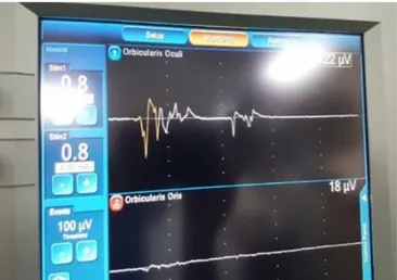
Ethical Committee of Dicle University Faculty of Medicine.(02.10.2019/196). Furthermore, a drilling system was used in synchronization with neuromonitoring. The amplitude of the stimulation current of the device used in this study ranged from 0.5 to 1 mAp. The amplitude of the stimulation current can be varied in 0.1 mA increments. This device was used for mastoid and cochlear implant surgeries at amplitudes of 0.8 mAmp and 0.5 mAmp, respectively. Facial movements were evoked by stimulation via superficial sensors placed on the same side of the face (on the rims of the mouth and eye, and the sternal part of the neck as ground electrodes). After detecting these activities, the electrodes transmitted them to the main device in the form of electrical current. These were displayed as sound or action potentials in the dashboard panel of the monitoring device, allowing us to indirectly track the nerve activities (Figure B).
Figure A: Image of patient with facial stimulator inserted
The referred surgical landmarks were the digastric ridge, the lateral wall of the attic, the lateral semicircular canal, fossa incudis, the stapes and its tendon, the pyramidal process (at the second turn level), the level of the short arm of the intact incus, the cochleariform process, the mastoid and tympanic segments of the facial nerve, and the level of the geniculate ganglion. The 171 patients who underwent surgery, of the remaning 30 were randomly
selected otologic cases that were monitered. The remaining 30 otologic surgery patients were not monitored and compared statistically for the duration of the operation and the emerging complications. In this study, monitoring patients using as stimulator prop were used.
Figure B: Monitor image of action potentials obtained by nerve
stimulation
RESULTS
Of the 171 patients (89 females, 82 males) who underwent otologic and neuro-otologic surgery, the youngest patient was 2 years old and the oldest was 58 years old. Gender distribution, the type of the surgery, and the numerical data of the patients have that presented in Table 1.
Table I: Gender distribution, the type of the surgery, and the
numerical data of the patients.
Surgical Type Female Male Total
Cochlear Implant 83 78 161
Neuro-otologic surgery 6 4 10
810 Of the study patients; no responses were detected in seven despite the stimulation. Response occurred only after increasing the amplitude of the stimulation current in two patients. In the remaining 162 patients, the responses were detected. Of the seven patients, in whom no responses were detected; electrode contact was lost in three, nerve fatigue was found in two, and muscle-relaxant use for anesthesia was detected in one patient; while, no causes were identified in one patient. Despite the presence of responses, a transient postoperative paresis was observed in one patient. In cochlear implant patients, the mean duration of surgery was 2.35 hours without monitoring; whereas, it was 1.45 hours in monitored patients. There were no significant differences in the rate of emerging complications.
There are some additional results detected in our study although they were not predetermined as study objectives. These are as follows: A stimulation current in the range from 0.8 to 1 mAmp applied at a 1 mm bone thickness is required to stimulate the nerve. It requires approximately from 0.1 to 0.2 mAmp current to stimulate the exposed nerve in the temporal bone. When the nerve is covered with a thin layer of bone, the threshold of stimulation can be increased to 0.5 mA. Stimulation occurs at a current intensity of 0.5 mA in the pontocerebellar angle.
DISCUSSION
The key findings of our study are increased safety of the surgery and shortened duration of operation accompanied by a safe surgical procedure. An interesting finding is that no differences were observed in the complication rates The major probable reasons for this conclusion of low complication rates in the absence of monitoring can be the routine preoperative evaluation of the tomography images of the patients and the experience of the surgeon parallelling to the learning curve of the
surgical anatomy. Furthermore; based on the radiologic imaging findings, selection of the patients without a deviant facial nerve course in the respective surgical site can be a major factor leading to the lack of difference in the complication rates. Facial nerve monitoring is not an all-in-one remedy and it may not replace the anatomical knowledge. As Prass10 stated in
1996, "an inappropriate setting, equipment failure or user errors can lead to more untoward consequences compared to potential adverse consequences of no facial monitoring". Monitorization not only creates a secure surgical site but it can also save time for the surgeon; as we observed in our study. For instance, a thorough anatomical knowledge of the facial nerve course is essential during the removal of the granulation tissue in the tympanic sinus or facial recess. Monitoring can ensure complete elimination of the pathologic tissue without damaging the facial nerve. Thereby, it can allow for the removal of the pathological tissue without needing repeat surgery. A synchronized drilling system with neurostimulation will not only signal during the operation when the facial nerve course is approached but it will also contribute to accomplishing a fine surgical procedure, taking the anatomical landmarks into account. Some factors affect stimulation-related signs becoming manifest during neuromonitoring. The major one is the use of muscle relaxants. It should be ensured that the effects of curare-like muscle relaxants used for anesthesia induction are completely resolved. Supporting this recommendation, in one of our patients, no activities were observed in response to neurostimulation at the identified area of the facial nerve course.
However, we encountered no complications when we completed the surgical procedures depending on our estimations of the possible course of the facial nerve. Following this case, the issue was solved after informing the anesthetists that no muscle relaxants should be
used for anesthesia or, if their use is needed, the doses should be appropriately adjusted. Secondly, the contact of the electrodes with the facial nerve stimulation areas and fatigue, occurring due to multiple neural stimulations, may reduce the capacity of the nerve to be stimulated. Facial nerve monitoring does not eliminate the requirement for a thorough anatomical knowledge of the surgeon. However, the use of facial nerve monitoring will allow for extending the margins of the surgical site and enable an active removal process of the pathology. Another key finding of our study is our observation that the amplitude of the stimuli was similar to the reports in the literature regardless of whether a stimulator alone or one synchronized with a drilling system was used11.
During the excision of the granulation tissue; traction of the nerve due to a facial nerve engagement with the drill, the aspirator tip, or the pick elevator; or facial nerve stimulation evoked by a generated electric environment in the pontocerebellar angle due to the irrigation solution may induce a warning signal in the monitor12,13. The reason for finding low
threshold levels of stimulation in the inner ear or the pontocerebellar angle may probably be the absence of the nerve sheath of the facial nerve in these regions. Facial contractions may occur due to the contact of the probe with the superior vestibular nerve in the presence of vestibulo-facial anastomotic fibers. However, we did not have the chance to experience it as we did not perform any vestibular neurectomies. It is reported that even the cerebrospinal fluid can transmit an electrical stimulus. Even though it is reported that this effect is minimal, the probability of this factor should be considered during surgery. In acoustic tumor excision procedures, facial nerve functions may diminish proportionally with the tumor size. This type of cases elevates the neurostimulation threshold as high as 0.8 mA. A lack of neurostimulation despite a
stimulation current of 3 mAmp may suggest a disruption in nerve integrity. In summary, neuromonitoring is associated with many advantages. Guntinas et al. conducted a study about the parotid gland surgery and they reported similar results to those of our study in regards to the functional results and the duration of the surgery14.
Different approaches to facial nerve monitoring are available. Babin et al.15 developed a feline
facial nerve model to evaluate the safety of repetitive facial nerve stimulations. To the facial nerve of the cats, they applied 3 stimulation currents of 1 mA per second during one hour. They reported that a transient loss of sensitivity occurred for a few minutes after the cessation of the stimuli; however, they reported that no permanent changes occurred in the sensitivity of the facial nerve based on the findings of their experimental study. Particularly over the last 10 years, thousands of patients were monitored with electrophysiologic techniques, including the intraoperative stimulation; however, no evidence was reported to support any harmful effects of neurostimulation15-17. Thereby;
despite the lack of objective evidence, many surgeons are convinced that facial nerve monitoring is useful during otologic surgery and it is feasible to use it in routine otologic procedures18,19. Kassam et al.20 reported that
the risk of facial paralysis was reduced in association with the use of neuromonitoring in parotid gland surgery.
In conclusion that this technological method may contribute significantly to obtaining electrophysiologic data, as well as ensuring the safety of the operation and reducing the complication rates.
Declaration of Conflicting Interests: The
authors declare that they have no conflict of interest.
Financial Disclosure: No financial support
812
REFERENCES
1. Green JD, Shelton C, Brackmann DE. Iatrogenic facial nerve injury during otologic surgery. Laryngoscope. 1994; 104: 922-6.
2. Marina MB, Mazita A, Fadzilah I, et al. Iatrogenic facial nerve palsy: lessons to learn. Singapore Med J. 2009; 50: 1154-7.
3. Silverstein H. Adaptor for continuos Stimulation (SACS) with the WR-S8 monitor stimulator. Otolaryngol Head Neck Surg. 1990; 103: 493-6.
4. Schuring AG. Iatrogenic facial nerve injury. Am J Otol. 1988; 9: 432-3.
5. Nilssen EL, Wormald PJ. Facial nerve palsy in mastoid surgery. J Laryngol Otol. 1997; 111: 113-6.
6. Selesnick SH, Lynn-Macrae AG. The incidence of facial nerve dehiscence at surgery for cholesteatoma. Otol Neurotol. 2001; 22: 129-32.
7. Wang H-M, Lin J-C, Lee K-W, et al. Analysis of mastoid findings at surgery to treat middle ear cholesteatoma. Arch Otolaryngol Head Neck Surg. 2006; 132: 1307-10. 8. Delgado TE, Bucheit WA, Rosenholtz HR, et al.
Intraoperative monitoring of facila muscle evoked responses obtained by intracranial stimulation of the facila nerve: a more accurate technique for facila nerve dissection. Neurosurgery. 1979; 4: 418-21.
9. Roland PS, Meyerhoff WL. Intraoperative electrophysiological monitoring of the facial nerve: Is it standard of practice? Am J Otolaryngol. 1994; 15: 267-70.
10. Prass RL. Iatrogenic facial nerve injury: the role of facial nerve monitoring. Otolaryngol Clin North Am. 1996; 29: 265-75.
11. Dr. Nuri Özgirgin, Dr. Tuncay Özçelik, Dr. Selçuk Aksoy, Dr. Barkın Gürcan. Fasiyal sinir monitorizasyonu. K.B.B. ve Baş Boyun Cerrahisi Dergisi.1993; l: 205-8.
12. Silverstein H., Smouha EE., Jones R. Routine intraoperative facial nerve monitoring during otologic surgery. The American Journal of Otology.1988; 9: 269-75.
13. Leonetti JP, Matz GJ, Smith PG. Facial nerve monitoring in otologic surgery: clinical indications and intraoperative technique. Ann Otol Rhinol Laryngol. 1990; 99: 911-8.
14. Guntinas-Lichius O, Eisele DW. Facial Nerve Monitoring. Adv Otorhinolaryngol. 2016; 78: 46-52. 15. Babin RW, Ryu JH, McCabe BF. Responses to step and
repeated impulse accelerations in second order vestibular neurons of the cat. Am J Otolaryngol. 1980; 1: 385-923.
16. Boublata L, Belahreche M, Ouchtati R, et al. Int Vestibular Schwannomas Surgery Operated By Retrosigmoid Transmeatal Approach in Semi-sitting Position with Intraoperative Facial Nerve Monitoring. World Neurosurg. 2017; 103: 231-40.
17. Yingling CD, Gardi JN. Intraoperative monitoring of facial and cochlear nerves during acoustic neuroma surgery. 1992. Neurosurg Clin N Am. 2008; 19: 289-315.
18. Chen L, Chen L, Liu L, et al. Vestibular schwannoma microsurgery with special reference to facial nerve preservation. Clin Neurol Neurosurg. 2009; 111: 47-53. 19. Yingling CD, Gardi JN. Intraoperative monitoring of facial and cochlear nerves during acoustic neuroma surgery. 1992. Neurosurg Clin N Am. 2008; 19: 289-315.
20. Kassam S Chegini S,M Kumar. Pitfalls in monitoring of the facial nerve during operations for parotid cancer. British Journal of Oral and Maxillofacial Surgery.2017; 55: 863–4.