Smad2 and Smad4 gene mutations in hepatocellular carcinoma
MC Yakicier*
,1, MB Irmak
1, A Romano
1,3, M Kew
2and M Ozturk*
,11Department of Molecular Biology and Genetics, Bilkent University 06533 Bilkent, Ankara, Turkey; 2University of The
Witwatersrand, Johannesburg, South Africa
TGF-b is a negative regulator of liver growth. Smad family of genes, as mediators of TGF-b pathway, are candidate tumor suppressor genes in hepatocellular carcinoma (HCC). We studied 35 HCC and non-tumour liver tissues for possible mutations in Smad2 and Smad4 genes. Three tumours displayed somatic mutations; two in Smad4 (Asp332Gly and Cys401Arg) and one in Smad2 (Gln407Arg) genes. All three mutations were A:T ? G:C transitions suspected to result from oxidative stress as observed in mitochondrial DNA. These observation demonstrate that TGF-b pathway is altered in hepatocellular carcinoma.
Keywords: hepatocellular carcinoma; Smad2; Smad4; TGFb; tumor suppressor genes; somatic mutation
Introduction
Hepatocellular carcinoma (HCC), one of the ten most frequent cancers world-wide, is associated with well de®ned viral and non-viral etiological factors. Chronic infection with hepatitis B (HBV) and hepatitis C (HCV) viruses, and oral intake of a¯atoxins are the major causes of HCC, but the molecular mechanisms underlying the malignant transformation of hepato-cytes are largely unknown (Ozturk, 1995). Long latent period (20 ± 30 years) between viral infections and the development of HCC suggests the multi-step nature of hepatocarcinogenesis (Ozturk, 1995). Genetic studies provide sucient evidence for this hypothesis. Allelo-type studies indicate that many chromosomal regions (i.e. 1p, 1q, 4q, 5q, 6q, 8p, 8q, 9p, 10q, 11p, 13q, 14q, 16p, 16q, 17p) undergo structural changes in HCC (Nagai et al., 1997; Piao et al., 1998; for review see Grisham, 1996). Some of these chromosomal changes are associated with dierent tumour stages, suggesting that they are associated with the tumour progression (Tsuda et al., 1990). The critical genes located at these chromosomal regions are mostly unknown. To date only a few genes including p53, p16, mannose-6-phosphate/insulin-like growth factor II receptor (M6P/IGFIIR), b-catenin (Miyoshi et al., 1998; De la Coste et al., 1998) and cyclin D have been shown to be signi®cantly altered in HCC (for review see Grisham, 1996). Both the suspected genetic heterogeneity of HCC and the discordance between allelotype and mutation studies strongly suggest that many other genes undergo somatic mutations in these tumours.
Genes encoding for proteins involved in the control of hepatocyte growth (i.e. inhibitors of hepatocyte proliferation and the activators of apoptotic cell death) are potential tumour suppressor genes for HCC. Transforming growth factor-b (TGF-b) and activin are potent inhibitors of hepatocyte growth (Fausto et al., 1995). TGF-b is also known to induce apoptosis in both hepatocytes and HCC cell lines (Oberhammer et al., 1992; Gressner et al., 1997). Certain transformed cells, however, show defective response to TGF-b-induced growth inhibition including some liver-derived cell lines (Fynan and Reiss, 1993). Based on these observations, genes involved in TGFb signalling pathway are candidate tumour suppressor genes in HCC. For instance, M6P/IGFIIR gene was shown to be mutated in about 25% of HCCs displaying LOH at the M6P/IGFIIR locus (De Souza et al., 1995). Although the protein product of this gene has a broad range of cellular functions, it is suspected to be an activator of latent TGF-b in the liver. In addition, at least two other genes involved in HCC, namely cyclin D and c-myc genes appear to be among the ultimate targets of TGFb signalling pathway, as their expression level is modi®ed following treatment of dierent cell lines with TGFb (Alexandrow and Moses, 1995, Grisham, 1996).
Recently, the Smad family of proteins have been discovered as mediators of TGFb signalling pathway. Eight distinct members of Smad family have been identi®ed in vertebrates and at least two of them, namely Smad2 and Smad4 act as tumour suppressor genes in humans (Heldin et al., 1997; Baker and Harland, 1997). Smad4 and Smad2 are both involved in cytoplasmic signal transduction upon activation of TGF-b and activin receptors by their speci®c ligands (for review see Heldin et al., 1997). Missense mutations within carboxyl terminal eector domains of Smad2 and Smad4 have been identi®ed in dierent cancers including the cancers of pancreas (Hahn et al., 1996; Moskaluk et al., 1997), biliary tract (Hahn et al., 1998), colon (Eppert et al., 1996; Riggins et al., 1996; Takagi et al., 1996), lung (Uchida et al., 1996), as well as head and neck carcinomas (Kim et al., 1996). Smad2 and Smad4 appear to be the most critical targets of mutational inactivation because a study based on 167 tumour samples suggested that mutational inactivation of the other Smad genes does not account for the widespread resistance of cancer cells to TGF-b (Riggins et al., 1997). Based on these indications, we selected Smad2 and Smad4 (also called JV18-1 and DPC4 respectively) as candidate tumour suppressor genes in HCC and studied 35 primary tumours as well as six hepatoma cell lines for possible mutations. We demonstrate that both genes display somatic mutations in HCC, but the mutation frequency is low. We will discuss possible implications of these observations in
*Correspondence: M Ozturk and MC Yakicier
3Current address: Institute of Molecular Pathology, A-1030 Vienna,
Austria
Received 18 January 1999; revised 18 March 1999; accepted 18 March 1999
relation with the role of TGFb signalling pathway in hepatocellular carcinogenesis.
Results and Discussion
To investigate the potential involvement of the Smad2 and Smad4 genes in HCCs, we screened 35 HCC samples as well as six hepatoma cell lines for possible genetic alterations of these genes. Initially, six hepatoma cell lines were tested by RT ± PCR for the expression of Smad2 and Smad4 genes. All cell lines expressed both Smad2 and Smad4 genes and RT ± PCR
products did not show size alteration in the respective coding region (data not shown). Homozygous and large deletions of Smad2 and Smad4 genes have been reported for other cancers (Hahn et al., 1996, Riggins et al., 1996). But the hepatoma cell lines tested here did not show such alterations.
Next, we studied genomic DNA from 35 primary tumours. The majority of the previously identi®ed Smad2 and Smad4 gene mutations are located within the region coding for the highly conserved carboxyl terminal domains, corresponding to exon 8 ± 11 of both genes (Hahn et al., 1996; Riggins et al., 1996; Schutte et al., 1996, Hahn et al., 1998). In addition, two mutations, one located in exon 4 of Smad2 and another located in exon 2 of Smad4 aecting the N-terminal regions of Smad2 and Smad4 (Arg133Cys and Arg100Thr, respectively) proteins have been described in colon and pancreas cancers (Hata et al., 1997; Shi et al., 1997). To test whether HCCs display similar mutations, we studied exon 4 of Smad2, exon 2 of Smad4, in addition to exons 8 ± 11 of both genes by SSCP analysis. A total of seven HCC samples showed altered migration pattern (see Figure 1 for exon 8 alterations as an example). Four of these alterations (three in exon 8 and one in exon 10 of Smad4 gene) were also present in the non-tumour liver samples of the respective patients. DNA sequence analysis of these four tumours did not reveal any alteration in exonic sequences and immediately ¯anking intronic regions (data not shown) suggesting that constitutional polymorphisms aecting intronic sequences were present in these patients. The other three SSCP alterations were seen only in tumour DNA, but not in non tumorous liver DNA. Sequence analysis of tumour and non-tumour liver DNA samples from
Figure 1 SSCP analysis of Smad4 gene (exon 8) in hepatocellular carcinomas. T and N denotes tumour and matching non-tumour liver DNA, respectively. T19, T29 and T37, as well as their matching non-tumour liver DNAs (N18, N28 and N36, respectively) display abnormally migrating bands
Figure 2 SSCP and DNA sequence analyses of Smad2 (exon 10) and Smad4 (exons 8 and 9) genes indicate the presence of somatic mutations in three hepatocellular carcinomas. (a) SSCP analysis of tumours demonstrating mutant allelic shifts in tumours T49, T37 and T39 (the asterisk indicates the samples displaying band-shifts); (b) Sequence analysis of hepatocellular carcinoma (T) and non tumour liver tissue (N) DNAs of the samples identi®ed in a. Somatic single base-pair mutations (A:T ? G:C transitions) were underlined
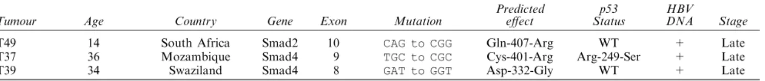
three patients revealed three dierent somatic muta-tions (Figure 2). Two tumour DNAs (T37 and T39) displayed missense mutations in Smad4 gene. An A? G transition at codon 332, leading to a Asp332Gly change was observed in T39. The other Smad4 mutation was a T?C transition at codon 401 leading to a Cys401Arg replacement in T37. One tumour DNA (T49) carried a transition mutation (A?G) at codon 407 of Smad2 gene leading to a Gln407Arg change (Figure 2).
All three of these somatic mutations were novel and have not been described previously. They all appear to be deleterious mutations because of the strong side chain charge changes in the replaced amino acid residues resulting from each mutation. Crystal struc-ture of C-terminal domain of Smad4 reveals that Asp 332 residue is located in the trimeric interface region and forms a hydrogen bond with His 371 (Shi et al., 1997). Because this interface region is crucial for trimeric complex formation, it is likely that Asp332Gly change, breaking the hydrogen bond, blocks the TGFb signal by preventing trimer forma-tion of Smad4 protein. The Cys401 residue of Smad4 is highly conserved between Smad family members. This residue is located within the b6 sheet which makes the hydrophobic core, the b sandwich of Smad4 protein. Amino acid change from cysteine to arginine will most probably aect Smad4 protein function by changing the conformation of the b sandwich. Gln407 of Smad2 is located in a region that is homologous to H4 helix domain of Smad4, also in trimeric interface region (Shi et al., 1997).
All three mutations are located in the MH2 domain of Smad proteins. These proteins consist of three structurally and functionally distinct domains. The amino and carboxyl terminal regions of Smad proteins are referred to as MH1 and MH2 domains, respec-tively. The central region serves as a linker domain between MH1 and MH2 domains. The MH2 domain of Smad 1, 2 and 3 mediates homomeric interactions and is responsible for heteromeric interactions with Smad4. The MH2 domain also called eector domain is able to mimic the activity of the full length proteins (Heldin et al., 1997). Thus it is very likely that the three mutations described here are not random mutations and that they either aect protein-protein interactions or destabilise the whole structure of the protein. Taken together, all three mutations described here appear to be deleterious mutations which may aect TGFb signalling pathway in these tumours.
The tumours described here have been previously examined for the presence of p53 gene mutations and HBV DNA (Unsal et al., 1994). As shown in Table 1, HBV DNA was present in all three tumours with Smad mutations. p53 was mutant (Arg249Ser) in T37 but not in two other tumours. Thus, it appears that Smad mutations described here are associated with HBV
infection and that there appears to be no correlation with p53 mutations. On the other hand, it is noteworthy that all three mutations described here aected A:T pairs with a consistent change to G:C pairs. This pattern was recently shown to be a common type of mitochondrial DNA mutation in colorectal tumours and explained by high level of reactive oxygen species (ROS) in mitochondria (Polyak et al., 1998). Similarly, Smad gene mutations observed here may be caused by high levels of ROS in the precursor cells of these tumours.
Previous studies showed that Smad4 is altered in a signi®cant portion of pancreatic (Hahn et al., 1996) and common bile duct cancers (Hanh et al., 1998) and a minority of colorectal cancers (Eppert et al., 1996; Takagi et al., 1996) but rarely in other tumours (Schutte et al., 1996). Smad2 gene alterations are also limited to a small fraction of colorectal and lung cancers (Riggins et al., 1996, 1997; Uchida et al., 1996. Our data suggest that, Smad2 and Smad4 gene mutations may contribute to a fraction of HCCs. Low prevalence of these alterations may be explained with genetic heterogeneity of HCC (Unsal et al., 1994). Indeed, Kawate et al. (1999) reported very recently that mutations of TGFb-receptor II, Smad2 and Smad4 mutations are rare in HCCs from Japan. In addition, Smad mutations in HCC could occur at late stages during tumor progression. In an animal model of compound heterozygosity for APC and Smad4, inactivation of Smad4 gene resulted in the malignant progression of the intestinal and colonic polyps initiated by LOH in the APC gene. This observation suggested the inactivation of Smad4 as a late event in carcinogenesis (Takaku et al., 1998).
In conclusion, we identi®ed Smad gene mutations in HCC at a frequency of *10%. The occurrence of these mutations provides evidence that TGFb pathway is altered in HCC. In conformation to our hypothesis, TGFb itself was identi®ed as a new form of tumour suppressor for liver and lung cancers in mice heterozygous for deletion of the TGFb gene (Tang et al., 1998). However, the low frequency of Smad gene mutations in human HCC also suggest that other genes of TGFb pathway may also be alterated in these tumours.
Materials and methods Tumour samples and cell lines
We analysed a total of 35 pair of DNA samples isolated from HCC and non-tumour liver tissues from patients living in dierent geographical regions including Mozambique (n=7), South Africa (n=11), China (n=8), Japan (n=6) and Germany (n=3). Characteristics of these tumours and methods for DNA isolation have been described previously (Unsal et al., 1994). We also analysed six established
Table 1 Smad gene mutations in hepatocellular carcinoma
Predicted p53 HBV
Tumour Age Country Gene Exon Mutation eect Status DNA Stage
T49 T37 T39 14 36 34 South Africa Mozambique Swaziland Smad2 Smad4 Smad4 10 9 8 CAG to CGG TGC to CGC GAT to GGT Gln-407-Arg Cys-401-Arg Asp-332-Gly WT Arg-249-Ser WT + + + Late Late Late 4881
hepatoma cell lines (HepG2, Huh7, Mahlavu, PLC/PRF/5, Hep3B and Focus). Genomic DNA from cell lines was isolated as described (Unsal et al., 1994). Total RNA from cell lines were isolated using Ultraspect RNA extraction kit (Biotecx) according to the manufacturer's directions. RT ± PCR
cDNAs were synthesised by MMLV reverse transcriptase (SuperScriptTM, Gibco) using oligo (dT)
12 ± 18 primers. The
entire coding region of Smad2 and Smad4 genes were ampli®ed using primers indicated in Table 2. PCR was performed in 50 ml reaction mixture containing 1 ml of the complementary DNA mix, 16Taq buer (MBI), 1.5 mM
MgCl2, 1 unit of Taq DNA polymerase, 20 pmol of each
primer and 10 mMeach of dNTPs. PCR conditions consisted
of 35 cycles of denaturation at 948C for 60 s, annealing at temperatures varying between 55 ± 568C (see Table 2 for primer sequences and annealing temperatures) for 30 s, followed by an extension at 728C for 90 s. The ®nal cycle included an additional 3 min extension at 728C.
SSCP analysis
PCR was performed using 50 ng genomic DNA, 16Taq buer (MBI), 1.5 mM MgCl2, 1 unit of Taq DNA
polymerase, 20 pmol of each primer, 40 mM each of dNTPs,
2 mCi [a-32P]dATP (1000 Ci/mmol, Amersham) in a ®nal
volume of 25 ml. PCR conditions consisted of 30 cycles of denaturation at 948C for 45 s, annealing at temperatures varying between 50 ± 608C (see Table 2 for primer sequences and annealing temperatures) for 30 s, followed by an extension at 728C for 1 min. The ®nal cycle included an additional 3 min extension at 728C. SSCP analysis of PCR products was done at both 48C and room temperature, on 10% of acrylamide gel with or without glycerol as well as on MDE gels (FMC Bioproducts). Gels were mounted on a 3M Whatman paper, dried and visualised by autoradiography. Sequence analysis
For the analysis of tumour PCR products, aberrantly migrating radioactive bands were recovered from SSCP gels in order to enrich for mutant DNA fragments before sequencing. Selected bands were cut with a razor blade, eluted into TE and reampli®ed as described, except that [a-32P]dATP was omitted. Non-tumour PCR products were
analysed directly following non-radioactive PCR amplifica-tion. PCR products were puri®ed using Quick PCR puri®cation kit (Qiagen). For sequencing reactions, dye terminator cycle sequencing ready reaction kit (ABI Prism) was used. Reaction products were then analysed using the ABI Prism 377 ¯uorescent DNA sequencer (Perkin Elmer). Identi®ed mutations were con®rmed by DNA sequencing of
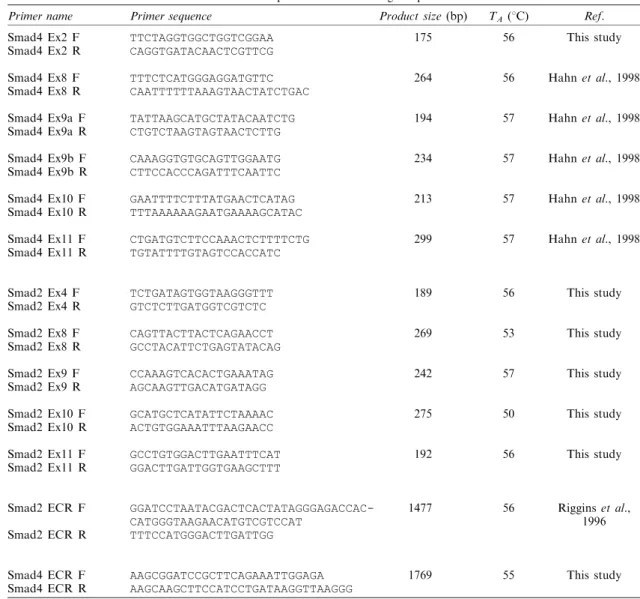
Table 2 List of primers and annealing temperatures
Primer name Primer sequence Product size (bp) TA (8C) Ref.
Smad4 Ex2 F
Smad4 Ex2 R TTCTAGGTGGCTGGTCGGAACAGGTGATACAACTCGTTCG 175 56 This study
Smad4 Ex8 F
Smad4 Ex8 R TTTCTCATGGGAGGATGTTCCAATTTTTTAAAGTAACTATCTGAC 264 56 Hahn et al., 1998
Smad4 Ex9a F
Smad4 Ex9a R TATTAAGCATGCTATACAATCTGCTGTCTAAGTAGTAACTCTTG 194 57 Hahn et al., 1998
Smad4 Ex9b F
Smad4 Ex9b R CAAAGGTGTGCAGTTGGAATGCTTCCACCCAGATTTCAATTC 234 57 Hahn et al., 1998
Smad4 Ex10 F
Smad4 Ex10 R GAATTTTCTTTATGAACTCATAGTTTAAAAAAGAATGAAAAGCATAC 213 57 Hahn et al., 1998
Smad4 Ex11 F
Smad4 Ex11 R CTGATGTCTTCCAAACTCTTTTCTGTGTATTTTGTAGTCCACCATC 299 57 Hahn et al., 1998
Smad2 Ex4 F
Smad2 Ex4 R TCTGATAGTGGTAAGGGTTTGTCTCTTGATGGTCGTCTC 189 56 This study
Smad2 Ex8 F
Smad2 Ex8 R CAGTTACTTACTCAGAACCTGCCTACATTCTGAGTATACAG 269 53 This study
Smad2 Ex9 F
Smad2 Ex9 R CCAAAGTCACACTGAAATAGAGCAAGTTGACATGATAGG 242 57 This study
Smad2 Ex10 F
Smad2 Ex10 R GCATGCTCATATTCTAAAACACTGTGGAAATTTAAGAACC 275 50 This study
Smad2 Ex11 F
Smad2 Ex11 R GCCTGTGGACTTGAATTTCATGGACTTGATTGGTGAAGCTTT 192 56 This study
Smad2 ECR F Smad2 ECR R GGATCCTAATACGACTCACTATAGGGAGACCAC-CATGGGTAAGAACATGTCGTCCAT TTTCCATGGGACTTGATTGG 1477 56 Riggins et al., 1996 Smad4 ECR F
Smad4 ECR R AAGCGGATCCGCTTCAGAAATTGGAGAAAGCAAGCTTCCATCCTGATAAGGTTAAGGG 1769 55 This study
Ex and ECR identify primers used for PCR ampli®cations of exons and the entire coding regions, respectively 4882
independent PCR products after cloning into pGEM-T plasmid (Promega).
Acknowledgements
This work was supported by grants from TUBITAK and TWAS. We thank Dr Marie Ricciardone and Ms Birsen
Cevher for help in DNA sequence analyses and Mrs Lut®ye Mesci for oligonucleotide synthesis. We also thank Dr SE Kern for providing us with exon-intron boundary sequence information for Smad4 gene.
References
Alexandrow MG and Moses HL. (1995). Cancer Res., 55, 1452 ± 1457.
Baker JC and Harland RM. (1997). Curr. Opin. Genet. Dev., 7, 467 ± 473.
De la Coste A, Romagnolo B, Billuart P, Renard CA, Buendia MA, Soubrane O, Fabre M, Chelly J, Beldjord C, Kahn A and Perret C. (1998). Proc. Natl. Acad. Sci. USA, 95, 8847 ± 8851.
De Souza AT, Hankins GR, Washington MK, Orton TC and Jirtle RL. (1995). Nat. Genet., 11, 447 ± 449.
Eppert K, Scherer SW, Ozcelik H, Pirone R, Hoodless P, Kim H, Tsui LC, Bapat B, Gallinger S, Andrulis IL, Thomsen GH, Wrana JL and Attisano L. (1996). Cell, 86, 543 ± 552.
Fausto N, Laird AD and Webber EM. (1995). FASEB J., 9, 1527 ± 1535.
Fynan TM and Reiss M. (1993). Crit. Rev. Onco., 4, 493 ± 540.
Grisham JW. (1996). Carcinogenesis, 18, 59 ± 81.
Gressner AM, Lahme B, Mannherz H-G and Polzar B. (1997). J. Hepatol., 26, 1079 ± 1092.
Hahn SA, Bartsch D, Schroers A, Galehdari H, Becker M, Ramaswamy A, Schwarte-Waldho I, Maschek H and Schmiegel W. (1998). Cancer Res., 58, 1124 ± 1126. Hahn SA, Schutte M, Shamsul Hoque ATM, Moskaluk CA,
da Costa LT, Rosenblum E., Weinstein CL, Fischer A, Yeo CJ, Hruban RH and Kern SE. (1996). Science, 271, 350 ± 353.
Hata A, Lo RS, Wotton D, Lagna G and Massague J. (1997). Nature, 388, 82 ± 87.
Heldin C-H, Miyazono K and Dijke PT. (1997). Nature, 390, 465 ± 471.
Kawate S, Takenoshita S, Ohwada S, Mogi A, Fukusato T, Makita F, Kuwano H and Morishita Y. (1999). Int. J. Oncol., 14, 127 ± 131.
Kim SK, Fan Y, Papadimitrakopoulou V, Clayman G, Hittelman WN, Ki Hong W, Lotan R and Mao L. (1996). Cancer Res., 56, 2519 ± 2521.
Miyamura T and Hirohashi S. (1990). Proc. Natl. Acad. Sci. USA, 87, 6791 ± 6794.
Miyoshi Y, Iwao K, Nagasawa Y, Aihara T, Sasaki Y, Imaoka S, Murata M, Shimano T and Nakamura Y. (1988). Cancer Res., 58, 2524 ± 2527.
Moskaluk CA, Hruban RH, Schutte M, Lietman SA, Symrk T, Fusaro L, Fusaro R, Lynch J, Yeo CJ, Jackson CE, Lynch HT and Kern SE. (1997). Diag. Mol. Pathol., 6, 85 ± 90.
Nagai H, Pineau P, Tiollais P, Buendia MA and Dejean A. (1997). Oncogene, 14, 2927 ± 2933.
Oberhammer FA, Pavelka M, Sharma S, Tiefenbacher R, Purchio AF, Bursch W and Schulte-Hermann R. (1992). Proc. Natl. Acad. Sci. USA, 89, 5408 ± 5412.
Ozturk M. (1995). Biology of hepatocellular carcinoma. In: Gastrointestinal Cancers Ð Biology, diagnosis, and ther-apy, Rustgi AK (ed). Lippincott-Raven Publishers: Philadelphia, pp. 511 ± 525.
Piao Z, Park CH, Park J-H and Kim H. (1998). Int J. Cancer, 75, 29 ± 33.
Polyak K, Li Y, Zhu H, Lengauer C, Willson JKV, Markowitz SD, Trush MA, Kinzler KW and Vogelstein B. (1998). Nat. Genet., 20, 291 ± 293.
Riggins GJ, Thiagalingam S, Rozenblum E, Weinstein CL, Kern SE, Hamilton SR, Wilson JKV, Markowitz SD, Kinzler KW and Vogelstein B. (1996). Nat. Genet., 13, 347 ± 349.
Riggins GJ, Kinzler KW, Vogelstein B and Thiagalingam S. (1997). Cancer Res., 57, 2578 ± 2580.
Schutte M, Hruban RH, Hedrik L, Cho KR, Nadasdy GM, Weinstein CL, Bova GS, Isaacs WB, Cairns P, Nawroz H, Sidransky D, Casero RA, Meltzer PS, Hahn SA and Kern SE. (1996). Cancer Res., 56, 2527 ± 2530.
Shi Y, Hata A, Lo RS, Massague J and Pavletich N. (1997). Nature, 388, 87 ± 93.
Takagi Y, Kohmura H, Futamura M, Kida H, Tanemura H, Shimokowa K and Saji S. (1996). Gastroenterology, 111, 1369 ± 1372.
Takaku K, Oshima M, Miyoshi H, Matsui M, Seldin MF and Taketo MM. (1998). Cell, 92, 645 ± 656.
Tang B, Bottinger EP, Jakowlew SB, Bagnall KM, Mariano J, Anver MR, Letterio JJ and Wake®eld LM. (1998). Nature Med., 4, 802 ± 807.
Tsuda H, Zhang W, Shimosato Y, Yokota J, Terada M, Sugimura T, Miyamura T and Hirohashi S. (1990). Proc. Natl. Acad. Sci. USA, 87, 6791 ± 6794.
Uchida K, Nagatage M, Osada H, Yatabe Y, Kondo M, Mitsudomi T, Masuda A, Takahashi T and Takahashi T. (1996). Cancer Res., 56, 5583 ± 5585.
Unsal H, Yakicier MC, Marcais C, Kew M, Volkmann M, Zentgraf H, Isselbacher KJ and Ozturk M. (1994). Proc. Natl. Acad. Sci. USA, 91, 822 ± 826.