Science
REVIEW
Cite this:Biomater. Sci., 2013, 1, 1119
Received 13th June 2013, Accepted 13th August 2013 DOI: 10.1039/c3bm60150a www.rsc.org/biomaterialsscience
Neural di
fferentiation on synthetic scaffold materials
Busra Mammadov, Melike Sever, Mustafa O. Guler* and Ayse B. Tekinay*
The potential of stem cells to differentiate into a variety of subgroups of neural cells makes stem cell differentiation and transplantation a promising candidate for neurodegenerative disorder therapies. However, selective differentiation of stem cells to neurons while preventing glial scar formation is a complex process. Mimicking the natural environment of neural tissue is pivotal, thus various synthetic materials have been developed for this purpose. The synthetic scaffolds can direct stem cells into a neural lineage by including extracellular factors that act on cell fate, which are mainly soluble signals, extracellu-lar matrix proteins and physical factors (e.g. elasticity and topography). This article reviews synthetic materials developed for neural regeneration in terms of their extracellular matrix mimicking properties. Functionalization of synthetic materials by addition of bioactive chemical groups and adjustment of physical properties such as topography, electroactivity and elasticity are discussed.
1.
Introduction
Neurodegeneration is the progressive loss of structure and function of nerve cells. Degeneration is a common pheno-menon in many neurodegenerative diseases and nerve injury. Neurodegeneration generally occurs due to the accumulation of misfolded aggregated proteins in some parts of the aging brain and, as a result, cell death and inflammatory damage occur in those areas in the brain.1 Clinically, there are
different types of neurodegeneration in different neurodegen-erative diseases. Therefore, there are some differences in the proximal triggers and pathological markers such as Lewy bodies in Parkinson’s disease, plaques and tangles in Alzhei-mer’s disease, demyelination in multiple sclerosis and motor-neuron death in ALS. On the other hand, despite different trig-gering factors, these diseases share some overlapping down-stream and secondary pathways such as neuroinflammation. Adult central nervous system cells have poor regeneration capacity, so any damage to the central nervous system might be permanent. Lost cells cannot be replaced with new func-tional ones, and remaining nerve cells cannot make new con-nections after injury due to the inhibitory properties of the extracellular matrix.2 Besides neurodegenerative disorders,
Busra Mammadov
Busra Mammadov graduated
from Department of Molecular Biology and Genetics at Middle
East Technical University
(METU) in 2006 and obtained her MS degree in Biology at
Fatih University in 2008.
Currently she is a PhD candidate at Institute of Materials Science and Nanotechnology, National Nanotechnology Research Center (UNAM), Bilkent University. She is working on development of synthetic scaffolds for neural regeneration and testing these scaffolds in vitro for induction of neural differentiation and in vivo for neural regeneration.
Melike Sever
Melike Sever graduated from Department of Molecular Biology and Genetics at Middle East Technical University (METU), Turkey in 2011. She obtained her MSc degree in Biochemistry from the same university in 2012. Currently, she is pursuing
her doctoral research in
Materials Science and Nanotech-nology at National Nanotechno-logy Research Center (UNAM), Bilkent University, Turkey. She is
continuing her research on
neural tissue regeneration at Nanobiotechnology Laboratory. Institute of Materials Science and Nanotechnology, National Nanotechnology
Research Center (UNAM), Bilkent University, Ankara, Turkey 06800. E-mail: [email protected], [email protected]
Published on 20 September 2013. Downloaded on 7/19/2018 6:14:32 PM.
View Article Online
traumatic injuries such as spinal cord injuries have destructive effects on motor, sensory, and autonomic functions. It gene-rally causes a permanent loss of sensation below the site of the injury. In the case of peripheral nerve injuries, cells in the per-ipheral nervous system have a regeneration capacity unlike the ones in the central nervous system.
Fully restoring the functional capacities of neurons after damage due to traumatic injuries or neurodegenerative dis-orders is still not possible. Although conventional therapies provide neural regeneration up to a certain level, they are not as efficient as desired. When the neural regeneration term is used, it refers to several different mechanisms to restore the functions of the degenerated neural tissue. It either occurs by the generation of new neurons from the progenitor cells resid-ing nearby the damaged area, the generation of new synapses by the neurons that survive after the damage or by the repair of the axons/myelin sheets around the axons to prevent sec-ondary cell loss after injury. It is more effective to combine these strategies in order to achieve functional regeneration of the nervous system. However, due to some intrinsic characteri-stics of the nervous system such as the unproliferative nature of neurons, the presence of progenitors in very localized areas in low numbers along with the upregulation of inhibitory elements upon injury leading to glial scar formation, the regeneration capacity of the central nervous system (CNS) is quite limited. This limited capacity of regeneration is aimed to be improved by materials designed for stem cell culture, di ffer-entiation and transplantation, which are called‘scaffolds’.
Stem cells are promising candidates for the treatment of neurological disorders since they can differentiate into neural cells when induced appropriately. Their differentiation can be enhanced by using bioactive materials, which can also be used as vehicles for cell transplantation to the damage site. In pre-vious studies, various synthetic materials have been analyzed as scaffolds in vitro and in vivo for their potential to induce neural differentiation and nerve regeneration.3–6This review is focused on synthetic materials developed for neural regene-ration including polymeric materials and self-assembled
systems, and their modifications to support neural
differentiation.
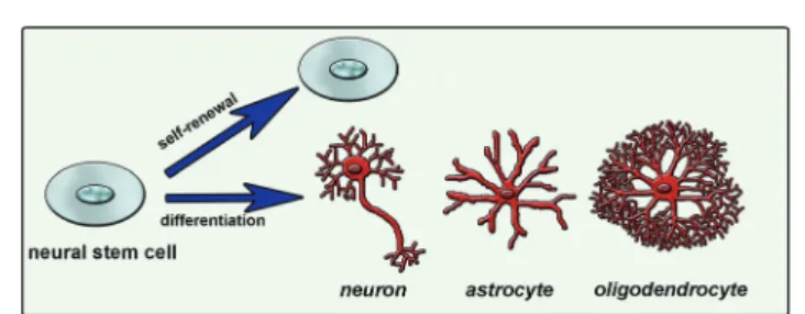
Natural cues for directing cell fate consist of different chemical, physical and biological signals in the extracellular matrix (ECM), neighboring cell interactions, soluble factors such as chemokines, growth factors and hormones, and the inherited potency of the cell.7 Combination of these factors and their interactions affects the ultimate cellular differen-tiation mechanisms (Fig. 1). Understanding this complex environment and mimicking it as efficiently as possible will enable the directing of cell differentiation in a desired manner. One way of mimicking the natural environment of cells in vitro is to supply soluble factors in the culture media. This approach is relatively simple but costly since it requires a high concentration of soluble factors. Also it is not very efficient since it does not mimic the mechanical properties of the cells’ natural environment. Cells are embedded in tissues in a three-dimensional (3D) manner and they can migrate or grow extensions such as axons and dendrites of neural cells. Physical properties of the 3D network including stiffness, roughness and pore size are also important and should be con-sidered while designing a synthetic system that mimics the native tissue. As conventional in vitro cell culture surfaces cannot provide an appropriate environment to cells, 3D scaffolds combined with soluble signals or bioactive signals
Mustafa O. Guler
Mustafa O. Guler is a professor of Materials Science and Nanotech-nology at Bilkent University. He received his PhD degree in chem-istry from Northwestern University in Evanston, IL, USA in 2006. After receiving his PhD, he had worked at the Institute for
Biona-notechnology in Medicine at
Northwestern University and
Nanotope Inc. in Chicago, IL, USA until 2008. His research is based on discoveries of nanostructures at the interface of chemistry, biology, and materials science.
Ayse B. Tekinay
Ayse B. Tekinay is a professor of Materials Science and Nanotech-nology at Bilkent University. She received her PhD degree in mole-cular biology at the Rockefeller University, New York, USA in 2006. After receiving her PhD, she continued her post-doctoral studies at the Rockefeller Univer-sity until 2009. Her research focus is nanobiotechnology, cell-extracellular matrix interactions, and use of extracellular matrix platforms for tissue regeneration and regenerative medicine. Fig. 1 Neural stem cell fate determination is mainly guided by extracellular matrix molecules, soluble factors and cell-to-cell interactions.
tethered to the surface are used to mimic the natural neural niche.
Natural extracellular matrix proteins or synthetic scaffolds have been used for developing 3D scaffolds to mimic the bio-logical, chemical and mechanical properties of natural matrix of the cells. Although natural proteins are biocompatible, it is not easy to functionalize these scaffolds with desired bioactive groups, or to manipulate their mechanical properties such as stiffness. Thus, these scaffolds might not be sufficient to direct the differentiation of cells especially into complex cells like neurons.8–12 On the other hand, synthetic materials can be synthesized with desired functionalities such as specific bioactive groups, stiffness and roughness in order to mimic the natural environment of cells.
2.
Approaches in the design of synthetic
materials for neural differentiation
A variety of different methods can be used to develop scaffold materials with desired properties, including the support of cell survival and the induction of differentiation into desired cell types. Besides changing the material type, nerve regeneration studies have also focused on designing scaffolds with incor-porated bioactivity for the induction of neurogenesis. One approach to induce differentiation is by adding soluble indu-cers while culturing cells on non-bioactive scaffolds. In this approach, a scaffold is a better environment for cell survival compared to a tissue culture plate. However, as the scaffold itself does not include any signal for differentiation, differen-tiation is induced solely by the soluble inducers added to the growth media. Soluble inducers commonly used for neural differentiation include neurotrophic factors (nerve growth factor (NGF), brain-derived neurotrophic factor (BDNF), neuro-trophin-3 (NT-3) etc.) and retinoic acid (RA).
Another approach is by embedding the soluble inducers
mentioned above into the scaffold in order to provide a
gradual release or by tethering these inducers to the surface of the scaffold through functional groups, in order to provide spatial organization to cells. In the case of gradual release, the scaffold is not bioactive but has a role in differentiation by releasing inducers at desired concentrations over a longer time period. When tethering the inducers to the surface, the scaffold is bioactive, however its bioactivity is not directly related to the differentiation process. Bioactive groups on the material’s surface are presented so that they bind to the soluble inducers and present them similar to the extracellular matrix components (such as heparan sulfate proteoglycans) presenting growth factors to the cells. Soluble factors can also be immobilized on material surfaces directly by covalent
attachment without the use of the growth factor affine
proteoglycans.13
Functionalization of the surface with bioactive signals is a method used for differentiation induction directed by scaffold. Bioactive groups can be either chemical groups inspired by the natural environment of cells in a specific tissue, like the brain,
or functional groups of natural inducers such as peptides found in neural differentiation-inducing proteins. Besides the addition of bioactive signals, scaffolds can also be functio-nalized by tuning their mechanical properties and physical morphology in order to support neural cell survival and di ffer-entiation. Since they present bioactive groups, these scaffolds can be used alone for the induction of differentiation, depend-ing on the complexity of the final cell type and potency of the starting cell for differentiation into desired cell phenotypes (Fig. 2). Such a scaffold can induce differentiation into
different neuronal subtypes. However, it might not be
sufficient for maturation of the induced cells into functional cells. Additional inducers can be added to the culture medium to promote maturation or the scaffold can be functionalized with multiple bioactive groups to overcome the maturation problem.
2.1. Use of synthetic scaffolds in combination with soluble inducers
Nonbioactive synthetic scaffolds can be used in combination with soluble inducers. These scaffolds combined with soluble inducers are not actively involved in the differentiation process; however, they support differentiation by providing a 3D environment for cells that is more similar to their natural environment when compared to two-dimensional (2D) tissue culture surfaces. Neurons can have outgrowths in all directions within 3D scaffolds, thus 3D scaffolds are more accurate rep-resentations of in vivo tissue architecture, compared to 2D scaffolds. They provide in vivo-like cell–cell interactions which increases cellular survival and leads to more realistic gene expression and cellular behavior. 2D cultures have less com-patibility with in vivo systems. For example, it has been shown that dopaminergic neurons isolated from an embryonic brain display longer viability in 3D systems when compared to monolayer 2D cultures.14On the other hand, nutrient depri-vation in 3D scaffolds causes more severe alterations in the expression of specific genes, cell proliferation and viability as well as productivity. Also, it was shown that mechanical inju-ries cause a more severe response in cells grown in 3D neural Fig. 2 Bioactive scaffold design for neural differentiation of stem cells.
cultures than 2D scaffolds even when they are subjected to the same strain and strain rate.15
These scaffolds do not have bioactive groups and they usually do not induce differentiation by themselves, except for when stiffness is used to control differentiation pathway.8–12
Hence, defining an optimal cocktail of soluble inducers such as growth factors plays an essential role in the success of differentiation; however, it is costly and complex. Inducers are often chosen by considering the extracellular signals that play a role in neural cell survival and differentiation in the central nervous system. This approach is beneficial in terms of the ease of material synthesis as the material is not further modi-fied and it is sufficient that the material does not interfere with cell survival. However, the lack of bioactivity makes the use of additional inducers, such as growth factors, inevitable. The selection of soluble inducers and their concentrations should be optimized to achieve the differentiation of cells cul-tured on nonbioactive scaffolds. Several different materials, mostly polymers, are used for the production of such scaffolds, examples of which are given below.
Nanofibrous poly-L-lactic acid (PLLA) scaffolds with high
porosity were used for differentiation of neural stem cells (NSCs) and found to support neurite outgrowth.16 Poly(ethyl-ene-co-vinyl alcohol) (EVAL) membranes are another type of polymeric scaffold used for neural cell culture. Rat NSCs cul-tured on these scaffolds differentiated into neurons and astro-cytes in the presence of basic fibroblast growth factor (bFGF). However, polyvinyl alcohol (PVA) scaffolds used in the same study did not support cell viability.17 Although they do not contain bioactive signals, such scaffolds provide an initial environment for cells to produce their own microenvironment. NSCs encapsulated in a biodegradable polyethylene glycol (PEG) hydrogel in the presence of bFGF were observed to produce fibronectin, an indication of the production of a suit-able microenvironment. These cells later differentiated into neurons and astrocytes, as demonstrated by immunohisto-chemistry and western blot analyses. Differentiated neurons were found to express synaptic protein synaptophysin and they were responsive to the neurotransmitter GABA (Fig. 3) indicat-ing the functionality of de novo differentiated neurons.18 Human bone marrow mesenchymal stem cells (MSCs) cultured on polyesters of 3-hydroxyalkanoate scaffolds in the presence of neural induction media (serum-free DMEM containing bFGF, IBMX, INDO and insulin) differentiated into neural cells
with a better efficiency when compared to those grown on
polylactic acid (PLA) scaffolds. In comparison, 3D scaffolds supported differentiation better than 2D polymer films and smaller pore sizes resulted in more effective differentiation, while scaffolds with larger pore sizes lead to a promotion of proliferation.19In addition to MSCs, NSCs were also found to effectively differentiate into neural cells on polyhydroxyalkano-ates (PHA) with better efficiency when cultured on 3D scaffolds rather than 2D films of the same polymers. Among the three different PHA scaffolds used, poly(3-hydroxybutyrate) (PHB),
copolymer of 3-hydroxybutyrate and 4-hydroxybutyrate
(P3HB4HB) and copolymer of 3-hydroxybutyrate and
3-hydroxyhexanoate (PHBHHx), PHBHHx was found to support neural differentiation of NSCs with better efficiency as demon-strated by higher levels ofβ-III tubulin expression by western blot and immunostaining.20 Microspheres composed of co-polymers of PHA were also used for neural cell culture. PC-12 cells, primary cortical neurons (CN), and neuronal progenitor cells (NPC) were cultured on poly(3-hydroxybutyrate-co-3-hydro-xyvalerate) (PHBV) microspheres along with the soluble growth factor, BDNF. PHBV microspheres supported the growth and proliferation of PC-12 cells and the differentiation of NPCs into neurons. However, the level of maturation of differen-tiated NPCs was lower when compared to the full maturation of CNs.21
Another nonbioactive polymeric scaffold for neural regene-ration was fabricated by ink-jet microdispensing
PLA/polyca-prolactone (PCL) in 80/20 proportions. In this study,
genetically modified human embryonic kidney cells (EcR-293 cells), which can mimic Schwann cells by secreting NGF upon induction, were utilized. A PLA/PCL scaffold was found to support cell adhesion and cell growth.22 Another genetically modified cell line used for neural differentiation was NSCs which were altered to express TrkC and neurotrophin-3 (NT-3). These cells differentiated on macroporous poly(lactic-co-glyco-lic acid) (PLGA) scaffolds without the use of additional soluble inducers. TrkC and NT-3 expression by NSCs favored neuronal
differentiation over astrocyte and oligodendrocyte
differentiation.23
Polymeric scaffolds fabricated by electrospinning have also been used for neural differentiation of stem cells. Polyurethane (PU) scaffolds with high porosity produced by electrospinning technique were used for the differentiation of human embryo-nic stem cells (hESCs) into neurons. The hESCs cultured on PU scaffolds were induced with a neural induction medium composed of Neuronal A basal medium supplemented with N2 and B27 supplements along with epidermal growth factor (EGF) and bFGF. Differentiated cells were positive for β-III tubulin, an early neural marker, MAP2ab, a mature neuron marker, and tyrosine hydroxylase (TH), a dopaminergic cell Fig. 3 Differentiation of NSCs into astrocytes (a) and neurons (b) in PEG hydro-gels. Expression of nestin by undifferentiated NSCs (c), synaptophysin by differ-entiated neurons (d) andfibronectin by neural cells (e) are also shown. Scale bar represents 10μm (a–d) and 50 μm (e). Reprinted with permission from ref. 18.
marker. Differentiated cells did not express glial fibrillary acidic protein (GFAP), indicating neural differentiation in the absence of astrocytic differentiation. The morphology of the differentiated cells, as demonstrated by scanning electron microscopy (SEM) images, also resembled neuronal mor-phology24(Fig. 4).
Soluble factors can be used in combination with extracellu-lar matrix proteins for a more efficient induction of neural differentiation. Poly(lactic-co-glycolic acid) (PLGA)/poly(L-lactic
acid) (PLLA) polymer scaffolds with pore sizes between
250–500 μm coated with fibronectin or Matrigel™ were used for neural differentiation of hESC by adding RA into the growth media. The PLGA/PLLA scaffold provided a biodegrad-able 3D matrix and fibronectin/Matrigel™ coating which, along with RA, served as neural differentiation inducers. The hESCs grown on these scaffolds formed rosette-like structures with a similar morphology to an embryonic neural tube and expressed nestin andβ-III tubulin. These structures were also found to express neurofilament (NF) by RNA analysis.25
Encapsulating differentiation inducer proteins in scaffolds provides sustained release over a long period depending on the scaffold material. Synthetic materials are commonly used to entrap neural inducer proteins to provide a gradual release for supporting neural differentiation. In one study, PLGA con-duits and NT-3 were combined to generate NT-3-loaded PLGA carriers and this scaffold was used to co-culture NSCs and Schwann cells. Sustained release of NT-3 from the scaffold lasted up to 4 weeks. Immunoreactivity against MAP2 showed the differentiation of NSCs into neurons. In addition, synaptic structures and myelin sheaths were detected in the co-culture by double-immunostaining and electron microscopy analyses. Synapses between cells were excitable and capable of releasing synaptic vesicles under depolarization conditions. These results indicated a positive effect of NT-3 release from PLGA on the differentiation of NSCs into neurons, the development of synaptic connections and the myelination of neurites.26
NGF-encapsulating scaffolds are also commonly used in neural cell culture. A biodegradable electrospun copolymer of ε-caprolactone and ethyl ethylene phosphate (PCLEEP) with
encapsulated NGF was used for the culture of PC-12 cells. Sus-tained release of NGF from the scaffold was observed for 3 months, resulting in neurite outgrowth of PC-12 cells.27 Sus-tained release of NGF from PCL was obSus-tained for 28 days by using PCL-bovine serum albumin (BSA)-NGF nanofibers that were also fabricated by electrospinning. The incorporation of BSA into fibers enhanced the sustained release and homo-geneous distribution of NGF when compared to PCL fibers without BSA incorporation.28Hydrogels of polymers are also used for sustained release of neural inducers. NGF-loaded lysine-incorporated poly(2-hydroxyethylmethacrylate) [p(HEMA)] hydrogels resulted in the slow release of NGF due to the posi-tive charge of the hydrogel provided by the lysine moieties. Dorsal root ganglion (DRG) neurons cultured in these hydro-gels extended much longer neurites when compared to the soluble NGF-treated cells.29 The p(HEMA) microporous gels with a gradient of immobilized NGF were used for PC-12 neurite outgrowth assay. In this study, neurites grew in the direction of higher concentration of NGF in the gradient.30
The sustained release of another neuronal induction factor, RA, was also obtained by encapsulating RA within aligned PCL nanofibers. MSC neuronal fate was affected by both nanofiber topography and controlled RA release. Without RA release, nanofiber topography was not sufficient to induce synaptophy-sin expression from MSCs, emphasizing the importance of the combined effect of topography and sustained release.31
Chemical conjugation of growth factors to electrospun nanofibers is also effective in inducing neural differentiation, even more effective than physical adsorption, which leads to burst release, as shown by Cho et al. In this study, amine-ter-minated PEG-poly(ε-caprolactone) conjugates were electrospun to obtain random and aligned nanofibers. NGF was chemically conjugated to free amine ends of PEG on the surface of the fibers. MSCs seeded on these NGF-conjugated scaffolds trans-differentiated into neural cells after 7 days, as evidenced by the expression of both immature (nestin and β-III tubulin) and mature (MAP-2) neural markers by RT-PCR and immunostain-ing analysis. The expression of neural markers was at the highest level on NGF-conjugated aligned scaffolds when com-pared to random fibers and NGF-adsorbed fibers.32
2.2. Physical, chemical or biological functionalization of scaffolds for promotion of neural differentiation
Chemically, physically and biologically functionalized
scaffolds can hold several characteristics of the natural environment of cells at the same time. Bioactivation can be achieved through modulating the scaffold by addition of small chemical groups inspired by specific chemicals found in different tissues (e.g. phosphate in bone) as well as by present-ing short peptide sequences on the surface of the scaffold that are functional domains of inducer proteins. Physical pro-perties of tissues including elasticity, stiffness, roughness and electrical conductivity are other important parameters that should be considered for scaffold functionalization. In this section, different approaches of scaffold functionalization for neural cell culture are explained in detail.
Fig. 4 Neural differentiation of hESCs on electrospun PU scaffolds. Differen-tiated cells expressβ-III tubulin (a), MAP2ab (b) and TH (c). (d) SEM micrographs of differentiated cells are demonstrated. Reprinted with permission from ref. 24.
2.2.1. Modification of scaffold to provide control over sub-strate stiffness. When designing a scaffold for neural differen-tiation, the mechanical properties of the scaffold should be designed to be optimal, near to that of brain tissue, which is below 1 kPa.33Cell differentiation caused by tissue elasticity is proposed to be driven by myosin-II motors and the same mechanism can be effective for cell responses to scaffold stiffness.34 Scaffolds produced by using this approach are
beneficial in that they provide similar mechanical signals to cells as those cultured in their natural environment. The mechanical stimulation of stem cells by culturing in such a scaffold can be directed into desired cell fates. However, the process of producing such scaffolds requires some extra care in order not to interfere with cell viability. Scaffolds with adaptable stiffness can be produced by adjusting the level of crosslinking agents. Most of the crosslinking agents are cyto-toxic by themselves, so an additional step is required to get rid of remaining agents after crosslinking of the scaffold to avoid a cytotoxicity problem.
In a pioneering study, polyacrylamide gels with elasticities between 0.1–1 kPa were coated with collagen I and used for
the direct induction of neural differentiation of hMSCs
without a requirement for any soluble factor. The hMSCs obtained a neuronal morphology (Fig. 5) with the expression of a wide array of neural markers, including commitment (nestin), early differentiation (β-III tubulin), and mature neural cell markers (NF and MAP2) as demonstrated by immunofluo-rescence, western blot and microarray analyses. After three weeks of culture on these soft substrates, hMSCs committed to neural cell fate irreversibly even under the influence of myo-genic and osteomyo-genic inducers.9
Laminin-coated methacrylamide chitosan (MAC) hydrogels with different stiffness values were also used for analysis of stem cell behavior with respect to changing stiffness. The pro-liferation and differentiation of NSCs were found to be
promoted on the softest scaffolds with elasticities less than 1 kPa (Fig. 6).10MAC hydrogels with a similar stiffness to brain tissue were also functionalized by IFN-γ and resulted in neural differentiation of NSCs more effectively than brain-derived neurotrophic factor (BDNF)-treated NSCs.35 Polydimethylsil-oxane (PDMS) substrates with differing stiffness produced by using varying proportions of cross-linking agents were also used for NSC culture. In this study, astrocyte differentiation occurred at the highest level on soft substrates while oligo-dendrocyte differentiation rate (induced by addition of thyroid hormone) was found to be independent of substrate stiffness. The number of differentiated neurons was also observed to be independent of stiffness, while maturation of these differen-tiated neurons was highly dependent on the degree of stiffness. Neurite length and expression of synaptic proteins were promoted on scaffolds with stiffness values near to that of brain tissue (Fig. 7).12
2.2.2. Electrically conductive scaffolds. Since neurons are electrically excitable cells, electrical conductivity is an impor-tant physical property to enhance neural cell activity.36 Provid-ing electrical conductivity allows electrical stimulation of the cells cultured within these scaffolds and this might be useful in terms of eliciting action potential by cells and improving synaptic connections. Electrical conductivity can be incorpor-ated into synthetic scaffolds by using conductive materials during synthesis. Aligned nanofiber scaffolds formed by elec-trospun PLLA blended with carbon nanotubes (CNT) were con-structed for this purpose. Both the conductivity of CNT and the alignment of the fibers were found to promote neural differentiation of mouse embryonic stem cells as shown by a higher expression of mature neuronal markers MAP-2 and neuron specific enolase (NSE).37Electrically active electrospun fibers were also produced from blends of poly(lactide-co-ε-caprolactone) (PLCL) and a conductive polymer, polyaniline
Fig. 5 Neural differentiation of hMSCs on soft polyacrylamide gel scaffolds. (a) Change in cell morphology when cultured on the substrate with low stiffness. Note the neural projection-like branches. (b) Expression of neural markersβ-III tubulin and NF by differentiated cells. (c) Western blot analysis of neural markers for cells grown on PA gels with different elasticities. Glass (GL) surface was used as control. Reprinted with permission from ref. 9.
Fig. 6 Differentiation of NSCs in neurobasal media on soft MAC hydrogel of <1 kPa substrate stiffness over 8 days of culture. Reprinted with permission from ref. 10.
Fig. 7 Differentiation of NSCs into neurons on tissue culture plate (a) and PDMS substrates with changing stiffness; 750 kPa (b) and 12 kPa (c). Reprinted with permission from ref. 12.
(PAni). PC-12 cells were cultured on these electrically active fibers and their viability, differentiation, and morphologies of neurite extensions were analyzed. The PLCL-PAni blends enhanced NGF-induced neurite outgrowth by PC-12 cells.
Growth-associated protein 43 (GAP-43) and β-III tubulin
expressions were also found to be higher in cells cultured on these nanofibers.38
In another study, glass surfaces coated with the electroac-tive silsesquioxane precursor N-(4-aminophenyl)-N′-(4′-(3-triethoxysilyl-propyl-ureido) phenyl-1,4-quinonenediimine) (ATQD) were covalently modified with cyclic RGD peptide. PC-12 cells were reported to extend neurites on these surfaces even in the absence of NGF. Addition of NGF further enhanced the level of neurite extension (Fig. 8).39
2.2.3. Effect of substrate topography. Surface geometry, topography and alignment are other physical parameters that affect neural differentiation. Modifying surfaces with a certain geometry or topography or providing alignment in the surface enhances cell orientation and improves the polarity, which is important in neural cell development. Besides, orienting themselves on the scaffold, neurons can also orient their neu-rites in the direction of alignments on the surface. Such an alignment is especially beneficial when considering such materials for regeneration of peripheral nerves whose align-ments are naturally guided by Schwann cells. Aligned nano-fiber substrates with immobilized signal proteins are found to induce neurite extension in several studies.
Aligned PLLA nanofibers produced by electrospinning tech-nique modified by bFGF and laminin immobilization via heparin interaction could efficiently induce neurite extension from DRG. Neurites of DRG cells were extended in the direc-tion of alignment, which has considerable importance for nerve bridging in clinical applications (Fig. 9).40Aligned PLCL nanofibers also induced DRG neurite alignment through the direction of fibers and coating PLCL fibers with multi-walled carbon nanotubes was found to lead to extension of much
longer neurites. CNTs used in this study were ionically modi-fied. This coating increased the hydrophilicity of the PLCL fibers, which was attributed to longer neurite production. In addition, the CNT coating provided electrical conductivity to the non-conducting PLCL fibers, making the environment more suitable for neural cells.41 Much longer neurites were
produced by PC-12 cells cultured on CNT-coated PLCL nano-fibers compared to the uncoated PLCL nano-fibers, similar to the response of DRG cells.42
Embryonic stem cells (ESCs) seeded on aligned electrospun PCL fibers exhibited neurite growth in the same direction as the alignment, similar to DRG cells on PLLA and PLCL fibers, after retinoic acid treatment (Fig. 10). In addition, the number of differentiated astrocytes was less on aligned PCL fibers com-pared to cells cultured on random PCL fibers.43 Nanoscale
ridge-groove patterns of polyurethane acrylate (PUA) fabricated by UV-assisted capillary force lithography were also used to direct selective neural differentiation of ESCs without the use of any soluble inducers. ESCs seeded on aligned PUA sub-strates differentiated into neural cells that express neural markers Tuj1, HuC/D and MAP2, but not GFAP, indicating the absence of astrocytes. Cells cultured on flat PUA surfaces didn’t express any of these neural markers. Some of the cells on the ridge-groove patterns even differentiated into serotoner-gic and GABAerserotoner-gic neurons as demonstrated by the expression of serotonin and GABA.44
Aligned electrospun nanofibers were also used for NSC differentiation. The direction of NSC elongation and neurite outgrowth was found to be parallel to the direction of PLLA fibers for aligned scaffolds (Fig. 11). Also, the rate of NSC differentiation was higher for PLLA nanofibers than that for microfibers, indicating that the aligned nanofibrous PLLA scaffold could have more potential use in neural tissue engi-neering than microscale aligned fibers.45Effects of PLLA
nano-fibers might vary with respect to different properties of these fibers such as fiber diameter or density. For example, when DRG cells were cultured on highly aligned PLLA electrospun fibers, the direction and extent of neurite extension and Schwann cell migration from DRG explants was found to be influenced significantly by fiber diameter. The length of neu-rites was shorter on small diameter fibers (293 nm) when Fig. 9 Neurite outgrowth from DRG cells on PLLA nanofibers demonstrated by neurofilament staining. DRG cells on (A) random nanofibers, (B) aligned nanofibers, and (C) aligned nanofibers immobilized with bFGF. Reprinted with permission from ref. 40.
Fig. 8 Neurite extension of PC-12 cells on electroactive surfaces. Tissue culture plate without (a), and with NGF (b), electroactive ATQD-RGD without (c) and with NGF (d) (50 ng mL−1). Reprinted with permission from ref. 39.
compared to the large diameter ones (1325 nm).46In another study, increasing the PLLA fiber density was found to be corr-elated to an increase in neurite density, without affecting the length of the extending neurites (Fig. 12).47The effect of topo-graphy on NSC differentiation has also been demonstrated with several other polymeric fibers as scaffolds. NSCs cultured on aligned poly(ε-caprolactone)/gelatin scaffolds exhibited neurite outgrowth parallel to the fiber direction. In this study, gelatin was found to further promote differentiation in addition to scaffold alignment.48
Neuritogenesis and major neurite (axon) formation was demonstrated to be accelerated in primary motor neurons cul-tured on electrospun PLLA nanofibers (0.6–0.8 μm diameter) when compared to PLLA films and glass substrates. However, there was no difference between random and aligned fibers in the acceleration of neurite formation, and the minor neurite density and neurites were shorter in cells grown on fibers.49
Besides the aligned polymeric fibers, nanogratings were
also used to produce aligned surfaces for stem cell
differentiation. Aligned PDMS nanogratings lead to neural differentiation of hMSCs, even in the absence of any soluble factor, as evidenced by upregulation of the neural markers nestin,β-III tubulin, MAP2 and synaptophysin. Cells seeded on flat PDMS showed significantly lower expression of these neural markers, while the addition of retinoic acid increased their expression levels. Also, nanogratings were found to induce neural differentiation better than micropatterned PDMS substrates.50Micropatterned PDMS surfaces coated with poly-L-lysine (PLL) and laminin can also serve as effective
neurite guidance surfaces for NSCs but the channel width should be properly defined for proper alignment and adequate neuron density. Smaller micropatterns were found to provide a perfect alignment, but hindered neurite development. On the other hand, larger micropatterns provided higher neuron density but neurites escaped out of the microchannel.51 Pre-cisely-sized micropatterned poly(methyl methacrylate) (PMMA) grooved scaffolds were used for mature astrocyte differen-tiation from radial glia-like cells (RGLC) in vitro without adding any soluble or substrate-adsorbed biochemical factors. RGLC were highly organized and the cells aligned along a 2 μm-patterned PMMA line. They expressed both nestin and Pax6, and generated different intermediate progenitors. These micropatterned surfaces also supported and directed axonal growth and neuronal migration.52
Surface topography also affects the resting membrane
potential and voltage-gated calcium channel responsiveness, which are important parameters in the determination of the functionality of a differentiated neural cell. Neural progenitor cells grown on polystyrene microbead arrayed substrates exerted more negative resting membrane potentials and higher voltage-gated calcium channel responsiveness when compared to cells grown on flat polystyrene surfaces, indicating that surface roughness can direct the differentiation of stem cells into more functional neural cells.53
Besides topography, surface geometry was also found to
have profound effects on cellular behaviors including
adhesion, proliferation and differentiation. Poly(ε-capro-lactone) nanowires fabricated by a solvent free nanoscale tem-plate technique were found to support PC-12 cell adhesion, proliferation and viability better when compared to smooth PCL surfaces. Cells were found to interact with PCL nanowires via their lamellopodia and filopodia, as evidenced by SEM imaging. Neural differentiation was evidenced by the presence of neurite extensions as well as NF and TH expression evi-denced by immunofluorescence analysis, while neurite exten-sion was not observed on a smooth PCL surface.54
Fig. 10 Neural differentiation of ESCs on random (a,b) and aligned (c,d) PCL fibers. Immunohistochemistry is performed for a neural cell marker, Tuj1. Reprinted with permission from ref. 43.
Fig. 11 SEM images of NSCs differentiated on aligned nanofibers (a), microfibers (b) and random nanofibers (c) of PCL. Arrows indicate sites of inter-action between cells and the scaffold. Reprinted with permission from ref. 45.
Fig. 12 Effect of fiber density on the number of neurites produced by DRG cells cultured on a. low density, and b. high density aligned electrospun PLLA scaffolds for five days. Immunohistochemistry is performed for neurofilament. Reproduced with permission from ref. 47.
2.2.4. Chemical modifications of the scaffolds. The incor-poration of specific chemical groups into scaffolds in order to mimic the abundance of some chemical moieties in the extra-cellular environment is another important modification for neural differentiation. Inspired by the abundance of certain chemical groups in specific tissues, substrates can be functio-nalized for the induction of NSC differentiation. Glass surfaces activated by hydroxyl (–OH), sulfonic (–SO3H), amino (–NH2),
carboxyl (–COOH), mercapto (–SH) and methyl (–CH3) groups
were used for NSC culture to deduce if any of these groups leads differentiation into a specific neural cell subtype. –SO3H
functionalized surfaces induced more oligodendrocytic di ffer-entiation, while NSCs grown on –COOH functionalized sur-faces were more prone to differentiate into astrocytes. Neuronal differentiation could take place only on amino-func-tionalized surfaces, however, the differentiation efficiency was low (Fig. 13).55
Another method of functionalizing polymeric scaffolds is through mimicking the chemical structure of key proteins in neural differentiation. Acetylcholine-like polymers were syn-thesized by click chemistry and free radical polymerization
from a bioactive unit mimicking acetylcholine, dimethylamino-methyl methacrylate (DMAEMA) and a bioinert unit, poly-(ethylene glycol) monomethyl ether-glycidyl methacrylate
(MePEG-GMA). Polymeric surfaces consisting of 1 : 60
(mol/mol) of MePEG-GMA to DMAEMA were found to support adhesion, normal morphology and neuronal outgrowth of hippocampal neurons effectively.56
The introduction of specific chemical groups on polymeric scaffolds can also be used for modification of surface tension, which in turn was found to affect neurite outgrowth. Embryo-nic cortical neurons that were cultured on electrospun PLLA and PLGA fibers treated with KOH to change surface tension grew longer neurites on the surfaces with the lowest tension, which were also the most hydrophobic scaffolds used in the study.57Chemical heterogeneity of the surface, which leads to surface disorder and the formation of local gradients in the surface free-energy, is another parameter that was found to have a role in neuritogenesis. PC-12 cells seeded on a self-assembled monolayer of alkylsiloxanes, which were highly dis-ordered with high levels of chemical heterogeneity, could extend neurites within 48 h even without NGF treatment.58
Fig. 13 Differentiation of NSCs on chemically functionalized glass substrates as demonstrated by expression of markers for astrocytes (GFAP), neurons (β-III tubulin) and oligodendrocytes (O4). Undifferentiated NSCs are shown by nestin expression. Reprinted with permission from ref. 55.
2.2.5. ECM proteins for scaffold biofunctionalization. Extracellular matrix proteins that are known to affect neural cell adhesion and differentiation can also be used for modifi-cation of scaffolds to incorporate bioactivity. As these proteins are active role players in the differentiation of cells in their native environment, their use in scaffolds is quite
advan-tageous in terms of improving differentiation efficiency.
However, since these proteins are isolated from animals, their use in scaffolds limits their clinical use.
In order to find an optimal combination of inducer mole-cules and biomaterials for NSC differentiation, a combinatorial protein display was carried out to screen mixtures of a variety of biomaterials with different soluble signals. Natural and syn-thetic matrices (fibronectin (FN), laminin (LN), PLL, RGD, IKVAV, PEI; poly(ethyleneimine)) containing different growth factors (EGF, NGF, CNTF, NT-3) were immobilized onto gold surfaces by photo-assisted patterning of an alkanethiol self-assembled monolayer as spots. NSCs were grown on these
spots, each with different combinations of matrices and
growth factors, and their differentiation was analyzed by immunohistochemistry. This study revealed FN/CNTF and
RGD/CNTF to be the most efficient inducers of astroglial
differentiation and LN/NGF, FN/NGF, RGD/NGF, FN/NT-3 and RGD/NT-3 as the most effective neuronal differentiation indu-cers among the studied combinations.59
In another study, methylcellulose (MC) scaffolds functio-nalized with laminin-1 (MC-x-LN1) were used for culturing primary murine neurospheres. MC-x-LN1 was found to enhance both NSC survival and maturation. In addition, lower levels of apoptotic activity were observed on MC-x-LN1 when compared to unmodified MC controls. The expression of neuronal and oligodendrocyte precursor markers showed a higher differentiation level on laminin-functionalized scaffolds when compared to cells on unmodified MC surfaces.60
Aerogels are also an important class of materials with tunable chemical, physical, and surface properties. Polyurea crosslinked silica aerogels (PCSA) were coated with PLL, base-ment membrane extract (BME), or laminin-1 and used for a culture of DRG neurons. Interactions of DRG neurons were tracked on PCSA and PLL, BME, and laminin-coated aerogels. In this study, laminin was found to be the most effective surface treatment for the attachment and growth of DRG neurons on the PCSA surface.61
Polymeric scaffolds can also be functionalized by producing fibers of a mixture of a polymer and an ECM protein by
electro-spinning technique. Electrospun blended PLLA/laminin
fibers promoted neural differentiation of PC-12 cells in the
presence of NGF significantly more efficiently than the
unblended PLLA nanofibers.62PLCL/collagen fibrous scaffolds fabricated by electrospinning were used for neural differen-tiation of MSCs. Neural induction was performed gradually, first culturing MSCs in pretreatment media composed of β-mercaptoethanol, EGF, and bFGF; and then in the neural
induction media consisting of N2 supplement,
β-mercap-toethanol, insulin, EGF, NGF, and BDNF. MSCs on PCL/col-lagen fibers gained a neuronal phenotype with multipolar
elongations along with the expression of neural markers NF and nestin (Fig. 14).63
2.2.6. ECM-derived short peptides for scaffold biofunctio-nalization. The presentation of short peptides that are func-tional domains of signal proteins on the scaffold surface is another approach for the induction of neural differentiation. This approach provides the differentiation-inducing properties of ECM proteins as these scaffolds present peptides that inter-act with cell surface receptors similarly to native ECM proteins. Besides, it also holds the advantage of producing completely synthetic yet bioactive scaffolds without the risk of pathogenic contaminations caused by the use of animal-derived proteins.
In such a study, functional domains of ECM proteins were produced as a fusion protein to the Escherichia coli outer mem-brane protein, OmpA, and attached to gold-coated surfaces. These bioactive surfaces effectively induced neurite extension and branching of neurites in PC-12 cells, as well as di fferen-tiation of NSCs.64Differentiation of fetal NSCs was promoted
on superporous p(HEMA-AEMA) hydrogels modified with
laminin-derived IKVAV, when compared to unmodified
p(HEMA-AEMA). NSCs cultured on IKVAV-p(HEMA-AEMA) scaffolds expressed higher levels of β-III tubulin, NF and synap-tophysin.65 Polyamide electrospun nanofibers covalently
attached to neuroactive tenascin C-derived peptides were found to promote neuritogenesis and neurite extension in primary neurons isolated from several different regions of brain, when compared to those cultured on unmodified poly-amide scaffolds and PLL-coated glass cover slips.66
In another study, poly(HEMA-co-AEMA) nerve guidance channels were produced by a fiber templating technique and modified with laminin-derived peptides (YIGSR and IKVAV). Primary chick DRG cells cultured in these channels could effectively extend neurites. There was no statistical difference Fig. 14 Neural differentiation of MSCs on PLCL/collagen scaffolds. SEM images of differentiated (a) and undifferentiated (b) MSCs. Neurofilament (green) and nestin (red) expressions of differentiated MSCs are shown in (c) and (d) respect-ively. Reprinted with permission from ref. 63.
in terms of neurite length between the cells in these channels and those on a PLL/laminin positive control surface, indicat-ing that laminin-derived epitopes are as efficient as laminin itself in the induction of neurite outgrowth.67
Self-assembled peptide scaffold systems contain functional domains found in ECM proteins or other neural differentiation inducer proteins, which bind to cell surface receptors. Peptide amphiphile (PA) nanofibers with cell specific signals can be produced by synthesis of the epitope of a signal protein and linking this peptide to an alkyl tail along with hydrophobic amino acids. PA molecules form nanofibers in aqueous solu-tions by self-assembly through charge neutralization and hydrogen bonding. These nanofibers present epitopes on their surface, directly available to cell surface receptors. PA nano-fibers with laminin epitope (IKVAV) were found to induce selec-tive differentiation of neural progenitor cells into neurons while suppressing astrocyte differentiation.68 PA nanofibers
can present multiple epitopes at the same time. PA nanofibers, formed from laminin-derived IKVAV-PAs along with growth factor affine heparan sulfate-mimicking PAs, stimulated PC-12 differentiation cooperatively. Neurite outgrowth was promoted when cells were cultured on scaffolds presenting both func-tional groups. In addition, cells were found to extend neurites on these scaffolds even in the presence of inhibitory chondroi-tin sulfate proteoglycans.69 Heparan sulfate-mimicking PAs were found to have affinity towards NGF, providing an increase in the local concentration of NGF in close vicinity of cells. More importantly, NGF does not lose its bioactivity after inter-action with these PA nanofibers, which was evident from its inductive effect on neurite outgrowth (Fig. 15).70
The PA nanofibers were also used in a spinal cord injury (SCI) model in mice. IKVAV-PA injection led to a reduction in astrogliosis and cell death while increasing the number of oligodendroglia at the site of injury. IKVAV peptide alone was not able to promote recovery, which showed that both the nanofiber structure of the PA and the IKVAV sequence were required.71The anatomical basis of the behavioural recovery coming from the injection of IKVAV-PA was analyzed in a separ-ate study and the major factor for this improvement was found to be the increased density of serotonergic fibers close to the lesion.72
The mechanical properties of PA nanofiber scaffolds can also be modified by using different proportions of signal
incorporated PAs and non-bioactive but mechanically more stable molecules. Such an approach leads to the formation of more stable and stiffer gels.73In addition, their gelation kine-tics can be modified by changing the amino acid sequences that are important for structural properties of the fibers. By including more hydrophilic and bulky amino acids, gelation can be slowed down without disrupting bioactivity, which can be important for in vivo applications such as injections.74
The PA gels can also be used for efficient delivery of biologi-cal molecules to the treatment site. One such example is the delivery of Sonic hedgehog (SHH) via monodomain gels con-taining aligned PA nanofibers. SHH protein has important roles in peripheral nerve regeneration. SHH signalling was inhibited in rats and their cavernous nerve was crushed leading peripheral nerve damage to form the animal model in this study. SHH was then delivered to the crushed cavernous nerve by the PA gel and promoted regeneration, suppressed penile apoptosis and improved erectile function. Such a treat-ment might be crucial in regeneration of the cavernous nerve in prostatectomy and diabetic patients where SHH levels are decreased.75 A hybrid matrix approach with the combination of type I collagen and peptide amphiphile nanofibers was also shown to support neuronal survival, morphogenesis, and fine control over dendrite and axon growth of Purkinje cells (PC). While collagen provided a favorable mechanical support, the laminin epitope concentration was adjusted to control the matrix bioactivity by using PAs. Therefore, this system enabled the adjustment of laminin epitope density to control bioacti-vity without affecting its structural integrity.76
Self-assembled peptide nanofibers produced from alternat-ing basic, hydrophobic and acidic amino acids (e.g., RADA16) are also used for neural cell culture. They were initially reported to support the adhesion, neurite extension and synapse formation of PC-12 cells as well as primary neural cells.77These scaffolds better enhanced neural differentiation when they were functionalized with epitopes of ECM proteins (laminin, collagen, and fibronectin) and bone marrow homing
peptides (BMHP1, and BMHP2).78 RADA scaffolds were also
synthesized by incorporating an FGL motif, a synthetic FGF receptor derived from a neural cell adhesion molecule (NCAM). DRG neurons cultured on these scaffolds extended much longer neurites when compared to bare (RADA)16 scaffolds, and the number of cells extending the neurites was
higher in FGL-incorporated scaffolds.79 When (RADA)16
scaffolds were mixed with laminin for biofunctionalization, NSC differentiation was enhanced in 3D culture.80(RADA)16-I scaffolds were also successful in axonal regeneration of an in vivo acute optic tract injury, and provided visual recovery.81
Peptide nanofibers can also be used as delivery vehicles for the sustained delivery of cytokines. (RADA)16 scaffolds modi-fied by the addition of negatively or positively charged amino acids were used for sustained delivery of vascular endothelial growth factor, bFGF, and BDNF. Positively charged growth factors could be released more readily from positively charged scaffolds, while they were released slowly from negatively charged scaffolds.82 Glycine spacers were shown to increase
Fig. 15 Immunostaining of PC-12 cells againstβ-III tubulin (a) and Syn1 (b) on NGF-treated heparan sulfate-mimicking nanofibers. Panel (c) shows merged images ofβ-III tubulin and Syn1 on the same cells. Reprinted with permission from ref. 70.
both the stability and functional motif exposure of the nanofi-bers. Nanofibers with more glycine residues incorporated between the self-assembling sequence (RADA)16-I and the bio-active sequence (PFSSTKT, derived from BMHP1) supported NSCs adhesion, proliferation and differentiation more effec-tively than those with shorter spacers.83
Short peptide epitopes derived from ECM components were also used to produce peptide-modified silica thin gels for neural differentiation induction. Embryonic carcinoma stem cells were cultured on laminin-derived YIGSR, fibronectin-derived RGD and tenascin-fibronectin-derived NID peptide-modified sur-faces in the presence of retinoic acid. RGD/YIGSR-modified substrates induced longer neurite formation, while the RGD/ YIGSR/NID substrate resulted in an increased number of neu-rites per field.84
Mussel adhesive protein-inspired immobilization strategies are also useful in the attachment of bioactive signals to organic and inorganic materials for the modification of scaffolds. Growth factors and adhesion peptides containing amine and thiol groups were immobilized onto the polydop-amine (PD)-coated polymer substrates for NSCs differentiation. ECM protein-derived adhesion peptides (fibronectin [RGD] and laminin [YIGSR]) and neurotrophic factors (NGF and glial cell line-derived neurotrophic (GDNF)) were immobilized by using this strategy. These scaffolds promoted differentiation and proliferation of human fetal brain-derived NSCs and human induced pluripotent stem cell-derived NSCs.
Enhance-ment of neuronal differentiation of human fetal NSCs in
PS-PD substrates was revealed by immunostaining and qRT-PCR analyses of Tuj1. Immunostaining and qRT-PCR of astrocyte marker GFAP demonstrated that immobilization of GDNF, C(K)GGYIGSR, and KGGRGD enhanced glial differen-tiation (astrocyte lineage) of human NSCs on the PS-PD substrates.85
Nanofiber-like viruses can also be used for the presentation of cell surface receptor binding epitopes on the scaffold surface. M13 phages were genetically engineered to express IKVAV and RGD peptides as their coat proteins at high density and they were drop-cast in order to produce aligned nano-fibrous scaffolds. NSCs could easily differentiate into neurons on these viral scaffolds and produced neurites in the same direction as the alignment of the viral nanofibers.86
3.
Identification methods for de novo
differentiated neurons
The methods used in the evaluation of the effect of culture conditions (i.e. scaffold or medium components) are critical for the assessment of the effectiveness of the biomaterials. Analyses of already differentiated neurons or differentiation of neural progenitor cells are simpler due to the limited cell fates and end with more reliable results, however, analysis of the differentiation of stem cells from other tissues requires more effort. Use of more than one method should be preferred in order to avoid unreliable results. For instance, although the
distinct morphology of neural cells is useful to get an idea about the identity of the differentiated cell, it might be mis-leading if used alone. Cellular toxicity is known to cause cell body shrinkage along with some extensions that might be con-fusing due to its resemblance to neural cell morphology.87 Hence, morphological observations should be supported with the expression of lineage-specific genes and functional tests to verify that the differentiated cell is able to conduct action potentials. Several methods are used for the analysis of different aspects of neural differentiation. Morphological observations are mainly based on optical microscopy images that are useful in terms of neurite outgrowth analysis. A variety of softwares are available for neurite outgrowth-based measure-ments from optical microscopy images. Among these softwares, Image J is the most commonly used one. To evaluate the effect of scaffolds on neurite outgrowth, the total length of neurites per image,12,28,29,39–43,45–47,49,51,56,57,62,64,66,67,69,70,76,77,79,83,86 mean number of neurites extended by a single cell,38,49,58,64 number of branches in a specified area,64and percentage of cells extending neurites27,28,30,38,49,60,66,69,79 are commonly measured by using Image J. The imaging of cells with electron microscopy is also useful in morphological analysis of di fferen-tiated cells as it provides more detailed images at higher magnifications. Growth cone morphology and the morphology of the extended neurites can be easily observed in SEM images.16,19,20,24,39,45,48,53,54,61–63,69,78,86Morphology of synapses can be analyzed in fine detail by transmission electron microscopy imaging.26
Since morphological observations alone are not sufficient to define the differentiation status, they can be supported with molecular information such as the expression of marker genes. There are various lineage-specific marker genes defined for neural progenitor cells, neurons, astrocytes and oligo-dendrocytes, and some of these even specify the subtypes of neurons.88 Table 3 gives a list of the most commonly used marker genes and analysis methods. Expression of these genes can be analyzed both at the RNA level (by microarray and RT-PCR methods) and protein level (by western blot, immuno-staining and ELISA methods). It is important to check protein levels, even if mRNA levels rise indicating differentiation. mRNA levels, although informative, might be misleading in some cases where gene expression levels at the transcriptional and translational levels differ.89,90
By using morphological observations along with expression analysis of various marker genes, a differentiated cell’s identity can be defined. However, in order to verify whether a neuron is capable of conducting action potentials and making synapses like its natural counterparts, additional tests are required to verify its functionality. Electrophysiological recordings of single cells by patch clamp is quite useful for such an ana-lysis.76Commercially available calcium-binding dyes can also be used to analyze stimulation based on calcium ion flux in differentiated neurons. Stimulation by neurotransmitters or high extracellular K+ ion concentrations leading to depolariz-ation results in an increase in the intracellular calcium ion levels. Fluorescence upon binding of intracellular calcium to
Table 1 Synthetic materials used as substrates for neural cell culture and differentiation
Substrate Cells used Ref.
Acetylcholine-like polymers Hippocampal neurons 56
Glass PC-12, NSC 39, 55
Gold PC-12, NSC 64
M13 phages (nanofiber-like viruses) NSC 86
Methacrylamide chitosan hydrogels NSC 10, 35
Methylcellulose Primary murine neurospheres 60
Peptide amphiphile nanofibers Neural progenitor cells, PC-12, Purkinje cells 68–76
Peptide nanofibers (RADA16) PC-12, NSC, DRG 77–83
Polyacrylamide gels MSC 9
Polyamide nanofibers Primary neurons from brain 66
Polycaprolactone (PCL) fibers ESC, MSC, EcR-293 cells 22, 31, 43
Polydimethylsiloxane (PDMS) NSC, MSC 12, 50, 51
Poly(ethylene-co-vinyl alcohol) NSC 17
Polyethylene glycol (PEG) NSC 18, 56
Polyhydroxyalkanoates (PHA) MSC, NSC 19, 21
Poly(lactic-co-glycolic acid) (PLGA) fibers NSC, ESC, cortical neurons 23, 25, 26, 57
Poly(lactide-co-ε-caprolactone) (PLCL) PC-12, DRG, MSC 38, 41, 42, 63
Poly-L-lactic acid (PLLA) ESC, MSC, NSC, DRG, cortical neurons 16, 25, 37, 38, 40–42, 45, 46, 57, 63
Polydopamine substrate NSC 85
Poly(methyl methacrylate) (PMMA) Radial glia-like cells 52
Poly(2-hydroxyethylmethacrylate) (p(HEMA)) DRG cells, PC-12, NSC 37, 65, 67
Polyurea cross-linked silica aerogels DRG 61
Polyurethane scaffolds ESC 24, 29, 44, 61
Poly(ε-caprolactone) PC-12, MSC, NSC 27, 28, 32, 48
Silica thin gels Embryonic carcinoma cells 84
Table 2 Functionalization of synthetic substrates for improved neural cell culture and differentiation
Substrate Functionalization Cells used Result Ref.
Modification for providing control over substrate stiffness
Polyacrylamide gels Stiffness in 0.1–1 kPa range, coating with collagen I
MSC Neuronal morphology along with neural
marker expressions
9 Methacrylamide
chitosan hydrogels
Stiffness modification and coating with laminin
NSC Proliferation and differentiation is
promoted on softest scaffolds
10 Methacrylamide
chitosan hydrogels
Stiffness modification and functionalization with IFN-γ
NSC Neural differentiation 35
PDMS Stiffness modification NSC Differentiation into astrocytes and
maturation of differentiated neurons highest on softest substrates
12
Providing electrical conductivity by modifications
PLLA Electrospinning blending with carbon
nanotubes
ESC Neural differentiation is promoted 37
PLCL Electrospinning blending with PAni PC-12 Neurite outgrowth is enhanced 38
Glass Silsesquioxane precursor ATQD covalently
attached to RGD peptide
PC-12 Neurite outgrowth even in the absence of
NGF
39
Providing alignment in substrate
PLLA fibers Aligned fibers NSC, DRG NSC differentiation. Neurite extension in
the direction of alignment, both for NSCs and DRGs
45, 46
Aligned fibers modified with bFGF and laminin immobilization by heparin interaction
DRG Neurite extension in the direction of
alignment
40
PLCL nanofibers Aligned fibers coated with carbon
nanotubes
DRG Longer neurites growing in the direction of
alignment
41
PCL fibers Aligned fibers ESC After RA treatment, lower number of
astrocytes on aligned fibers along with neurite extension in the direction of alignment
43
Polyurethane acrylate Nanoscale ridge-groove patterns ESC Selective neural differentiation even in the
absence of any soluble factor
44 Poly(ε-caprolactone) Aligned fibers produced by
electrospinning blending with gelatin
NSC Differentiation is promoted, neurite
extension in the direction of alignment 48
PDMS Aligned nanogratings MSC Upregulation of neural markers 50
Aligned channels by micropatterning and coating with poly-L-lysine and laminin
Alignment of neurites differs with channel width
51
the dye can be imaged with confocal microscopy in a time series to obtain a plot of calcium spikes over time.18,53Another fluorescent imaging method developed as a functionality test
for neurons is based on the release of synaptic vesicles upon depolarization of the cell membrane by high extra-cellular K+ ion concentrations. A fluorescent dye, (N-3-Table 2 (Contd.)
Substrate Functionalization Cells used Result Ref.
PMMA Microgrooved patterns Radial glia-like cells Cell alignment along with directed axonal
growth
52
PA nanofibers Aligned PA nanofibers releasing SHH In vivo study for
cavernous nerve regeneration
Cavernous nerve regeneration, suppression in penile apoptosis and improvement in erectile function
75
Chemical modifications
Glass Introduction of chemical groups;–OH,
–SO3H,–NH2,–SH, –COOH, –CH3
NSCs Differentiation to oligodendrocytes on
–SO3H-modified surface, astrocytes on –COOH-modified surface; neurons only on –NH2-modified surface
55
Acetylcholine like polymers
Synthesized by using acetylcholine mimicking dimethylaminomethyl methacrylate
Hippocampal neurons
Cells adhere, preserve normal morphology and extend neurites
56
PLLA or PLGA fibers Modification of surface tension by KOH treatment
Cortical neurons Longer neurites on surfaces with lowest tension
57
Use of ECM proteins for biofunctionalization
Methylcellulose Laminin-1 functionalization Primary murine
neurospheres
NSC survival and maturation 60
PLLA fibers PLLA/laminin fibers produced by
electrospinning blending
PC-12 Neural differentiation is enhanced 62
PLCL PLCL/collagen fibers produced by
electrospinning blending
MSC Neuronal phenotype and expression of NF
and nestin
63 Polyurea cross-linked
silica aerogels
Poly-L-lysine/basement membrane extract/ laminin-1 coating
DRG Better attachment and growth of DRG on
laminin-coated surface
61 Peptide nanofibers
(RADA16)
Mixing with laminin NSC Differentiation is improved in 3D culture 80
Use of ECM-derived short peptides for biofunctionalization
Gold ECM proteins fused to E. coli outer
membrane protein
PC-12, NSC Neurite outgrowth and branching 64
Peptide amphiphile (PA) nanofibers
Laminin-derived epitope (IKVAV) incorporation
Neural progenitor cells
Selective differentiation into neurons, suppression of astrocyte differentiation
68
PA nanofibers IKVAV incorporation In vivo study for SCI
treatment
Reduction in astrogliosis and cell death, increase in oligodendroglia cell number at injury site
71, 72
PA nanofibers Heparan sulfate-mimicking epitope
incorporation
PC-12 Neurite outgrowth provided by NGF affinity
of nanofibers
70
PA nanofibers IKVAV incorporation along with heparan
sulfate-mimicking epitopes
PC-12 Neurite outgrowth is promoted by
cooperative effect of the two epitopes
69
PA nanofibers Mixing collagen I with PAs carrying
laminin-derived epitopes to produce a hybrid matrix
Purkinje cells Improved mechanical support and
adjustment of laminin epitope density is provided for control over dendrite and axon growth
76
Peptide nanofibers (RADA16)
Epitopes derived from laminin, collagen, fibronectin and bone marrow homing peptides are incorporated
NSC Neural differentiation is enhanced 78
Peptide nanofibers (RADA16)
FGL motif (a synthetic FGF receptor derived from NCAM) is incorporated
DRG Higher percentage of cells extend neurites
and neurites are much longer
79
Silica thin gels Modified with laminin (YIGSR)/
fibronectin (RGD)/tenascin (NID)-derived epitopes
Embryonic carcinoma cells
Longer neurite outgrowth on RGD/YIGSR, increased number of neurites on RGD/ YIGSR/NID
84
PS Immobilization of fibronectin (RGD),
laminin (YIGSR)-derived peptides and NGF, GDNF on polydopamine-coated PS
NSC Enhancement in differentiation 85
M13 phages (nanofiber-like viruses)
Genetic engineering for expression of IKVAV and RGD epitopes on phage surface
NSC Neural differentiation and outgrowth of
neurites in the same direction of phage alignment
86
Polyamide nanofibers Covalently attached Tenascin–C-derived peptides Primary neurons from brain Neurite outgrowth 66 p(HEMA-co-AEMA) nerve guidance channels
Modified with laminin-derived peptides (YIGSR and IKVAV)
DRG cells, NSC Neurite outgrowth of DRG, differentiation of NSC
65, 67
Table 3 Lineage-specific markers for identification of nervous system cells
Marker Function Ref. Methods used for analysis of expression
NSC markers
Nestin Intermediate filament 91 Immunostaining12,17,18,20,25,32,43,44,50,52,53,55,59,60,63,65,78,83,85
Western blot9,52 RT-PCR10,19,32,37,50
PAX6 Transcription factor
controlling progenitor cell identity and neural fate
92, 93 Western blot52 RT-PCR10
Neuron markers
β-III tubulin Microtubule protein
specifically expressed in early stages of neural commitment. Recognized by Tuj1 antibody
94 Immunostaining9,10,12,16,18,20,23–25,31,32,35,38,43,49–53,55,59–61,64,65,68,70,76,78,80,83–86 Western blot9,18,20,23,26,38
RT-PCR10,12,19,32,37
Neurofilament (NF) Intermediate filament
found in axons 95 Immunostaining9,40,45–47,49,54,57,62,63,65 Western blot9 RT-PCR25,50 Microtubule-associated protein-2 (MAP2) Provide stability to microtubules in dendrites 96, 97 Immunostaining21,23,24,26,31,32,37,44,49,50,56,76
MAP2ab: specific for mature neurons
Western blot9 MAP2c: expressed both in
neurons and neural progenitors
RT-PCR31,32,37,50
Tau Provides stability to
microtubules in axons
97 Immunostaining56
Neuron-specific enolase (NSE)
Glycolytic enzyme 98, 99 Immunostaining17
RT-PCR10,37 Neuronal nuclear protein
(NeuN)
DNA binding nuclear protein found in mature neurons. Recently identified as Fox3 gene product acting as tissue specific splicing factor
100–102 Immunostaining76
Doublecortin (DCX) Microtubule binding
protein. Expressed in progenitor cells committed to neuronal; sharply reduced expression after expression of mature neural markers (i.e. NeuN)
103 RT-PCR10
Synaptophysin Presynaptic vesicle
membrane glycoprotein
104 Immunostaining18,31,50,65,69,70,76
Synapsin1 Presynaptic
vesicle-associated phosphoprotein
105 Immunostaining23,26
Growth-associated protein 43 (GAP43)
Localized in growth cone, GAP43 guides axonal growth and modulates formation of new connections
106 Western blot38
Tyrosine hydroxylase (TH) Rate-limiting enzyme of catecholamine biosynthesis. Essential for dopamine synthesis
107 Immunostaining24,54,80
RT-PCR50
Astrocyte markers Glial fibrillary acidic protein (GFAP)
Intermediate filament 108 Immunostaining10,12,17,18,20,21,23,24,26,31,35,43,44,50–52,55,59,60,68,78,83,85 Western blot18,26,52
RT-PCR10,12,19,31,50
S100B Calcium binding protein 109 RT-PCR10
Oligodendrocyte markers Galactosylcerebroside (GalC)
A glycolipid in myelin 110 Immunostaining10,83