NOVEL NEAR-IR PHOTOSENSITIZERS FOR PHOTODYNAMIC THERAPY & DESIGNING HEAVY ATOM FREE PHOTOSENSITIZERS
FOR THE PHOTODYNAMIC THERAPY
A THESIS SUBMITTED TO
THE GRADUATE SCHOOL OF ENGINEERING AND SCIENCE OF BİLKENT UNIVERSITY
IN PARTIAL FULFILLMENT OF THE REQUIREMENTS FOR THE DEGREE OF
DOCTOR OF PHILOSOPHY IN
MATERIALS SCIENCE AND NANOTECHNOLOGY
By Bilal Kılıç March 2016
ii
NOVEL NEAR-IR PHOTOSENSITIZERS FOR PHOTODYNAMIC THERAPY & DESIGNING HEAVY ATOM FREE PHOTOSENSITIZERS FOR THE PHOTODYNAMIC THERAPY
By Bilal KILIÇ March 2016
We certify that I have read this thesis and that in my opinion it is fully adequate, in scope and in quality, as a dissertation for the degree of doctor of philosophy.
Engin U. Akkaya (Advisor)
Mehmet Bayındır
iii
Zeynel Seferoğlu
Serdar Atılgan
Approved for the Graduate School of Engineering and Science:
Levent Onural
iv
ABSTRACT
NOVEL NEAR-IR PHOTOSENSITIZERS FOR PHOTODYNAMIC THERAPY & DESIGNING HEAVY ATOM FREE
PHOTOSENSITIZERS FOR THE PHOTODYNAMIC THERAPY Bilal Kılıç
Ph.D. in Materials Science and Nanotechnology Advisor: Engin U. Akkaya
March 2016
Photodynamic therapy (PDT) is a promising and emerging noninvasive treatment modality for the treatment of a great number of malignant and non-malignant diseases. Therapeutic strategy in PDT is performed by combining a photosensitizer which can potently and specifically kill diseased cell and a light source, preferentially near-IR light (650-800 nm). In spite of the fact that numerous studies have been published and notable progress has been made in tumor biology and photochemistry which let us to design novel therapeutic agents, the number of the new therapeutic agents which have been recently got approval for clinical application are relatively low. We aimed to produce novel photosensitizers. In the chapters 3, 4 and 5 BODIPY dyes which are amenable to functionalization have been used to produce a novel series of BODIPY-based photosensitizers. We synthesized and characterized a series of novel BODIPY-based photosensitizers. The first part describes novel near-IR BODIPY-BODIPY-based photosensitizers. In the second part, the main goal was to design and synthesize heavy atom free BODIPY-based photosensitizers. In this respect, a new modification of the BODIPY dyes, novel orthogonal BODIPYs were synthesized and their computational analysis were done. A cell culture assay with cancer cell lines was performed in order to demonstrate photocytotoxicity of the orthogonal BODIPY. The third study underlines the importance of micelle-embedded BODIPY-based photosensitizers for photodynamic therapy.
v ÖZET
FOTODİNAMİK TERAPİ İÇİN KIZIL ÖTESİ IŞIĞI SOĞURABİLEN YENİ FOTODUYARLAŞTIRICI VE AĞIR ATOM İÇERMEYEN
FOTODUYARLAŞTIRICILARIN TASARLANMASI Bilal Kılıç
Malzeme Bilimi ve Nanoteknoloji, Doktora TEZ DANIŞMANI: ENGİN UMUT AKKAYA
Mart 2016
Fotodinamik terapi kanserli ve kötü huylu olmayan birçok hastalığın tedavisinde kullanılan yaygın ve umut vaat eden bir tedavi yöntemidir. Tedavi yöntemi, hasta hücreyi seçici ve etkili bir şekilde öldüren bir fotoduyarlaştırıcı ve bir ışık kaynağının, tercihen yakın kızıl ötesi (650-800 nm), kullanılması ile gerçekleştirilir. Son yıllarda yayınlanan çok sayıda çalışmaya ve bizlere yeni fotouyarıcılar dizayn etmemize olanak sağlayan tümör biyolojisindeki ve fotokimyadaki gelişmelere rağmen, klinik uygulamaları için onay alan terapi ajanı sayısı oldukça azdır. Bu tezde yeni fotoduyarlaştırıcılar sentezlemeyi hedefledik. Üçüncü dördüncü beşinci bölümlerde çeşitlendirilmeye oldukça uygun olan BODIPY boyar maddeleri kullanılarak bir seri yeni BODIPY temelli fotoduyarlaştırıcılar sentezlendi ve karakterize edildi. Birinci bölüm yeni yakın kızıl ötesi ışığı soğurabilen BODIPY temelli fotoduyarlaştırıcıları içermektedir. İkinci kısım ise, ağır atom içermeyen BODIPY temelli fotoduyarlaştırıcıların dizaynı ve sentezini içermektedir. Bu doğrultuda BODIPY boyar maddelerinin yeni bir türevi olan, bir birine dik BODIPY türevleri sentezlenmiş ve bu boyar maddelerin teorik çalışmaları da yapılmıştır. Bir birine dik BODIPY türevlerinin ışık sitotoksisitesini göstermek amacıyla hücre kültürü deneyleri gerçekleştirilmiştir. Misel içerisine yerleştirilmiş BODIPY temelli fotoduyarlıştırıcıların önemine ise üçüncü çalışmada vurgu yapılmaktadır.
vi
ACKNOWLEDGEMENTS
I would like to express my heartfelt thanks and deepest appreciation to my supervisor Prof. Dr. Engin U. Akkaya for his unremitting support, infinite patience, encouragement and guidance during the course of this research. I am also heartily grateful to him for demonstrating us how to become a good scientist and mentor. Without his guidance and cheerfulness and positive attitude towards me this thesis would not have been possible. I will never forget his support and personality throughout my life.
I am deeply grateful Prof. Dr. Özdemir Doğan for his significant advices and discussions.
I would like to also appreciatively acknowledge my PhD thesis committee members, Assoc. Prof. Dr. Zeynel Seferoğlu and Assoc. Prof. Dr. Serdar Atılgan for their grateful support and positive attitudes towards me.
I owe special thanks to Dr. Ruslan Guliyev and Dr. Nisa Yesilgül for his support, advices from the very beginning.
I would like to express my special thanks to Dr. Safacan Kölemen, Dr. Ziya Köstereli, Dr. Seda Demirel, Bilal Uyar and Ahmet Atılgan for their partnership in my PhD studies.
My gratitude also goes to our present and previous group members H. Eser Iden, Merve Türkşanlı Kaplan, Gizem Çeltek, Şeyma Öztürk Guliyeva, Dr. Fazlı Sözmen, Tugrul Nalbantoglu, Tuba Yaşar, Ahmet Bekdemir, Elif Ertem Bekdemir, Tuğçe Durgut, Tuğçe Karataş, Cansu Kaya, Ceren Çamur, Darika Okeava, Hale Atılgan, Jose Bila and rest of the SCL (Supramolecular Chemistry Laboratory) members for great friendships, grateful collaborations, and wonderful atmosphere in the laboratory.
vii
I also owe grateful thanks to Dr. Lale Doğan, Dr. Dicle Güç for their notable efforts during the cell culture studies.
I would like to thank to Dr. Yavuz Dede and Selin Duman Muhammed Buyuktemiz for their helpful discussions, collaborations and computational studies
I would like to express my special thanks to UNAM (Institute of Materials Science and Nanotechnology) for their support.
I would like to thank to TÜBİTAK (The Scientific and Technological Research Council of Turkey) for financial support
Finally, the most special thanks go to my family for their unremitting support, love and understanding. Without their support and belief none of this would have been possible.
viii
LIST OF ABBREVIATIONS
BODIPY : 4,4-difluoro-4-bora-3a,4a-diaza-s-indacene
CASSCF : Complete active-space self consistent field methodology CHCl3 : Chloroform
DCM : Dichloromethane
DS-TR : Doubly substituted-tetra radicalic DFT : Density functional theory
ISC : Intersystem crossing
HOMO : Highest occupied molecular orbital LUMO : Lowest unoccupied molecular orbital MS : Mass spectrometry
NMR : Nuclear magnetic resonance
NOON : Natural orbitals and occupation numbers PDT : Photodynamic therapy
PS : Photosensitizer SS : Singly substituted
ix
TABLE OF CONTENTS
INTRODUCTION ... 1 BACKGROUND ... 3 2.1 Photodynamic Therapy ... 3 2.1.1 General Information ... 32.1.2 History of the Photodynamic therapy ... 5
2.1.3 Working Principle of PDT ... 9
2.1.4 Photodynamic Effect and Cell Death Mechanism ... 11
2.1.5 Light in Photodynamic Therapy ... 12
2.2 Photosensitizers ... 14
2.2.1 Photosensitizers based on BODIPY Dyes ... 14
2.2.2 Photosensitizers based on other Dyes ... 26
2.3 Application of PDT in medicine ... 30
NOVEL NEAR-IR PHOTOSENSITIZERS FOR PHOTODYNAMIC THERAPY ... 32
3.1 Objective ... 32
3.2 Introduction ... 33
3.3 Result and Discussion ... 35
3.4 Conclusion ... 48
3.5 Experimental Procedures ... 50
Designing Heavy Atom Free Photosensitizers for the Photodynamic Therapy . 59 4.1 Objective ... 60
4.2 Introduction ... 60
4.3 Result and Discussion ... 62
4.4 Conclusion ... 78
4.5. Experimental Procedures ... 79
Designing Micelle-embedded BODIPY based photosensitizers for the Photodynamic Therapy ... 94
5.1 Objective ... 95
5.2 Introduction ... 96
5.3 Result and Discussion ... 98
x
CONCLUSION ... 139 BIBLIOGRAPHY ... 141
xi
LIST OF FIGURES
Figure 1 : Chemical structure of porfimer sodium ... 7
Figure 2: Chemical Structure of the temoporfin ... 8
Figure 3 : Type 1 and Type 2 reaction in photodynamic therapy ... 10
Figure 4 : Absorption spectra of major intracellular absorbers Copyright 2000, John Wiley and Sons. Reprinted with permission from ref ( 49 ) ... 13
Figure 5 : Molecular structures of BODIPY and its precursors. ... 16
Figure 6 : Halogenated BODIPYderivative ... 17
Figure 7: Molecular structure of the BODIPY-based photosensitizer ... 18
Figure 8 : Molecular structures of the BODIPY based photosensitizers (5, 6 and 7) ... 19
Figure 9 : Molecular structure of the heteroaryl-fused BODIPY based photosensitizer (Compound 8) ... 19
Figure 10: Molecular structure of the heteroaryl-fused BODIPY-based photosensitizer (Compound 9) ... 20
Figure 11 : Molecular structures of the water soluble styryl-substituted BODIPY derivatives ... 21
Figure 12: Fluorescence microscope images of acridine orange (AO) and propidium iodide (PI) stained K562 cells, ... 22
Figure 13 : Molecular structure of iodinated styryl-substituted BODIPY derivative ... 22
Figure 14 : Molecular structure of the halogenated aza-BODIPY based photosensitizer ... 23
Figure 15: Molecular structure of the tetraiododibromo aza-BODIPY based photosensitizer ... 24
Figure 16 : Molecular Structure of the glutathione activated BODIPY ... 25
Figure 17 : Molecular Structure of the Lutetium texaphyrins ... 27
Figure 18 : Synthetic pathway of target compounds 4 and 5 ... 34
Figure 19: Synthetic pathway of target compounds 6 and 7 ... 35
xii
Figure 21 : The absorbance and emission spectra of the meso-CF3-substituted
BODIPY ... 37
Figure 22 : Absorbance spectrum of the target molecule TM1 in CH2Cl2 ... 38
Figure 23 : Absorbance spectrum of trap molecule diphenylisobenzofuran (DPBF) in dichloromethane under light and Photosensitizer (Target molecule 1) ... 40
Figure 24: The absorbance spectra of the Target molecules 1 and 2 in CH2Cl2 (TM1 and TM2) ... 41
Figure 25 : Absorbance spectrum of trap molecule diphenylisobenzofuran (DPBF) in dichloromethane under light and PS ( Target molecule 2) ... 42
Figure 26 : Photobleaching of DPFB ... 43
Figure 27 : Absorbance spectrum of trap molecule diphenylisobenzofuran (DPBF) in dichloromethane under light and PS (Target molecule 3) ... 45
Figure 28 : Absorbance spectrum of trap molecule diphenylisobenzofuran (DPBF) in dichloromethane under light and PS (Target molecule 4) ... 47
Figure 29 : The absorbance spectrum of the trap molecule in CH2Cl2. There is no decrease in absorbance spectrum of the trap molecule diphenylbenzoisofuran under the light irridiation of 890 nm LED. ... 48
Figure 30 : Synthesis of the meso-CF3 substituted BODIPY ... 51
Figure 31 : Synthesis of the iodinated meso-CF3 substituted BODIPY ... 52
Figure 32 :Synthesis of the brominated meso-CF3 substituted BODIPY ... 53
Figure 33 : Synthesis of the Target Molecule 1 ... 54
Figure 34 : Synthesis of the Target Molecule 2 ... 55
Figure 35 : Synthesis of Target Molecule 3 ... 57
Figure 36 : Synthesis of Target Molecule 4 ... 58
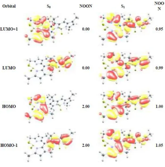
Figure 37 : Frontier molecular orbitals. HOMO (left) LUMO (right), of the parent BODIPY core at B3LYP/6-31G(d,p) level of theory. Surfaces are plotted at 0.2 a.u. Copyright 2011, John Wiley and Sons. Reprinted with permission from ref (102) ... 62 Figure 38: Frontier orbital plots and natural orbital occupation numbers
(NOON) for the dimer 3. a, S0 state. b, S1 state. a, b, Calculations were done at CAS(6e in 6o)/cc-pVDZ level. Surfaces are plotted at 0.2 a.u. All four orbitals
xiii
in both states retain characteristics of Bodipy frontier orbitals and NOONs prove tetraradicalic character of S1. Copyright 2011, John Wiley and Sons.
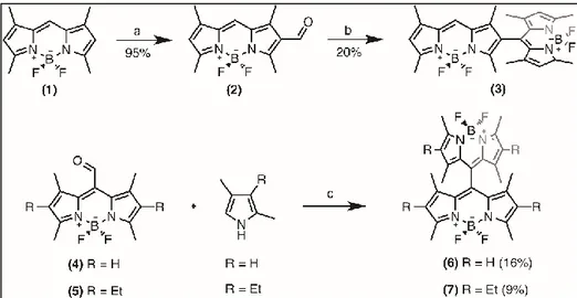
Reprinted with permission from ref (101). ... 64 Figure 39 : Structures of the dimeric Bodipys. a) X-ray diffraction structure of the orthogonal 8,2’ dimer (3). b) Optimized geometry of orthogonal 8,8’ dimer (6) at CAS(6e in 6o)/cc-pVDZ level. Dihedral angle between the two Bodipy units is very close (± 0.5o) to 90o in both dimers. Copyright 2011, John Wiley and Sons. Reprinted with permission from ref (101). ... 65 Figure 40 : Relative energies (eV) of equilibrium structures of S0, S1, and T1 states of 3. Vertical transitions computed at CAS(6e in 6o)/cc-pVDZ level ... 66 Figure 41: S1 wave function of bis-BODIPYs 3 and 6 ... 67 Figure 42 : Synthesis of the target photosensitizers. a) POCl3, DMF,
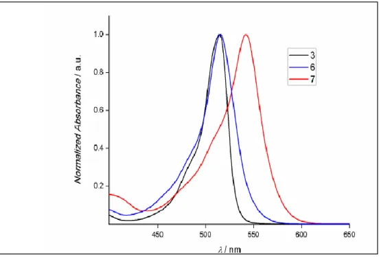
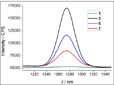
ClCH2CH2Cl, 50oC; b) 2,4-dimethylpyrrole, trifluoroacetic acid (TFA), CH2Cl2, p-chloranil, Et3N, BF3·OEt2; c) TFA, CH2Cl2, p-chloranil, Et3N, BF3·OEt2. Copyright 2011, John Wiley and Sons. Reprinted with permission from ref (101). ... 69 Figure 43: Absorbance spectra of the synthesized molecules 3, 6 and 7 in CHCl3 ... 70 Figure 44 : Singlet oxygen phosphorescence with sensitization from BODIPY derivatives: 1 (green), 3 (blue), 6 (black) and 7 (red) in CHCl3 at equal
absorbances (0,2) at the wavelength of the maximum of their respective absorbances. Copyright 2011, John Wiley and Sons. Reprinted with
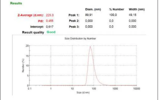
permission from ref (101). ... 71 Figure 45 : Comparative singlet oxygen generation experiment. Absorbance decrease of DPBF at 414 nm with time in dichloromethane in the presence of BODIPY photosensitizers: (3) (black), (6) (blue), (7) (red) and reference photosensitizer methylene blue (green). Copyright 2011, John Wiley and Sons. Reprinted with permission from ref (101). ... 73 Figure 46 : Size distribution of compound micellar compound 3. Cremophor EL was used for micelle formation ... 74 Figure 47 : Singlet oxygen generation experiment in aqueous solution. Decrease in Absorbance spectrum of trap molecule (anthracene derivative) in the presence
xiv
of 1.69 μM compound 3 in phosphate buffer saline (PBS). Details are given in singlet oxygen measurements part. Copyright 2011, John Wiley and Sons. Reprinted with permission from ref (101). ... 75 Figure 48: Photocytotoxicity of the sensitizer 3 as demonstrated by MTT assay. Cell suspensions (K562, human erythroleukemia cells) were seeded in 96-well flat-bottom plates and varying concentrations of the sensitizers were added into each well. Cells were kept either in dark, or under illumination with a green (520 nm) LED array at 2.5 mW/cm2 fluence rate for a period of 4 h at 370C in a humidified incubator containing 5% CO2. Copyright 2011, John Wiley and Sons. Reprinted with permission from ref (101). ... 76 Figure 49 : Photocytotoxic activity of the dimeric BODIPY 3 visualized via confocal microscopy. Cell suspensions (K562, human erythroleukemia cells) were seeded in 24-well plates. a, Cells in Control 1 (upper plates) wells were incubated in dark for 24 hours; b, Cells in Control 2 wells (second row plates) were incubated with 500 μl/well Cremophor-EL solubilized (3) (at a final concentration of 164 nM) and kept in dark for 24 hours in the same incubator, c, Cells in Control 3 well were illuminated for 4 h without the sensitizer and incubated for a further 20 h in dark at the incubator. d, Cells in Control 4 (bottom row of plates) well were illuminated for 4 h after addition of 164 nM (3) and incubated for a further 20 h in dark at the incubator. Cells in wells were collected after 24 h and imaged at 40x magnification. Live cells are
preferentially stained with acridine orange (AO, green) (a), whereas dead cells are preferentially stained with the dye propidium iodide (PI, red) due to increased cellular permeability (b). Copyright 2011, John Wiley and Sons. Reprinted with permission from ref (101) ... 77 Figure 50: Reaction of singlet oxygen with 1,3-Diphenylisobenzofuran ... 82 Figure 51 : Reaction of singlet oxygen with 2,2'-(Anthracene-9,10- ... 83 Figure 52: Decrease in absorbance of DPBF in dichloromethane in the presence of compound 3 in medium. Details are given in singlet oxygen measurements part. Copyright .2011, John Wiley and Sons. Reprinted with permission from ref (101). ... 85
xv
Figure 53: Decrease in absorbance of DPBF in dichloromethane in the presence
of ... 86
Figure 54: Decrease in absorbance of DPBF in dichloromethane in the presence of compound 7 in medium. Details are given in singlet oxygen measurements part ... 87
Figure 55: Decrease in absorbance of DPBF in dichloromethane in the presence of MB in medium. Details are given in singlet oxygen measurements part. Copyright . 2011, John Wiley and Sons. Reprinted with permission from ref (101). ... 88
Figure 56 : Packing diagrams of compound 3. Dashed lines indicate inter H-bond between C2 and F4 atoms. Copyright . 2011, John Wiley and Sons. Reprinted with permission from ref (101). ... 90
Figure 57 : Synthesis of the 4-decyloxybenzaldehyde ... 98
Figure 58 : Synthesis of the meso-substituted BODIPY ... 99
Figure 59 : Synthesis of the brominated meso-substituted BODIPY ... 101
Figure 60 : Reaction scheme of trap molecule with singlet oxygen ... 101
Figure 61 : Absorbance spectrum of trap molecule 2,2'-(anthracene-9,10-diylbis(methylene)dimalonic acid in water under light and Photosensitizer (Target molecule 1) ... 102
Figure 62 : Synthesis of the iodinated meso-substituted BODIPY ... 105
Figure 63 : Absorbance spectrum of trap molecule 2,2'-(anthracene-9,10-diylbis(methylene)dimalonic acid in water under light and Photosensitizer (Target molecule 2) ... 106
Figure 64 : Synthesis of the target molecule 3 ... 107
Figure 65 : Absorbance spectrum of trap molecule 2,2'-(anthracene-9,10-diylbis(methylene)dimalonic acid in water under light and Photosensitizer (Target molecule 3) ... 108
Figure 66 : Absorbance spectrum of trap molecule 2,2'-(anthracene-9,10-diylbis(methylene)dimalonic acid in water without photosensitizer ... 109
Figure 67 : The emission spectrum of the LED source ... 110
Figure 68 : Size distribution of compound micellar compound 3 (First Target Compound). Cremophor EL was used for embedding the dye. ... 111
xvi
Figure 69 : Size distribution of compound micellar compound 4 (Second Target
Compound). Cremophor EL was used for embedding the dye. ... 112
Figure 70 : Size distribution of compound micellar compound 6 (Third Target Compound). Cremophor EL was used for embedding the dye. ... 113
Figure 71 : : Zeta potential measurement of micellar compound 4 . Cremophor EL was used for embedding the dye. ... 114
Figure 72: : Zeta potential measurement of micellar compound 3 . Cremophor EL was used for embedding the dye. ... 115
Figure 73 : Zeta potential measurement of micellar compound 6 . Cremophor EL was used for embedding the dye. ... 116
Figure 74 : Normalized absorbance spectrum of compound 3 in CH2Cl2 ... 118
Figure 75 : The normalized fluorescence emission of Compound 3 in CH2Cl2119 Figure 76 : Normalized absorbance spectrum of compound 4 in CH2Cl2 ... 120
Figure 77 : The normalized fluorescence emission of Compound 4 in CH2Cl2121 Figure 78 : Normalized absorbance spectrum of compound 4 in CH2Cl2 ... 122
Figure 79 : The normalized fluorescence emission of Compound 4 in CH2Cl2123 Figure 80 : The normalized absorbance spectra of the compounds 3, 4, and 6 in CH2Cl2 ... 124
Figure 81 : The normalized fluorescence emission of Compound 3 , 4 and 6 in CH2Cl2 ... 125
Figure 82 : Comparative singlet oxygen generation experiment. Absorbance decrease of trap molecule at 378 nm with time in aqueous solution in the presence of BODIPY photosensitizers: (3) (black), (4) (red), (6) (blue) ... 126
Figure 83 : Absorbance decrease of trap molecule at 378 nm with time in aqueous solution in the presence of Compound 3 ... 127
Figure 84: Absorbance decrease of trap molecule at 378 nm with time in aqueous solution in the presence of Compound 6 ... 128
Figure 85 : Synthesis of compound 1 ... 131
Figure 86 : Synthesis of compound 2 ... 132
Figure 87 : Synthesis of compound 3 ... 134
Figure 88 : Synthesis of compound 4 ... 135
xvii
xviii
LIST OF TABLES
Table 1 : Clinically Approved Photodynamic therapy agenst1,81. ... 28 Table 2. Comparative spectroscopic properties of BODIPY compounds. ... 72 Table 3 : Characteristics of micellar compounds 4, 5 and 8. Cremophor EL was used for embedding the dye ... 116 Table 4 : Ф(1O
2) values of photosensitizer ... 117 Table 5 : Comparative spectroscopic properties of BODIPY compounds ... 125
1
CHAPTER 1
INTRODUCTION
Cancer also known as a malignant tumor is a challenging and terrifying disease and its most widely used treatment modalities are chemotherapy, radiation therapy and surgery. These treatment modalities frighten patients due to the side effects that up to know could not be obviated. A novel treatment modality, Photodynamic therapy that has been used clinically for around 25 years has been emerged and widely used as an alternative method for treatment of the malignant and nonmalignant diseases. Photodynamic therapy has numerous advantages compared to the conventional treatment modalities owing to the fact that it is noninvasive and selective treatment method. Over the past decade, there have been great number studies showing the application of PDT for the treatment of various diseases and there is an important impetus to produce novel and efficient photodynamic therapy agents. The only know side effects of the current photodynamic therapy agents, mostly porphyrin based photosensitizers is the skin photosensitization. To overcome this drawback, novel photosensitizers that absorb light in the near-IR region of the electromagnetic spectrum, where the deepest tissue penetration is available, are required to perform more effective therapeutic action.
With this consideration in mind, we design and synthesized a novel series of BODIPY based photosensitizers which absorb light in the therapeutic window of the electromagnetic spectrum (650-800 nm). BODIPY dyes are pertinent to functionalization on their core. Furthermore, BODIPY dyes have high fluorescence quantum yields, straightforward synthetic methods and high
2
absorption coefficients. Therefore, we introduced novel BODIPY-based photosensitizers in Chapter 3.
The choice of the photosensitizer plays an important role in the photodynamic therapy. It is a well-known fact that incorporation of the heavy atoms to the core of the BODIPY confer inter system crossing (ISC), yielding cytotoxic singlet oxygen. However, an effective photosensitizer should be non-toxic in the absence of light or has low dark-toxicity. Incorporation of the heavy atoms gives rise to an increase in dark toxicity of the photosensitizers. We have been at pains to find out a method in order to produce cytotoxic single oxygen without introducing singlet oxygen to the core of the BODIPY which alleviate the dark toxicity of the photosensitizers. In chapter 4, we introduced novel BODIPY derivatives which have orthogonal arrangement. This arrangement has a doubly substituted tetra radical state (DS-TR) that has a relationship with S1→T1 intersystem crossing, resulting in generation of cytotoxic singlet oxygen. In the chapter 5, we targeted Bodipy derivatives with heavy atom substitutions which would facilitate intersystem crossing. In addition, to ensure that these compounds would prefer a micellar structure as opposed to bulk aqueous solution, we incorporated long alkyl chains at the meso positions (C-8) of the Bodipy dye
3
CHAPTER 2
BACKGROUND
2.1 Photodynamic Therapy
2.1.1 General InformationPhotodynamic therapy (called PDT, photoirradiation therapy, phototherapy, or photochemotherapy) is one of the most promising and minimally invasive treatment modality for certain cancers such as bladder cancer, skin cancer, gynecological, head and neck cancer, gastrointerstinal cancer cervical cancer and gastric cancer and for many other certain diseases like psoriasis, basal cell carcinoma1. Furthermore, PDT is used as a treatment modality in cardiology2. The PDT strategy is based on the preferential localization of the certain photosensitizers in tumor tissues upon systematic administration. After the administration of the photosensitizer into the body, the sensitizer is activated by red or near infrared (NIR) which leads to generation of reactive oxygen species (ROS) including singlet oxygen (1O
2) and thus irreversibly damaging tumor cells. These PSs are harmless in the absence
of light and the phototoxicity of these photosensitizers mainly relies on the generation of the singlet oxygen, which is converted from its triplet state to its singlet state by the exited-state dye. Photosensitizing agents, which is also known as photosensitizer, are administrated locally or intravenously
Moreover, the photosensitizer is selectively absorbed and retained within the targeted tissues3. When the target tissue is illuminated by a certain light source at an appropriate wavelength, especially in the therapeutic window, the nearby normal tissues which contain little amount of photosensitizer or no photosensitizer are prevented from photodamage and injury.
4
Over the past decade, significant progress has been made in the development of novel photosensitizers4. However, the efficacy and properties of these materials must be improved in order to meet treatment requirements.
The first property relevant to the PS is its absorption maximum which determines its efficiency with regard to the singlet oxygen generation. It is a well-known fact that photosensitization relies on the absorption of light of the correct wavelength by a photosensitizer
A convenient photodynamic therapy agent should have absorption maxima in the spectral region 700- 900 nm which is known as therapeutic window. Because of the fact that below 700 nm light array cannot penetrate deeply into the tissue and water into the body begins to absorb light above 900 nm, a well-designed photosensitizer should absorb light in the therapeutic window5.
Another key issue influencing the photodynamic capability of a photosensitizer is the efficiency of the singlet oxygen generation. Singlet oxygen is one of the most important parameters for PDT which is very reactive and causes cell death.
5 2.1.2 History of the Photodynamic therapy
Although light has been used a therapeutic agent for thousands of years in order to heal malignant and non-malignant diseases, photodynamic therapy was initially observed more than hundred years ago6,7. Light was widely used by ancient Egyptian, Indian and Chinese civilization as a treatment modality for various diseases such as psoriasis, rickets and skin cancer8.
In 1900, Oscar Raab has showed that the use of organic photosensitizer acridine orange in combination with day light gives rise to the cytotoxic effect on infusoria7. In the same year, scientist J.Prime has discovered that patients suffered from dermatitis when they were exposure to the sunlight after treatment which was executed by using eosin9.
In 1901, Niels Finsen has reported that smallpox and cutaneous tuberculosis was treated by using light. In 1903, he was awarded Nobel Prize for his work on phototherapy10 .
In the same year, Herman Von Tappeiner, director of the Institute of Pharmacology at the University of Munich and A. Jesionek, the dermatologist, have demonstrated that skin tumors successfully treated by using eosin which is applied topically in the presence of light. It was then named as photodynamic action. The term “photodynamic” was firstly used by von Tappeiner and A. Jodbauer in 190711.
In 1911, W. Hausmann has reported that paramecium and red blood cells were treated with light and hematoporphyrin which was a derivative of the most widely studied photosensitizers, porphyrins, in the mid-nineteenth century. He also reported that the treated mice showed skin reactions, such as extensive edama and erythema12.
In 1913, the German physician Friedrich Meyer-Bertz conducted a study with hematoporphyrin to treat a human. He delivered the photosensitizer, 200 mg hematoporphyrin, into his hand intravenously and he suffered from pain and
6
swelling after the exposure to the sun light. It was the first reported photodynamic therapy experiment on humans13.
In 1942, the German scientist Auler and Banzer reported that hematoporphyrin in the malignant tissues has higher fluorescence compared to the surrounding tissues and these malignant tissues were killed by necrosis after illumination with a powerful quartz lamp14.
In 1955, Dr Samuel Schwartz reported that he developed a new hematoporphyrin derivative (HPD)15. It was shown that HPD was twice as phototoxic as hematoporphyrin16. This compound, HPD, was used by Richard Lipson and coworkers in their studies17. They demonstrated that hematoporphyrin derivatives can be used as a diagnostic tool16.
In 1972, first in vivo study was accomplished by I. Diamond and their coworkers. They reported by performing PDT to treat tumors which was implanted in rats, tumor growth was inhibited for approximately 20 days18.
In 1975, Thomas Dougherty and colleagues reported that they managed to eradicate malignant tissues in mice by using hematoporphyrin derivative (HpD) and red light.
J.F.Kelly and colleagues published a study in the same year. It revealed that bladder carcinoma in mice could be treated by using HPD in combination with light19.
Clinical trials with HPD were initiated in 19761. Kelly and colleagues performed a successful study which was the first human trials with hematoporphyrin derivative. This most widely used photosensitizer was used to treat recurrent bladder cancer. Malignant tissue growth was slowed down and these tissues were killed by necrosis. Following this study, Dougherty and coworkers treated 25 patients by using similar approach20. Y.Hayata and coworkers reported that PDT was used to treat obstructing malignant tissues, lung cancer. The result of this trial was not impressive but only around 7% of tumor of the patients was eradicated successfully21.
7
In 1984, J.S. Mc Caughan and coworkers reported that PDS was used as a treatment modalitiy for the oesphageal cancer22. Only one year after this breakthrough study, gastric cancer was cured with PDT by Y.Hayata and coworkers23.
Thereafter, photodynamic therapy was also used for the treatment of the pancreatic carcinoma24, cholangioma25, mesothelium carcinoma26 , skin carcinoma27, head and neck tumors28 , and brain tumors29 . According to these breakthrough studies, it was revealed that PDT treatment could be effectively applied on early-stage cancers30 .
In the late 1980s, there have been a great number of studies on PDT that results in microvascular collapse, subsequently this mircrovascular collapse gives rise to the severe tissue hypoxia and anoxia31,32.
In 1993, the most frequently used photosensitizer porfimer sodium (Photofrin), a purified hematoporphyrin derivative, was used to treat bladder cancer and its approval was reported in the same year in Canada. It was the first photosensitizer which was for approved for photodynamic therapy (Figure 1)33.
8
In the following years, other photosensitizers have been reported which employed in the photodynamic milieu and they were FDA approved. One of them is temoporfin (Foscan) which gained its approval in Europe and is used for the treatment of the head and neck tumor, brain tumor and skin carcinoma (Figure 2)34.
9 2.1.3 Working Principle of PDT
Photodynamic therapy includes various photophysical processes. Figure 3 graphically demonstrates the process of light absorption and energy transfer, which is essential for the photodynamic therapy. After the activation of the photosensitizer upon absorption of the light transforms the photosensitizer from its ground state (1PS) to its excited single state (1PS*
,Fig. 3) .This is a short-lived (nanoseconds) species and may decay from this excited state to ground state and during this decay the fluorescence emits. We can use this property clinically for photodetection35. The PS can undergo electron spin conversion to its long –lived (microseconds) triplet state (3PS*). Because of the fact that the loss of energy by emission of light (phosphorescence) is a spin forbidden process the photosensitizer triplet state has a long lifetime.
Because of the dependence on a long-lived triplet excited state, the synthesis of the photosensitizers are encouraged in order to improve this feature usually by attaching the heavy halogens to the photosensitizers, i.e. via the heavy atom effect. In this case we can obtain better results in terms of therapeutic effect.
Reactive oxygen can be generated through two types of reaction. In the type 2 reaction, the excited state photosensitizer reacts with molecular oxygen, yielding singlet oxygen. In the type one reaction the excited photosensitizer can react with a substrate either by proton or by electron transfer and form radical or radical ions. After the formation of these radicals, they can react with oxygen and produce oxygenated products36,37.
The second type reaction illustrates that the energy of the excited photosensitizer can be transferred to oxygen and the singlet oxygen, which is a cytotoxic species, is formed38.
The ratio between these two types of reactions is concentrations and oxygen dependent.
10
Owing to the fact that reactive oxygen species are highly reactive and have short half-life, only tissues in close proximity to the location, where reactive oxygen species are produced, are treated with photodynamic therapy.
As a result, the activated photosensitizer and energy transfer result in apoptosis and necrosis of targeted either malignant or nonmalignant tissue.
Because of the fact that the effectiveness of the nearly all photosensitizers depends on oxygen, photodynamic action of the photosensitizers does not happen in the area of tissues where they lack oxygen39.
Location of photodamage belongs to the type of the photosensitizer. Therefore, a great photodynamic agent would be diversely gathered in the target tissue. It is worth to address that specifity of destruction of cancer cells can be achieved by selective application of the photosensitizer, selective uptake of the photosensitizer, high quantum yields of singlet oxygen, which is produced during reactions we discussed above, and by the selective exposure of malignant tissue to the active light40. There are numerous drawbacks of photodynamic therapy.
11
One of them is the prolonged skin photosensitization41. Another one is that the patient needs to stay away from the sunlight exposure for a certain time (1-4 weeks).
Generally, efficiency of the photodynamic therapy and cytotoxic effect depends on administration of the photosensitizer, light exposure doses, interval between illumination of the tissues by a certain light source and administrations of the photosensitizer into the body, light source and type of photosensitizers42.
2.1.4 Photodynamic Effect and Cell Death Mechanism
Photodynamic therapy has three major components. These are photosensitizers, clinically known as drug, light source and oxygen. To obtain photodynamic action and to generate cytotoxic oxygen, singlet oxygen, all of these three components are required. Reactive singlet oxygen gives rise to the cellular destruction by means of direct cellular damage, vascular shutdown, activation of immune response system. After the generation of the reactive singlet oxygen, it directly reacts with cellular components, such as nucleic acids, lipids and proteins43. As a result, oxidative stress occurs, causing cellular destruction.
There are two types of cellular death. One of them is apoptosis and another one is necrosis. The type of the cellular death depends on localization of the photosensitizer. It is also dependent upon the quantity of the reactive singlet oxygen44. Patterson et al. has reported that photosensitizers which localized especially in the mitochondria or endoplasmic reticulum mostly gives rise to the apoptosis. On the other hand, photosensitizers that localized in lysosomes or in the plasma membrane is likely to cause necrosis45. Photodynamic therapy can also cause cellular destruction by means of acute local inflammation46.
There are a number of factors that affect efficiency of the treatment. Localization of the photosensitizer is one of the most widely known factors that determine
12
efficiency of the photodynamic therapy action. In order to improve efficacy of the treatment, a great number of study have been performed and published in recent years. To obtain better localization in the cancerous cells, selectivity of the photosensitizers should be improved. Photodynamic therapy with localized and targeted photosensitizer is a smart treatment option owing to the fact that only targeted cells are treated without any side effect.
By designing and synthesizing photosensitizer through conjugation to different type of targeting peptides, targeting for photosensitizers uptake into cancerous cells has been accomplished47. Other approaches that are used to improve localization of the photosensitizers are conjugation of small targeting ligands and protein conjugation. These types of photosensitizers are commonly known as third generation photosensitizers48.
In recent years, selectivity of the photosensitizers and designing novel activatable photosensitizers has been the focus of the extensive research4.
Akkaya group reported that near-IR absorbing BODIPY-based photosensitizers were selectively activated once they were inside the cells. In this study, they synthesized a series of BODIPY based photosensitizers that are in caged form, i.e. they cannot produce singlet oxygen. However, once these BODIPY-based chromophores are inside the cells, they become activated because of the fact that cancer cells have high intracellular glutathione concentration, photosensitizers become active after the quencher has been cleaved by intracellular glutathione3. 2.1.5 Light in Photodynamic Therapy
Light is one of the three essential components of the photodynamic therapy as it is essential for the life
It is a well-known fact that UV-light radiates at short-wavelength of the electromagnetic spectrum and it is a high-energy radiation. Also, it is an undeniable fact that all human beings are exposed to the UV-light during their daily life. Therefore, some precautions have to be taken against the UV-light during our daily life. This high energy ray is capable of remove electrons from
13
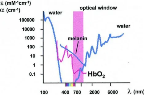
DNA and serious damage can occur, including tumor formation. During exposure to the UV-light, skin behaves like a protective coating. Melanin absorbs UV-light in order to impede its effect and the color of our skin is changed since the pigmentation occurs since melanin absorbs light in the region of the electromagnetic spectrum where sunlight radiates (Figure 4).
On the other hand, there are another biomolecules absorbing UV-light in the skin, such as hemoglobin and amino acids which contain aromatic molecules in their chemical structure49.
Intracellular absorption is therefore especially important considering the activation of the photosensitizer under a certain light source and at a certain wavelength. As a result, selection of the light source and its wavelengths range are of great importance in terms of skin sensitivity during the treatment of photodynamic therapy action. Long-wavelength light is essential to the sufficient tissue penetration due to the intracellular absorption and light scattering. In the
Figure 4 : Absorption spectra of major intracellular absorbers Copyright 2000, John Wiley and Sons. Reprinted with permission
14
last decade, considerable effort has been devoted to delivering of light to the tumor site, including interstitial and superficial illumination.
It was reported in the literature that photosensitizer, ALA, was used for treatment of the squamous cell carcinoma. It was applied topically and light source was superficially delivered to the tumor. For treatment of the thicker cancerous tissues, it can be necessary to administrate the photosensitizers into the body intravenously and light is required to deliver via implanted optical-fiber light sources.
2.2 Photosensitizers
15
As mentioned in the previous chapter, photosensitizer is one of the key components of the photodynamic therapy. In order to generate more effectively singlet oxygen which is another key component of the photodynamic therapy, the selection of an appropriate photosensitizer is of paramount importance. In recent years, production of the novel and effective photosensitizers has been the focus of extensive research4. There are a great number of clinically approved photosensitizers, such as HPD-photofrin, verteporfin, Foscan etc. However, there has not been yet any approval for the use of BODIPY-based photosensitizers in clinical treatment, but BODIPY-based photosensitizers have important features that will most probably provide clinical approval for their use in photodynamic therapy application in the near future3.
One of the most widely used photosensitizers, photofrin has a number disadvantages. Since its activation by light occurs at ca. 630 nm, light rays cannot penetrate into tissue more than few millimeters. Besides that, illumination of the skin at this wavelength gives rise to the painful edema which is known as photosensitivity50.
Due to the abovementioned drawbacks of phorphyrin-based photosensitizers, researchers have extensively attempted to develop novel photosensitizers and many studies have been focused on design and synthesis of the new photosensitizers in recent times51.
It is a well-known fact that a photosensitizer should be nontoxic in the absence of light and have low side effects and absorption especially at long wavelengths of the electromagnetic spectrum for deeper tissue penetration. Furthermore, photosensitizers should be resistance to photobleaching.
BODIPY (4, 4-difluoro-4-bora-3a, 4a-diaza-s-indacene) dyes have attracted much attention as a photosensitizer over last decade owing to its characteristics which are particularly required for the effective photosensitizers and photodynamic
16
action. In 1968, BODIPY was for the first time synthesized and reported by Treibs and Kreuzer52.
BODIPY dyes have attracted much attention as photosensitizers and fluorescent molecular sensors over the past few decades because of the fact that they have sharp absorption, high fluorescence quantum yields, high excitation coefficients, light to dark toxicity ratios53 and high stability54. Since BODIPY dyes have distinctive characteristics, they have been used for the application as solar cells55, fluorescent imaging probe56 and chemical sensor.57
Furthermore, BODIPY dyes are amenable to the modifications on their core (Figure 5) . Their absorption and fluorescence emission can be easily tuned. By utilizing these properties of the BODIPY dyes, their derivatives have been synthesized and different derivatives have developed over the past two decade by using substitution on the BODIPY core, by reacting an aldehyde with methyl groups on the 3 and 5-position of the BODIPY core (Knovenagel condensation reaction) 58 and by displacing the fluorine atoms with boron atoms on the 4-position of the BODIPY core59. Another modification option has been applied at the 8-position (meso) of the BODIPY core.60,61 Prasad and coworker have reported that meso-phenyl and meso-naphtalenyl substituted BODIPY derivatives
Figure 5 : Molecular structures of BODIPY and its precursors.
17
were synthesized by utilizing the distinctive feature of the BODIPY dyes which is straightforward synthesis and functionalization.62
All of these distinctive properties of the BODIPY dyes have inspired efforts to develop BODIPY based photosensitizers.63
Nagano and coworkers have shown that a halogenated BODIPY derivative can generate cytotoxic singlet oxygen. It was the first BODIPY which was reported as a photosensitizer in 2005. In this work, they introduced iodine atoms to the core of BODIPY in order to achieve ISC for the production of singlet oxygen. After the synthesis and characterization of the diiodo-BODIPY (3), singlet oxygen quantum yield was measured by using a trap molecule which is 1,3-diphenylisobenzofuran (DBPF) (Figure 6).
Cell culture studies were performed by using HeLa cells. According to the results of singlet oxygen generation experiments with cell culture, this halogenated BODIPY derivative is photocytotoxic under the light illumination.64
In 2010, Burgess and coworkers have reported a study. This work presents another new BODIPY based photosensitizer. In this study, a functional group, ethylene-carboxylic acid was introduced to the meso-position of the BODIPY unit. After synthesis and characterization of the halogenated and meso-substituted BODIPY
Figure 6 : Halogenated BODIPYderivative
18
derivative (4), cell culture studies were carried out. It is a noteworthy fact that this BODIPY derivative has high singlet oxygen quantum yield and high-to-dark phototoxicity (Figure 7).
HSC-2 cells were treated with this BODIPY-based photosensitizer under the illumination of light source at wavelength 525nm for 2 hours. It was shown that photosensitizer 4 was delivered in the mitochondria and gave rise to the cell death via apoptosis.53
In 2012, Arbeloa and colleagues have prepared a BODIPY-based photosensitizer by introducing iodine atoms at the 2- and 6-positions and 3- and 5-positions of the BODIPY unit. (Compound 5,6 and 7)
Figure 7: Molecular structure of the BODIPY-based photosensitizer
19
It was shown that attachment of the iodine atoms to the 3-and 5- position of the
BODIPY cores give rise to an increase in fluorescence of the molecule and they have high singlet oxygen quantum yield. It was experimentally proved that these photosensitizers are highly stable against light.65
Suzuki and coworkers reported that they synthesized and characterized heteroaryl-fused BODIPY derivatives (Figure 9 and 10). In this study, singlet oxygen generation experiments of the BODIPY-based photosensitizers were performed by using DPBF. It was found that they have high singlet oxygen yield.
Figure 8 : Molecular structures of the BODIPY based photosensitizers (5, 6 and 7)
Figure 9 : Molecular structure of the heteroaryl-fused BODIPY based photosensitizer (Compound 8)
20
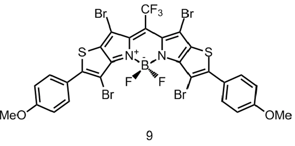
These novel photosensitizers (8 and 9) have absorption maxima in the near-IR range of the electromagnetic spectrum. The absorption maximum of the compound 8 is at 720 nm and its emission is at 754 nm. The absorption maximum of the compound 9 is at 766 nm and it emission is at 820 nm. Furthermore, it has been shown that these BODIPY derivatives can be used as imaging probe and photosensitizer at the same time due to fact that they are enough fluorescent for the imaging purpose even if they were brominated.66
Akkaya and coworkers reported a pioneering study in 2007 by synthesizing 3 novel water soluble styryl-substituted BODIPY derivatives67. In this study, it was shown that introduction of the styryl groups to the 3-and 5-position of the BODIPY core gives rise to the spectacular red shifts in the absorption maximum of the related BODIPY dye. These photosensitizers have absorption maximum in the 590-660 nm region. Water solubility of these photosensitizers was improved by introducing amphiphilic triethylenglycol groups to the BODIPY molecules (Figure 11).
The singlet oxygen generation experiments were performed by using 1,3-diphenylisobenzofuran (DPBF) which is the most widely used singlet oxygen trap molecule.
Figure 10: Molecular structure of the heteroaryl-fused BODIPY-based photosensitizer (Compound 9)
21
Cell culture experiments were carried out by using standard MTT assay (K562 Cells). The experiment performed in the dark and with photosensitizer exhibited no cell death. Besides that, experiment carried out in the dark without photosensitizer did not demonstrate any cytotoxic result either. Fluorescence microscope images showed that BODIPY based photosensitizer is effective photodynamic therapy agent (Figure 12).
Figure 11 : Molecular structures of the water soluble styryl-substituted BODIPY derivatives
22
In 2011, Ng and colleagues reported a similar study. A series of BODIPY-based photosensitizers have been synthesized67. They are similar to the photosensitizers which were reported by Akkaya group in 2006. In this work, iodine atoms were incorporated to the core of the BODIPY instead of bromine atoms which was used by Akkaya group for the heavy atom effect (Figure 13).
Halogenated
aza-BODIPY based
Figure 12: Fluorescence microscope images of acridine orange (AO) and propidium iodide (PI) stained K562 cells,
Live cells were preferentially stained with AO (green) and dead cells with PI (red) due to increased cellular permeability
Copyright 2000, John Wiley and Sons. Reprinted with permission from ref ( 67 )
Figure 13 : Molecular structure of iodinated styryl-substituted BODIPY derivative
23
photosensitizers were reported by O’Shea et al68. They synthesized and characterized a new series of photodynamic therapy agents which can be excited in the therapeutic window of the electromagnetic spectrum (650-800nm, beyond the absorptivity region of body tissue) (Figure 14).
After the synthesis and characterization of the BODIPY dyes, in vitro cellular uptake was investigated. Singlet oxygen generation efficiency of the photosensitizers was evaluated. 1,3-diphenylisobenzofuran (DPBF) was used as a trap molecule for the singlet oxygen generation experiments. In vivo assays and human tumor cell lines have prepared for the evaluation of the cytotoxicity of the compound 11. Activation of sensitizer resulted in the generation of the photoinduced cytotoxic singlet oxygen, causing cell death via apoptosis and necrosis69.
Figure 14 : Molecular structure of the halogenated aza-BODIPY based photosensitizer
24
In 2010, another aza-BODIPY based photodynamic therapy agents were reported by Ramaiah and coworkers. In this work, iodine atoms were introduced to the BODIPY cores in order to improve intersystem crossing efficacy (Figure 15). As a result, it was found that the target compounds were highly cytotoxic and promising singlet oxygen generators.
The absorption maximum of the compound 12 is at 666 nm and its emission is at 694 nm. As a result of the heavy atom incorporation, up to 70 % singlet oxygen generation efficiency was acquired.
In 2015, a glutathione activated BODIPY based photosensitizer (13) was reported by Akkaya et al 70(Figure 16). The singlet oxygen generation of this BODIPY based photosensitizer can be turned ON/OFF by using intracellular glutathione. It is a noteworthy fact that this BODIPY based photosensitizer can discern between concentrations in normal and malignant cells.
Figure 15: Molecular structure of the tetraiododibromo aza-BODIPY based photosensitizer
25
Figure 16 : Molecular Structure of the glutathione activated BODIPY
26 2.2.2 Photosensitizers based on other Dyes
There are a great number of photosensitizers based on various chromophores. As mentioned in the previous section of this chapter, the first clinically approved photosensitizer is Photofrin and it’s a porphyrin derivative (Table 1). In 1993, it was clinically approved in Canada and it is now most widely used photosensitizers for the photodynamic therapy application in many countries including USA. Even photofrin is a most widely used photosensitizer in the photodynamic milieu, it has some drawbacks. In order to overcome side effects and deficiencies of this most widely used photosensitizer, development of the novel photosensitizers have been the focus of extensive research. In recent years, many photosensitizers have been extensively developed and reported71.
5-Aminolaevulinic acid (5-ALA) is another clinically approved photosensitizer for the photodynamic therapy application. It is a porphyrin precursor (Protoporphyrin IX) and mainly used for the treatment of skin and bladder cancer. Clinical use of the ALA was reported in 1990 by Kennedy coworkers. It was shown that skin reaction after the treatment did not last as long as photofrin did.72 Texaphyrins, expanded porphyrins, are a new class of photosensitizers was reported in 1988 by Sessler et al.73.They have many of features in common with
porphyrins, such as aromatic molecular structure and colorfulness. They have mainly absorption in the near-IR region of the electromagnetic spectrum. There are five coordinating nitrogen atoms in their central core which confer them to react with the lanthanides in more stable manner.
In 2000, clinical applications of two texaphyrin derivatives, the gadolinium (III) and lutetium (III) were reported by J.L. Sessler and R.A. Miller74. The latter one, Lutetium (III) derivative is water soluble and has absorption maximum at 732 nm in the near-IR region of the electromagnetic spectrum (Figure 16). Its administration is performed intravenously by using injection. At this wavelength, light can deeply penetrate into the mammalian tissue.
It is found that this texaphyrin derivative, Lu-Tex, has high singlet oxygen quantum yield. Furthermore, it shows low dark toxicity and lower skin
27
photoinduced cytotoxicity compared to the Photofrin because of the fact that this photosensitizers can leave the human body faster than Photofrin.75,76 Phase trials of these photosensitizers have been performed for the treatment of breast cancer and atherosclerosis vascular diseases.
In 1998, cadmium coordinating texaphyrin , Cadmium (II) texaphyrin, derivatives was reported by Maiya et al. The absorption maximum of one of these derivatives was at 864 nm77.
Chlorin based photosensitizers have attracted much attention due to their potential as a putative cytotoxic singlet oxygen generator and they can make conjugation
28
with proteins. These features endow the chlorins potential photodynamic therapy agents78.
A novel series of chlorin based photosensitizer reported by Shan et al. in 200679. Bacteriochlorins are another group of photosensitizers absorbing lights in the near-IR range of the electromagnetic spectrum. In 2006, a series of bacteriochlorins were reported by Sharonov et al. They have absorption maximum between 760 and 830 nm, at longer wavelengths compared to the photofrin and chlorins80.
29
Photosensitizer Structure Wavelength Approved Trial Type of
Cancer HPD-Pormier
sodium (Photofrin)
Porphyrin 630 nm Worldwide Cervical,
Endobronchi al, Esophageal, Gastric,bladd er,brain.ovari an
ALA (Levulan) Porphyrin 635 nm Worldwide Skin, head
and neck, gynaecologic al 5-ALA-methylesther (Metvix)
Porphyrin 635 nm Europe Basal cell
carcinoma, bladder
5-ALA-benzylesther (Benzvix)
Porphyrin 635 nm Europe Gastrointesti
nal
5-ALA-hexylester (Hexvix)
Porphyrin 375-400 nm Europe Diagnosis of
bladder tumors
Verteporfin Chlorine 690 nm Worlwide Basal cell
carcinoma, pancreatic Temoporfin
(Foscan)
Chlorine 652 nm Europe Head and
neck, lung,
brain, skin,
bile duct
SnEt2 (Purlytin) 664 nm USA Skin, breast,
prostate Kaposi’s sarcoma Motexafin Lutetium (Lutex)
30
Padoporfin (TOOKAD)
Bacteriochlorin 762 nm USA Prostate
Silicon phtalocyanine (Pc4)
Phatolocyanine 675 nm USA Cutaneous T-Cell lymphoma Ce6-PVP (Fotolon), Ce6 derivetives (Radachlorin,Ph otodithazine Chlorin 660 nm Belarus, Russia Nasopharyng eal, Sarcoma, brain 2.3 Application of PDT in medicine
31
In the last decade the application of photodynamic therapy has increased and acquired growing acceptance around the world. Cancer is one of the most problematic disease and many people have suffered from various cancer types like cancers of the lung82, gastrointestinal tract83, bladder84, head and neck region85, prostate86 and nonmelanoma skin cancers and actinic keratosis.87,88,89,90,.91,92
Photodynamic therapy gives complete response in a high percentage of patients. There are also ex vivo procedure which has been used for treating bone marrow and hematopoietic stem cell grafts.93,94,95.96
Photodynamic therapy is also used as a treatment modality for the nonmalignant diseases such as age related macular degeneration and psoriasis97. Furthermore, some successful PDT treatment of the atherosclerosis plaque has been reported in recent years80
32
NOVEL NEAR-IR PHOTOSENSITIZERS FOR PHOTODYNAMIC THERAPY
Kilic, B.; Yesilgül, N. ; Akkaya, E. U.
33
In this project, four different types of novel BODIPY based photosensitizers were designed and synthesized which fulfill a great number of criteria for the PDT in terms of singlet oxygen generation, low dark toxicity, tissue penetration and solubility. All target compounds were synthesized by using standard BODIPY chemistry. Owing the fact that BODIPY dyes have remarkable features, such as high quantum yields, good solubility, photostability etc., BODIPY dyes were used for the design and synthesis of the target compounds as promising near-IR photosensitizer for the PDT.
3.2 Introduction
By considering the important parameters, which is essential for an ideal photosensitizer, our aim was to synthesize novel near-IR photodynamic therapy agents which will fulfil the requirement for an ideal photosensitizer and maximize the efficiency of the method.
The reason why we have used BODPY dyes ( 4,4-difluoro-4-bora-3a,4a-diaza-s-indacene) in order to obtain an ideal photosensitizer is that they have many distinctive properties such as high fluorescence quantum yield, photostability, long wavelength absorption and emission, high molar absorption coefficient and their simple synthesis method59.
The chemical and photochemical stabilities of BODIPY dyes, which are higher than those of many other chromophores, are other important factors that we took into consideration for the design of the photosensitizers. Modifications on the BODIPY core can be carried out by introduction of different groups which afford red-shifted BODIPY derivatives98,3. It was shown that the strongly electron-withdrawing group CF3 on the meso position of the BODIPY coreresulted in a large bathochromic shift compared to that of the congeners with aryl substituents in this position99.
34
By taking account the above information, we have designed meso-CF3-substituted BODIPY dyes having aryl substituents. The usual BODIPY molecules have absorption maximum at around 500 nm, which is not suitable for an effective light penetration into the tissue61. Thus, we have designed a general effective route to a novel family of the meso-CF3-3,5-diaryl-BODIPY dyes which is composed of 4 different target BODIPY molecules. These molecules have heavy atom functionalization on the 3, 5-positions of their BODIPY cores. This functionalization gives rise to the effective singlet oxygen generation for serving as a potential photodynamic therapy agent.62,100
Nagano et al. have recently shown that substitution of the BODIPY with iodine gives rise to a radical increase in the intersystem crossing efficiency62. On the other hand, several studies have recently shown that BODIPY dyes without heavy atom functionalization can be used as effective photosensitizers providing that they are synthesized as orthogonal dimers.101,102
Synthetic pathway for the target compounds is shown below (Figure 18 and Figure 19)
35 3.3 Result and Discussion
In this study, we have designed four different novel photosensitizers and they
were successfully synthesized and characterized. The TM1 (Compound 4) and TM2 (Compound 5) were obtained from the common precursor meso-CF3 -substituted and brominated BODIPY. The TM3 (Compound 6) and TM4 (Compound 7) were also prepared from another common precursor meso-CF3 -substituted and iodinated BODIPY.
First, we synthesized 2-methyl pyrrole via Wolf-Kischner reduction starting with pyrrole-2-carboxaldehyde (Figure 20).
36
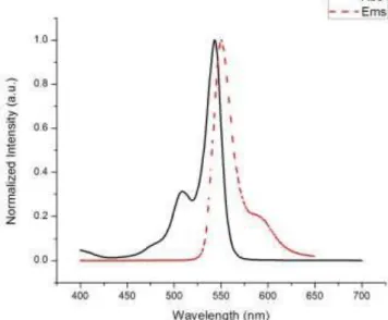
After the synthesis of the 2-methyl pyrrole, we synthesized meso-CF3-substituted BODIPY. The absorption λmax of meso-CF3-substituted BODIPY is at 543 nm and its emission is at 550 nm (Figure 21). After the completion of the synthesis of the meso-CF3-substituted BODIPY, well-known heavy atoms Iodine and Bromine were introduced to the BODIPY cores because of the fact that intersystem crossing of a photosensitizer is an important parameter for PDT action.
During the synthesis of the iodinated meso-CF3-substituted BODIPY we encountered a synthetic problem. The Iodination of the meso-CF3-substituted BODIPY with iodic acid and I2 was the problematic step and did not work. Following another procedure, we managed to synthesize the iodinated meso-CF3 -substituted BODIPY in good yields. Synthesis of the iodinated BODIPY by using this procedure was straightforward.
Since our aim was to design and synthesize photosensitizers for PDT presenting absorption maxima especially in the near-IR region of the spectrum, we introduced electron donating terminal groups to the BODIPY cores via Knoevenagel condensation which allows the extension of the conjugation at the same time.
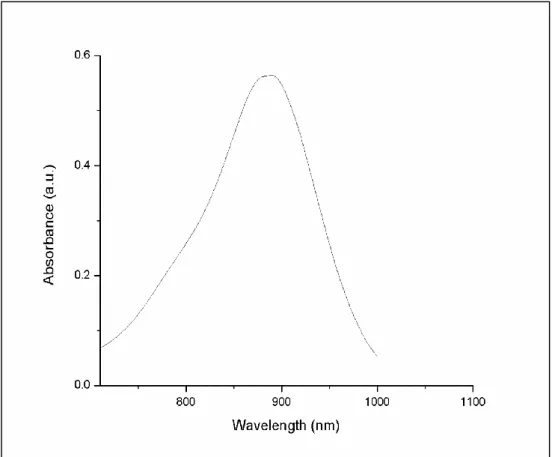
As has been mentioned above, the first target molecule TM1, compound 4, has been synthesized via Knoevenagel condensation and the 1H NMR, 13C NMR spectra and high resolution mass spectrum (HRMS) of the target compound have been evaluated. We obtained absorption spectrum of the target molecule TM1
37
using a Varian spectrophotometer. It has absorption maximum at 890 nm(Figure 22). The attachment of the electron donating group 4,4-dimethylaminobenzaldehyde to the meso-CF3-substituted and brominated BODIPY gives rise to a spectacular 304 nm red shift.
Figure 21 : The absorbance and emission spectra of the meso-CF3-substituted BODIPY
38
Figure 22 : Absorbance spectrum of the target molecule TM1 in CH2Cl2
After the completion of the synthesis of the target molecule TM1, we searched for the suitable trap molecule in order to determine the ability of the target molecule TM1 to convert ground state triplet oxygen to the reactive and cytotoxic singlet oxygen. We opted for the compound diphenylisobenzofuran (DPBF) as a trap molecule in order to assess singlet oxygen measurements. This trap molecule is ideal when the measurement is carried out in organic media.
39
We carried out the measurement in order to demonstrate the singlet oxygen generation capacity of the target molecule TM1 by using 1,3-diphenylisobenzofuran as a trap molecule and when examined carefully it has been shown that singlet oxygen producing ability of the target compound is rather good.
40
Air saturated DCM was obtained by bubbling the air for 5 minutes. Excitation of the target molecule was carried out with a near-IR light emitting diode (LED) source. The decrease of the absorbance band of the trap molecule at 410 nm was monitored.
This decrease of the absorbance band is caused by the oxidation of the trap molecule with reactive oxygen species such as singlet oxygen.
The experiment was performed at the concentration of 2x10⁻⁶ M of PS and 50x10 -6 M of DPBF over a period of 50 min. For the first 15 minutes sample was kept at dark, then irradiated at 890 nm light for 35 minutes. Absorbance was measured in 5 minutes intervals (Figure 23).
As noted above, the second target molecule TM2, compound 5, has been synthesized via Knoevenagel condensation and the 1H and 13C NMR spectra and high resolution mass spectrum (HRMS) of the target compound have been evaluated. We obtained absorption spectrum of the target molecule TM2 using a Varian spectrophotometer. It has absorption maximum at 886 nm (Figure 24). The attachment of the electron donating group 4, 4-dimethylaminobenzaldehyde to the meso-CF3-substituted and iodinated BODIPY gives rise to a spectacular 290 nm red shift.
Figure 23 : Absorbance spectrum of trap molecule diphenylisobenzofuran (DPBF) in dichloromethane under light and Photosensitizer (Target molecule 1)
41
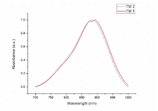
Figure 24: The absorbance spectra of the Target molecules 1 and 2 in CH2Cl2 (TM1 and TM2)
42
Maximum absorbance wavelength of target molecules TM1 and TM2 is determined to be 886 and 890 nm respectively (Figure 24). All wavelengths of our target compounds correspond to near IR region is very appropriate for the PDT applications because of the fact that it is the region of the electromagnetic spectrum where the deepest penetration of the light into the mammalian tissues is occurred
We carried out the same measurement in order to demonstrate the singlet oxygen generation capacity of the target molecule TM2 by using 1,3-diphenylisobenzofuran as a trap molecule
Figure 25 : Absorbance spectrum of trap molecule
diphenylisobenzofuran (DPBF) in dichloromethane under light and PS ( Target molecule 2)