DOI: 10.5455/annalsmedres.2018.09.172 2019;26(1):51-5
The relationship between preoperative and postoperative
neutrophil-lymphocyte and platelet-lymphocyte ratio with
post-dural-puncture headache in patients undergoing
cesarean section
Mehmet Sargin1, Mehmet Selcuk Uluer2, Mahmut Sami Tutar2
1Selcuk University, Faculty of Medicine Department of, Anesthesiology and Reanimation, Konya, Turkey 2Konya Training and Research Hospital, Department of, Anesthesiology and Reanimation, Konya Turkey Copyright © 2019 by authors and Annals of Medical Research Publishing Inc.
Abstract
Aim: This study aims to evaluate whether there is a possible relationship between the preoperative and early postoperative period
neutrophil-to-lymphocyte ratio and platelet-to-lymphocyte ratio and post-dural-puncture headache in patients undergoing cesarean section.
Material and Methods: Two hundred twenty pregnant women scheduled to undergo elective cesarean section under spinal
anesthesia, were studied. Patiens demografic data and blood count parameters were noted. Blood was sampled from a peripheral vein for neutrophil/lymphocyte ratio ve platelet/lymphocyte ratio. The time points for sampling blood were as follows: Preoperative; 1 day before and postoperative; within 6-12 hours after cesarean section. The patients were questioned for possible occurrence of spinal anesthesia induced headache on the first and seventh postoperative days. Post-dural puncture headache was evaluated according to the International Classification of Headache Disorders (ICHD-II) diagnostic criteria.
Results: A total of 220 patients were enrolled in the study and 217 patients completed the investigation. Post-dural puncture
headache was detected in 78 patients and the incidence was 35.9%. The measurements of laboratory parameters were statistically similar between two groups (P > 0.05).
Conclusion: Our study results showed no relationship between the neutrophil-to-lymphocyte ratio and platelet-to-lymphocyte ratio,
and post-dural-puncture headache in the preoperative and also, early postoperative period in patients who undergoing cesarean section.
Keywords: Post-Dural-Puncture Headache; Neutrophil-Lymphocyte Ratio; Platelet-Lymphocyte Ratio; Cesarean Section.
Received: 04.09.2018 Accepted: 15.09.2018 Available online: 18.10.2018
Corresponding Author: Mehmet Sargin, Selcuk University, Faculty of Medicine Department of, Anesthesiology and Reanimation,
Konya, Turkey, E-mail: [email protected]
INTRODUCTION
Although spinal anesthesia is the most popular and common anesthesia technique in cesarean section, it has various complications such as post-dural-puncture headache (PDPH) (1,2). Typically, it is exacerbated by movement with a throbbing nature, and is accompanied by photophobia and blurry vision (3). Unfortunately, the incidence of PDPH is higher in parturients compared to other patients (4,5). PDPH, the incidence of which varies from 0.5 to 52%, is one of the most significant complications after spinal anesthesia (6,7). Therefore, management of spinal anesthesia is very important for obstetric anesthesiologists. Understanding the underlying mechanism of PDPH is essential to manage
the preoperative prophylactic preventive (i.e., medication, needle choice, bevel direction, orientation of the needle in entry) and therapeutic options. The mechanism of PDPH is not clear, but leakage of cerebrospinal fluid (CSF) from the dural hole is the traditional theory. Although the precise mechanism of PDPH has not been elucidated, some high-risk factors for developing PDPH have been found, including female sex, young age, the needle size and design, the needle bevel direction and a previous history of PDPH (6,8).
The identification of neutrophil/ lymphocyte ratio (NLR) and platelet/lymphocyte ratio (PLR) is an extremely useful method in evaluating inflammatory response (9-14). They are evaluated through blood parameters. The
neutrophil-to-lymphocyte ratio (NLR) and platelet-to-lymphocyte ratio (PLR) have been utilized as prognostic and predictive markers in many studies.
In the present study, we aimed to evaluate the possible relationship between NLR and PLR and PDPH during preoperative and early postoperative period in patients who underwent cesarean section.
MATERIAL and METHODS
Institutional ethics committee approval and written consent from the patients were obtained for the study. Two hundred twenty pregnant women, gestational age 38-40 week, between the ages of 19-45 yr, ASA physical status I, scheduled to undergo elective CS under spinal anesthesia, were studied.
Exclusion criteria were contraindication to neuraxial anesthesia or known allergy to bupivacaine, spinal puncture failure, or a need for additional intraoperative analgesia, body mass index (BMI) >35 kg/m2, general anesthesia or epidural anesthesia, multiple gestation, emergency CS and patients with chronic headache history. Patients with hematological, infectious, or inflammatory diseases or severe renal or hepatic disease, pregnancy-induced hypertension, preeclampsia, eclampsia, gestational diabetes, macrosomic infants, intrauterine growth retardation and small for gestational age were also excluded from the present study.
All patients were expected to fast 6-8 hours before CS, and no one premedicated. Routine monitors (consisting of a pulse oximeter, 3-lead ECG and a non-invasive blood pressure cuff) were applied. Following prehydration with Ringer’s lactate solution 500 mL, spinal anesthesia was induced with hyperbaric bupivacaine 10-12.5 mg via a 25 G Quincke-tip spinal needle in the sitting position at the L3–4 or L4-5 vertebral level using a midline approach by an anesthesiologist with more then 5 years experience. Patients were then positioned in a 10˚ left-lateral tilt. Oxygen (4 l.min-1) was administered through a facemask. Surgery was initiated when the sensory block level reached at T4. Hypotension was defined as a decrease in SBP of >30% below baseline or to <90 mmHg and was treated by increasing the rate of crystalloid infusion. If hypotension persisted a bolus of iv ephedrine 5 mg was given. Bradycardia was defined as HR <60 beats/min and was treated with iv atropine 0.5 mg. Patiens demografic data [age, ASA (American Society of Anesthesiologist) physical status and Body mass index (BMI)], and other blood count parameters [White blood cell (WBC), Hemoglobin (Hb), Hematocrit (Htc), Platelet (Plt), Neutrophil, Lymphocyte] were also noted.
Blood was sampled from a peripheral vein for total leukocytic, neutrophil, lymphocyte, Platelet counts, NLR and PLR. The time points for sampling blood were as follows: Preoperative; 1 day before and postoperative; within 6-12 hours after CS. Neutrophil and lymphocyte
counts were derived from differential percentages of leukocytes measured by automatic cell counters (Sysmex XE-2100, USA).The calculation of NLR and PLR was done by one of our authors who was blinded to the samples from whichever group for the calculation of absolute counts of neutrophils, platelets and lymphocytes.
The patients were questioned for possible occurrence of spinal anesthesia induced headache on the first and seventh postoperative days. A telephone followup call was used if the hospital stay was shorter than 7 days. PDPH was evaluated according to the International Classification of Headache Disorders (ICHD-II) diagnostic criteria (15). Intensity of headache and puncture pain were assessed on a scale of 0 to 10, where 0 means no pain and 10 the worst possible pain (0 no, 1–3 mild, 4–6 moderate, 7–10 severe). In addition, hearing loss, tinnitus, photophobia, nausea and vomiting were recorded. Any treatment for the headache was recorded. Post dural puncture headache evaluations was performed by a different anesthesiologist blinded to the study.
Statistical analysis
Statistical analyses were performed with SPSS 15.0 software (SPSS Institute, Chicago, IL, USA). The compliance of variables with normal distribution were evaluated visually (histograms and probability plots) and analytically (Kolmogorov-Smirnov test). Descriptive analyses for normally distributed variables were given in mean and standard deviation. The comparisons in both groups were carried out using the Student t-test and Pearson Chi-Square test.p<0.05 value was considered statistically significant.
RESULTS
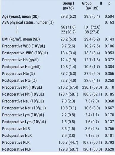
A total of 220 patients were enrolled in the study and 217 patients completed the investigation. Three patients were excluded from the study because they could not be reached via telephone. PDPH was detected in 78 patients and the incidence was 35,9%. Patients’ demographic and clinical characteristics are summarised in Table 1 and there were no significant differences between the groups regarding age, BMI, ASA physical status (P=0.504, P=0.143 and P=0.163, respectively).
The measurements of laboratory parameters were statistically similar between two groups (P > 0.05). There were no significant differences between the groups regarding preoperative and postoperative WBC, Hb, Htc, platelet, neutrophil, lymphocyte, NLR and PLR values (P=0.106, P=0.953, P=0.372, P=0.384, P=0.356, P=0.258, P=0.110, P=0.185, P=0.368, P=0.667, P=0.170, P=0.137, P=0.766, P=0.101, P=0.793 and P=0.629, respectively). Preoperative NLR was 3.5 (1.5) in Group I and 3.6 (2.3) in Group II. Postoperative NLR was 7.9 (3.8) in Group I and 7.1 (2.9) in Group II. Preoperative PLR was 105.7 (44.7) in Group I and 107.7 (60.1) in Group II. Postoperative PLR was 129.8 (60.7) in Group I and 126.1 (50.0) in Group II.
Table 1. Patients’ demographic and clinical characteristics
Group I
(n=78) Group II (n=139) p Age (years), mean (SD) 29.8 (5.2) 29.3 (5.4) 0.504
ASA physical status, number (%) I II 56 (71.8)22 (28.2) 101 (72.6)38 (27.4) 0.163 BMI (kg/m2), mean (SD) 28.2 (5.3) 29.4 (6.2) 0.143 Preoperative WBC (103/µL) 9.7 (2.6) 10.2 (2.5) 0.106 Postoperative WBC (103/µL) 13.4 (3.4) 13.3 (3.4) 0.953 Preoperative Hb (gr/dl) 12.4 (1.9) 12.7 (1.8) 0.372 Postoperative Hb (gr/dl) 10.8 (1.4) 10.5 (1.7) 0.384 Preoperative Htc (%) 37.2 (5.3) 37.9 (5.0) 0.356 Postoperative Htc (%) 32.7 (4.0) 32.6 (4.1) 0.258 Preoperative Plt (103/µL) 216.2 (67.4) 230.1 (59.0) 0.110 Postoperative Plt (103/µL) 178.4 (58.1) 188.3 (52.1) 0.185 Preoperative Neu (103/µL) 7.0 (2.3) 7.3 (2.3) 0.368 Postoperative Neu (103/µL) 10.8 (3.1) 10.6 (3.0) 0.667 Preoperative Lym (103/µL) 2.2 (0.8) 2.4 (1.1) 0.170 Postoperative Lym (103/µL) 1.5 (0.5) 1.6 (0.7) 0.137 Preoperative NLR 3.5 (1.5) 3.6 (2.3) 0.766 Postoperative NLR 7.9 (3.8) 7.1 (2.9) 0.101 Preoperative PLR 105.7 (44.7) 107.7 (60.1) 0.793 Postoperative PLR 129.8 (60.7) 126.1 (50.0) 0.629
DISCUSSION
Post-dural-puncture headache is an iatrogenic complication of the spinal anesthesia. In several studies, its incidence has been reported to range less than 2% and over 50% (6,7). Factors which affect the PDPH incidence include sex and age of patient, pregnancy, previous PDPH history, needle size, shape of the needle tip, bevel orientation, median vs paramedian approach, type of the local anesthetic solution, number of lumbar puncture (LP) interventions, replacing the needle stylet, and the degree of experience of the clinician (16-22).
Although the PDPH mechanism is not fully known, the symptoms are mostly based on an excessive loss of cerebrospinal fluid (CSF) from the dural puncture region, which eventually leads to decreased CSF pressure (23). Decreased CSF pressure reduces the buffering capacity of CSF in the brain, thereby, resulting in traction in the structures which are sensitive to intracranial pain such as meningeal veins, upper cervical, and cranial nerves (23). Another suggested mechanism is the vasodilatation of the intracranial veins; following the rapid decline in the CSF volume, adenosine is secreted, leading to vasodilatation of the intracranial veins (24,25).
Classical clinical presentation of PDPH is a dull and throbbing bilateral headache related to the alterations in the posture (exacerbated with sitting and standing, relieved with lying down), which often develops within
seven days and disappears within 14 days of the lumbar puncture (26). The International Classification of Headache Disorders’ (ICHD) criteria for the diagnosis of PDPH are given in Table 2 (15).
Table 2.The international classification of headache disorders criteria for the diagnosis of PDPH
A. Headache that worsens within 15 minutes after sitting or standing
and improves within 15 minutes after lying down, with at least one of the following and fulfilling criteria C and D
1. Neck stiffness 2. Tinnitus 3. Hypacausia 4. Photophobia 5. Nausea
B. Dural puncture has been performed
C. Headache develops within five days after dural puncture D. Headache resolves either
1. Spontaneously within one week
2. Within 48 hours after effective treatment of the spinal fluid leak; usually by epidural blood patch
Treatment of PDPH can be discussed under five headings: pre-emptive consideration, conservative management, drug therapy, invasive treatment, and home-based work. To minimize the PDPH risk, preventive measures such as the choice of needle type, keeping repeated interventions to a lesser extent before and after the lumbar puncture must be taken. The second stage of the therapy involves conservative management, such as bed rest and fluid therapy (27). Such as analgesic agents, sumatriptan, caffeine, dexamethasone and hydrocortisone, gabapentin and cosyntropine can bu used in drug treatment (7,28-32). Invasive treatment should be considered for patients who are unresponsive to conservative management and medications within 48 hours. Epidural blood patch (EDBP), epidural saline or dextran 40 infusion, intrathecal catheter, fibrin glue, and surgical closure of the dura are the invasive methods which are used for the treatment of PDPH (7,24,26,33,34). The common feature of EDPB and intratechal catheter methods have been shown to increase the inflammatory response at the site of dural puncture, thereby, leading to the closure of the hole in the dura (26,33).
In the study, we evaluated whether the NLR and PLR, predictors of systemic inflammation, can be used as predictors for PDPH. Although it is a novel systemic inflammation marker in the evaluation of the systemic inflammation in similar diseases and in the evaluation of the relationship between various diseases and systemic inflammation (35-39), it is still not widely adopted in anesthesia and algology practices. Although the number of studies investigating the effect of these markers on anesthesia practice, two studies examined the effect of the use of anesthetic agents on NLR (40,41). In addition, another study investigated the relationship
between preoperative NLR and anesthesia risk scores, such as the American Society of Anesthesiologists (ASA) and Revised Cardiac Risk Index (RCRI) (42). In the aforementioned studies, propofol was shown to improve leukocytic changes, compared to desflurane in patients who underwent coronary artery bypass grafting (40). In another study, total intravenous anesthesia (TIVA) using propofol and remifentanil was found to yield more positive outcomes in terms of NLR and leukocytic alterations, compared to the inhalation anesthesia using sevoflurane (41). Differently from aforementioned studies, Venkatraghavan et al.(42) reported a strong correlation between preoperative NLR and anesthesia risk scores, indicating that NLR can be used as a predictive marker in the preoperative period in the anesthesia practice.
In another study which examined the effect of spinal and general anesthesia on NLR in patients who underwent cesarean section, the authors reported that postoperative NLR was significantly lower in spinal anesthesia than general anesthesia (43). Unlike our study, pre- and postoperative NLR values of the patients who underwent cesarean section under spinal anesthesia were higher. In addition, blood samples were collected two hours after the operation in the aforementioned study, while we collected samples 6 to 12 hours after the operation.
Furthermore, in the present study, we evaluated whether NLR and PLR could be used as predictive markers of PDPH in the preoperative and postoperative period in patients who underwent cesarean section. During the study planning, based on the literature data, we hypothesized that the inflammation of the hole in the dura after LP accelerated the closure of the hole and that inflammation could decrease the incidence of PDPH. However, we were unable to find any significant difference between the NLR and PLR values of the patients with and without PDPH in the preoperative and early postoperative period.
The main limitation of our study is the failure to collect blood at the same time from each patient in the preoperative and postoperative period.
CONCLUSION
In conclusion, our study results suggest no relationship between the preoperative and early postoperative period NLR and PLR with PDPH in patients who undergoing cesarean section. However, further large-scale studies are required to establish a definite conclusion.
Competing interests: The authors declare that they have no competing interest.
Financial Disclosure: There are no financial supports
Ethical approval: This work has been approved by the Institutional Review Board.
Mehmet Sargin ORCID: 0000-0002-6574-273X Mehmet Selcuk Uluer ORCID: 0000-0002-5699-8688 Mahmut Sami Tutar ORCID: 0000-0002-5709-6504
REFERENCES
1. Ahsan-ul-Haq M, Kazmi EH, Hussain Q. Analysis of headache in obstetric patients: experience from a outcome
of general versus spinal anaesthesia for cesarean delivery in severe preeclampsia with fetal compromise. Biomedica 2005;21:2127.
2. Chohan U, Hamdani GA. Post dural puncture headache. J Pak Med Assoc. 2003;53:359-67.
3. Rodriques AM, Roy PM. Post-lumbar puncture headache. Rev Prat 2007;57:353-7.
4. Wadud R, Laiq N, Qureshi FA, et al. The frequency of postdural puncture headache in different age groups. J Coll Physicians Surg Pak 2006;16:389-92.
5. Yazigi A, Chalhoub V, Madi-Jebara S, et al. Prophylactic ondansetron is effective in the treatment of nausea and vomiting but not on pruritus after cesarean delivery with intrathecal sufentanil-morphine. J Clin Anesth 2002;14:183-6. 6. Kuczkowski KM. Post-dural puncture headache in the
obstetric patient: an old problem. New solutions. Minerva Anestesiol 2004;70:823-30.
7. Turnbull DK, Shepherd DB. Post-dural puncture headache: pathogenesis, prevention and treatment. Br J Anaesth 2003;91:718-29.
8. Olufemi Babatunde Omole, Gboyega Adebola Ogunbanjo. Postdural puncture headache: evidence-based review for primary care. South African Family Practice 2015;57:241-6. 9. Cupisti K, Dotzenrath C, Simon D, et al. Therapy of suspected
intrathoracic parathyroid adenomas. Experiences using open transthoracic approach and video assisted thoracoscopic surgery. Langenbecks Arch Surg 2002;386:488-93.
10. Gogas J, Kouskos E, Mantas D, et al. Pre-operative Tc-99msestamibi scanning and intra-operative nuclear mapping: are they accurate in localizing parathyroid adenoma? Acta Chir Belg 2003;103:626-30.
11. Grisel JJ, Al-Ghawi H, Heubi CH, et al. Successful removal of an intrathyroidal parathyroid adenoma located by technetiumTc 99m sestamibiscan and ultrasound. Thyroid. 2009;19:423-5.
12. Yabuta T, Tsushima Y, Masuoka H, et al. Ultrasonographic features of intrathyroidal parathyroid adenoma causing primary hyperparathyroidism. Endocr J 2011;58:989-94. 13. Mazeh H, Kouniavsky G, Schneider DF, et al. Intrathyroidal
parathyroidglands: small, but mighty (a Napoleon phenomenon). Surgery 2012;152:1193-200.
14. Abdo A, Kowdley GC. An intrathyroidal parathyroid: thyroid in sampling prior to lobectomy. Am Surg 2012;78:818-9. 15. The international classification of headache disorders 2003.
2nd ed. Cephalgia 2004;24:1-60.
16. Lybecker H, Moller JT, May O, et al. Incidence and predic¬tion of postdural puncture headache. A prospective study of 1021 spinal anesthesias. Anesth Analg 1990;70:389-94. 17. Halpern S, Preston R. Postdural puncture headache and
spinal needle design. Metaanalyses. Anesthesiology 1994;8:1376-83.
18. Ross BK, Chadwick HS, Mancuso JJ, et al. Sprotte needle for obstetric anesthesia: decreased incidence of post dural puncture headache. Reg Anesth 1992;17:29-33.
19. Tarkkila PJ, Heine H, Tervo RR. Comparison of Sprotte and Quincke needles with respect to post dural puncture headache and backache. Reg Anesth 1992;17:283-7. 20. Janik R, Dick W. Post spinal headache. Its incidence
follow¬ing the median and paramedian techniques. Anaesthesist. 1992;41:137-141.
21. Naulty JS, Hertwig L, Hunt CO, et al. Influence of local anesthetic solution on postdural puncture headache. Anesthesiology 1990;72:450-4.
22. Shnider SM, Levinson G. Anesthesia for cesarean section. 2nd ed. Baltimore: Williams & Wilkins. 1987. pp. 159-78.
23. Morewood GH. A rational approach to the cause, prevention and treatment of postdural puncture headache. CMAJ 1993;149:1087-93.
24. Ghaleb A, Khorasani A, Mangar D. Post-dural puncture headache. Int J General Med 2012;5:45-51.
25. Hlongwane TN. What’s new in obstetric anaesthesia? S Afr Fam Pract 2012;54:11-3.
26. Ahmed SV, Jayawarna C, Jude E. Post lumbar puncture headache: diagnosis and management. Postgrad Med J 2006;82:713-6.
27. Sudlow CL, Warlow CP. Posture and fluids for preventing post-dural puncture headache. Cochrane Database Sys Rev 2002;2:CD0001790.
28. Pardo M, Sonner JM. Manual of anesthesia practice. Cambridge: Cambridge University Press; 2007. p. 832-84. 29. Erol DD. The analgesic and antiemetic efficacy of gabapentin
or ergotamine/caffeine for the treatment of postdural puncture headache. Adv Med Sci 2011;56:25-9.
30. Wagner Y, Storr F, Cope S. Gabapectin in the treatment of post-dural puncture headache: a case series. Anaesth Intsive Care 2012;40:714-8.
31. Kuczkowski KM, Eisenmann UB. Hypertensive encephalopathy mimicking postdural puncture headache in a parturient beyond the edge of reproductive age. Anesth Analg 2004;99:1873-4.
32. Hakim SM. Cosyntropin for prophylaxis against postdural puncture headache after accidental dural puncture. Anesthesiology 2010;113:413-20.
33. Afpel CC, Saxena A, Cakmakkaya OS, et al. Prevention of postdural puncture headache after accidental dural puncture: a quantitative systematic review. Br J Anesth 2010;105:255-63.
34. Gil F, García-Aguado R, Barcia JA, et al. The effect of fibrin glue patch in an in vitro model of postdural puncture leakage. Anesth Analg 1998;87:1125-8.
35. Sener A, Ciftci Sivri HD, Sivri S, et al. The Prognostic Value of The Platelet/Lymphocyte Ratio in Predicting Short-Term Mortality in Patients with Acute Pulmonary Embolism. Sakarya Med J 2015;5:204-8.
36. Çetin M, Kiziltunc E, Elalmış ÖU, et al. Predictive Value of Neutrophil Lymphocyte Ratio and Platelet Lymphocyte Ratio in Patients with Coronary Slow Flow. Acta Cardiol Sin 2016;32:307-12.
37. Yushun Zhang, Wei Wu, Liming Dong, et al. Neutrophil to lymphocyte ratio predicts persistent organ failure and in-hospital mortality in an Asian Chinese population of acute pancreatitis. Medicine (Baltimore) 2016;95:4746.
38. Ataş H, Çevirgen Cemil B, Işıl Kurmuş G, et al. Assessment of systemic inflammation with neutrophil-lymphocyte ratio in lichen planus. Adv Dermatol Allergol 2016;33:188-92. 39. Varım C, Atılgan Acar B, Uyanık MS, et al. Association
between the neutrophil-to-lymphocyte ratio, a new marker of systemic inflammation, and restless legs syndrome. Singapore Med J 2016;57:514-6.
40. Aldemir M, Doğan Bakı E, Adalı F, et al. Comparison of the effects of propofol anaesthesia and desflurane anaesthesia on neutrophil/lymphocyte ratios after coronary artery bypass surgery. J Turgut Ozal Med Cent 2015;22:165-70. 41. Kim WH, Jin HS, Ko JS, et al. The effect of anesthetic
techniques on neutrophil-to-lymphocyte ratio after laparoscopy-assisted vaginal hysterectomy. Acta Anaesthesiol Taiwan 2011;49:83-7.
42. Lashmi Venkatraghavan, Tan Ping Tan, Jigesh Mehta, et al. Neutrophil Lymphocyte Ratio as a predictor of systemic inflammation - A cross-sectional study in a pre-admission setting. F1000Res 2015;4:123.
43. Erbaş M, Toman H, Gencer M, et al. The effect of general and spinal anesthesia on neutrophil to lymphocyte ratio in patients undergoing cesarian section. Anaesth, Pain & Intensive Care 2015;19:485-8.