doi: 10.3389/fphar.2019.00466
Edited by: Marco Leonti, University of Cagliari, Italy Reviewed by: Dezs ˝o Csupor, University of Szeged, Hungary Guillermo Benítez, University of Granada, Spain *Correspondence: Andrei Mocan [email protected] Atanas G. Atanasov [email protected]
†These authors have contributed
equally to this work and share first authorship
Specialty section: This article was submitted to Ethnopharmacology, a section of the journal Frontiers in Pharmacology Received: 16 August 2018 Accepted: 12 April 2019 Published: 13 June 2019 Citation: Tewari D, Samoil ˘a O, Gocan D, Mocan A, Moldovan C, Devkota HP, Atanasov AG, Zengin G, Echeverría J, Vodnar D, Szabo B and Cri ¸san G (2019) Medicinal Plants and Natural Products Used in Cataract Management. Front. Pharmacol. 10:466. doi: 10.3389/fphar.2019.00466
Medicinal Plants and Natural
Products Used in Cataract
Management
Devesh Tewari
1†, Ovidiu Samoil ˘a
2†, Diana Gocan
2, Andrei Mocan
3* , Cadmiel Moldovan
3,
Hari Prasad Devkota
4, Atanas G. Atanasov
5,6* , Gokhan Zengin
7, Javier Echeverría
8,
Dan Vodnar
9, Bianca Szabo
10and Gianina Cri ¸san
31Department of Pharmacognosy, School of Pharmaceutical Sciences, Lovely Professional University, Phagwara, India, 2Department of Ophthalmology, Iuliu Ha¸tieganu University of Medicine and Pharmacy, Cluj-Napoca, Romania,3Department of Pharmaceutical Botany, Iuliu Ha¸tieganu University of Medicine and Pharmacy, Cluj-Napoca, Romania,4Graduate School of Pharmaceutical Sciences, Kumamoto University, Kumamoto, Japan,5Institute of Genetics and Animal Breeding, Polish Academy of Sciences, Jastrz ˛ebiec, Poland,6Department of Pharmacognosy, University of Vienna, Vienna, Austria, 7Department of Biology, Faculty of Science, Selcuk University, Konya, Turkey,8Departamento de Ciencias del Ambiente, Facultad de Química y Biología, Universidad de Santiago de Chile, Santiago, Chile,9Department of Food Science, University of Agricultural Sciences and Veterinary Medicine of Cluj-Napoca, Cluj-Napoca, Romania,10Department of Anatomy, Iuliu Ha¸tieganu University of Medicine and Pharmacy, Cluj-Napoca, Romania
Cataract is the leading reason of blindness worldwide and is defined by the presence of
any lens opacities or loss of transparency. The most common symptoms of cataract are
impaired vision, decreased contrast sensitivity, color disturbance, and glare. Oxidative
stress is among the main mechanisms involved in the development of age-related
cataract. Surgery through phacoemulsification and intraocular lens implantation is the
most effective method for cataract treatment, however, there are chances of serious
complications and irreversible loss of vision associated with the surgery. Natural
compounds consisting of antioxidant or anti-inflammatory secondary metabolites can
serve as potential leads for anticataract agents. In this review, we tried to document
medicinal plants and plant-based natural products used for cataract treatment
worldwide, which are gathered from available ethnopharmacological/ethnobotanical
data. We have extensively explored a number of recognized databases like Scifinder,
PubMed, Science Direct, Google Scholar, and Scopus by using keywords and
phrases such as “cataract”, “blindness”, “traditional medicine”, “ethnopharmacology”,
“ethnobotany”, “herbs”, “medicinal plants”, or other relevant terms, and summarized
the plants/phytoconstituents that are evaluated in different models of cataract and also
tabulated 44 plants that are traditionally used in cataract in various folklore medical
practices. Moreover, we also categorized the plants according to scientific studies
carried out in different cataract models with their mechanisms of action.
Keywords: medicinal plants, natural products, cataract, antioxidant, aldose reductase, lens opacity, MAPK
Abbreviations:AGE, advanced glycation end products; AKR1B1, aldo-keto reductase family 1, member B1; AR, aldose reductase; ATP, adenosine triphosphate; BSA, bovine serum albumin; Cx, connexin; EPHA2: FRSA, free radical scavenging activity; GPX, glutathione peroxidase; GSH, glutathione; IL, interleukin; iNOS, inducible nitric oxide synthase; LPO, lipid peroxides; MAPKs, mitogen-activated protein kinase; NADPH, nicotinamide adenine dinucleotide phosphate; PKC, protein kinase C; RLAR, rat lens aldose reductase; RNS, reactive nitrogen species; ROS, reactive oxygen species; SOD, superoxide dismutase; TGF-β2, transforming growth factor β2; TNF-α, tumor necrosis factor-α; UV, ultraviolet; VEGF, vascular
CATARACT: AN OVERVIEW
The crystalline lens lies behind the iris and represents the
dynamic part of the eye’s optical system, responsible for focusing
the image onto the retina. Cataract is defined by the presence of
any lens opacities or loss of transparency. The most common
symptoms of cataract are impaired vision, decreased contrast
sensitivity, color disturbance, and glare. Changes in the lens
may also serve as markers for systemic health and aging in
the over-all population (
Song et al., 2014
). According to the
type of lens opacities, cataract is classified into three classical
types: nuclear, posterior subcapsular, and cortical. These types
can also be associated with each other and if untreated, they
progress to total lens opacification. Some of the most common
causes for cataract in adults are age, diabetes, steroid use,
family history, or trauma. Congenital cataract has a significant
prevalence, also.
Cataract is the foremost reason of blindness worldwide in
spite of the technological advancements in eye surgery in the
last two decades. In 2010, there were around 32 million blind
people and 191 million were with poor vision. One in three
blind people suffered from cataract (
Khairallah et al., 2015
). The
World Health Organization (WHO) suggests that by 2020 the
number of blind people will reach 90 million globally (
Khairallah
et al., 2015
;
Taylor, 2016
). The strategy to fight this challenge
is costly, aiming human resource, infrastructure development,
and effective disease control. The latter is dependent on the
characteristics of the specific disease. Prevalence of cataract
increases with age, from 5% for patients of age 52–62 to
64% for patients over 70 years, in Europe (
Prokofyeva et al.,
2013
). Age is a non-modifiable risk factor involved in the
pathogenesis of cataract, hence the progressive aging of the
population is an alarming issue. Identifying modifiable risk
factors for cataract is imperative and may help to establish the
preventive measures.
The surgical treatment for cataract consists of cataractous
lens extraction and intraocular lens implant. It is the only
current treatment available in order for patients to recover their
visual function. This implies a significant cost and there is a
significant lack of access to surgery, especially in the developing
world. Despite good postoperative outcomes, complications are
possible following cataract surgery. Studies have suggested that
pseudophakia patients have a higher risk of retinal detachment.
Endophthalmitis has also been reported in 0.12% of the operated
cases (
Toh et al., 2007
). After the surgery, the mobility of the
lens is lost and correcting glasses are usually necessary. This will
only increase the expense and the discomfort for the patient and
society. Medical treatment would be a desired alternative.
The most primitive written reference to cataract surgery
was discovered in Sanskrit manuscripts dating back from the
5th century BCE. It was attributed to
Sushruta, a well-known
ancient plastic surgeon who described a procedure known as
couching, in which the cataractous lens was displaced with a
sharp tool to fall it into the vitreous cavity, clearing the visual
axis, though the vision was significantly blurred as there were
no corrective lenses or glasses (
Uhr, 2003
;
Sachdev, 2017
). Even
at the time of Mesopotamia (ca. 3,000–4,000 BCE) records
reveal that mysticism along with different animal products,
vegetables, and minerals were utilized for the treatment of devil
and spirits causing eye diseases. Hundreds of remedies were
also described during the Greek era (ca. 460–375 BCE) for
disorders of the eyes. Moreover, eye diseases are also described
anatomically by
Sushruta (as mentioned above), Galen and
various medicinal and surgical procedures were described for
the treatment of eye diseases (
Duke-Elder, 1962
;
Albert and
Edwards, 1996
;
Goodman, 1996
). In 1748, the introduction of
modern cataract surgery was done by Jacques Daviel in Paris,
in which the cataractous lens is removed from the eye. Later on
in 1753, Samuel Sharp of London presented the intracapsular
procedure, wherein the whole lens was removed by an incision
by put on thumb pressure. In 1867 silk sutures for cataract
surgery was originally described by Henry Willard Williams of
Boston (
Uhr, 2003
).
CATARACT – PATHOGENESIS
Various mechanisms have been associated with age-related
cataract pathogenesis. Lens opacities may appear due to changes
in the microarchitecture, caused by mutations, biomechanical,
or physical changes.
Mutations
Despite cataract being a multifactorial disease, sometimes
mutations alone can cause lens opacities and this usually leads
to congenital or pediatric cataract. Studies have presented more
and more evidence that genetic factors are also part of age related
cataract pathogenesis, raising the probability of molecular genetic
relations between lens development and aging (
Hejtmancik and
Kantorow, 2004
). Out of around 42 genes and loci that have
been found to underlie congenital forms of cataract, a few of
them have been linked with age associated cataract: EPHA2
(encodes a member of ephrin receptor of
protein-tyrosine-kinases), CRYAA, CRYGS (both encode lens proteins), FYCO1
(encodes a scaffolding protein which is active in microtubule
transport of autophagic vesicle), or TDRD7 (encodes an
RNA-binding protein). The mutation p.Gly18Val in CRYGS results
in a protein with normal structure in physiological conditions.
The alterations in its structure occur after thermal or chemical
injury. A similar mutation is Phe71Leu in CRYAA. The discovery
of mutations in genes coding for TDRD7, EPHA2, and FYCO1
has provided the initial evidence for the functional importance
of posttranscriptional mRNA regulation, ephrin signaling, and
the autophagy pathway, respectively, in human lens transparency
(
Shiels and Hejtmancik, 2015
).
Gene mutations underlying secondary forms of cataract could
also play part in age related cataract formation. A mutation in
gene on 17q of galactokinase 1 (GALK1) which is responsible
for encoding of the first enzyme in galactose metabolism, trigger
autosomal recessive GALK1 1-deficiency with hypergalactosemia
and cataract as a result of galactitol accumulation and osmotic
stress. A coding variation in GALK1 (p.A198V) generates enzyme
instability associated with amplified risk of age-related cataract in
the Japanese population (
Okano et al., 2001
).
Oxidative Stress
Oxidative stress is among the main mechanisms involved
in the development of age-related cataract. Oxidative stress
occurs when reactive compounds like the superoxide anion,
hydroxyl radicals, and hydrogen peroxide are not neutralized
by antioxidant enzymes and defense systems. Enzymes like
catalase, SOD, and GPX are crucial for the homeostasis of the
antioxidant system and ROS. When levels of ROS increase, this
denatures the lens nucleic acids, proteins, and lipids, leading
to mutations and cell apoptosis. Metabolic activities mostly
take place in the lens epithelium. The lens epithelium uses the
antioxidative enzymes in order to prevent damages caused by
oxidative stress. Studies suggest that the highest concentration
of SOD is in the lens epithelium (
Rajkumar et al., 2013
). These
enzymes are also present in other parts of the lens and play
a very important part in maintaining the lens clarity (
Chang
et al., 2013
). SOD is responsible for converting superoxide
anion into hydrogen peroxide, and then hydrogen peroxide
is transformed into water by catalase or GPX. SOD enzyme
activity is associated with cofactors like zinc, manganese, and
copper. However, a decreased level of cofactors in cataractous
lenses was not found. Experimental animal models show a
decreased level of glutathione in the nucleus, therefore there is a
higher susceptibility for oxidative damage and opacity formation
(
Giblin, 2000
). Studies have shown that serum and aqueous
humor levels of antioxidative enzymes are decreased in patients
with cataract. However, there was no significant difference among
different types of cataract and enzymes serum levels (
Ohia et al.,
2005
;
Wang et al., 2015
).
Crystallins Problems
Crystallins, the major structural lens proteins have an imperative
role in the lens transparency and acquire post-translational
alterations during cataract formation, which lead to protein
insolubility, aggregation and loss of lens transparency. Out of
the three major crystallins,
α-, β-, and γ-, α crystallins exhibit
chaperone like activity, preventing them to aggregate. The
chaperone activity is reduced in cataractous lenses. Prolonged
hyperglycemic conditions increase the chances of crystallins
deterioration (
Reddy et al., 2014
). Calcium activates
calcium-binding proteins triggering changes in the shape and charge
of the proteins. Elevated levels of calcium appear to induce
proteolysis of crystallins by calpain, an intracellular cysteine
protease. Activation of calpain, an intracellular cysteine protease,
leads to proteolysis of the lens proteins. In order for calpains
to activate, a high level of calcium is required (
Obrosova et al.,
2010
). Studies demonstrate that the privation of an endogenous
inhibitor of calpain, named calpastatin, could be linked to the
initial changes that cause cataract (
Nakajima et al., 2014
). Some
antioxidants have been reported to regulate calcium influx in
selenite induced cataracts, for instance the flavonoid fraction of
Brassica oleracea (
Vibin et al., 2010
).
Protein Structures
Alterations in the protein structure are also determined by
UV exposure. Studies have shown that UVB generates more
damage than UVA and that damages are prevented by the lens
filters. After UV radiations, proteins suffer chemical reactions
resulting in aggregations, decreasing the transparency of the
lens (
Cetinel et al., 2017
). The crystalline lens is particularly
exposed to phototoxic damage, because it absorbs most of UV
radiation, together with cornea. The main association is with
cortical cataract, most of the absorption occurring at the posterior
surface of the lens. UV radiation can generate free radicals
including oxygen-derived species, that cause lipid peroxydation
of cellular membranes or can damage DNA directly (
Youn et al.,
2011
).
In vivo, induced cataract has no absolute threshold for
UV exposure. UV induced cataract for
in vivo exposure at
UV-300 nm has a continuous dose-response function (
Söderberg
et al., 2016
). UV radiation data from Eurosun library implied that
rates of cataract were higher in regions with higher ambient UV-B
radiation levels (
Delcourt et al., 2014
).
MEDICINAL PLANTS AND NATURAL
PRODUCTS USED AGAINST CATARACT
Opacity of the lens is triggered by free radicals in most of the
cases (
Varma et al., 1984
;
Thiagarajan and Manikandan, 2013
).
Severe oxidative stress also leads to the protein modifications
by free radicals, and several natural products from plants are
helpful in the prevention of proteins insolubilization, which
may delay the opacity of lenses (
Bhadada et al., 2016b
). Natural
compounds constituting of antioxidant or anti-inflammatory
secondary metabolites could be viewed as potentially optimal
anticataract agents as antioxidant effect is among the major
mechanisms for prevention of cataract in most of the cases,
however, not all the plants possessing antioxidant potential could
have anticataract properties. The role of plant polyphenols in
anti–cataractogenic activities is also studied in the comprehensive
manner either
in vitro or in vivo (
Rooban et al., 2009, 2011
;
Kim
et al., 2011c
;
Wang et al., 2011
;
Sasikala et al., 2013
;
Sunkireddy
et al., 2013
;
Ferlemi et al., 2016
).
Although there is substantial basic and applied research
in the field of cataract management by natural products,
mostly ethno-pharmacological/ethnobotanical research, there
are not many review papers available about the activity
analysis of natural products against different cataract models.
One paper focused on antioxidant containing plants against
cataract was found with 41 plants investigating anti-cataract
activity (
Thiagarajan and Manikandan, 2013
). Although there
are few ethnopharmacological surveys and their reviews available
(
Maregesi et al., 2017
), there is no detailed review available on
the activities of different plants extracts and natural products in
cataract models.
METHODOLOGY AND HYPOTHESIS
In this work, we attempted to gather and document the widely
scattered information from various preclinical investigations
and ethnopharmacological reports. We searched several web
databases namely, Scifinder, ScienceDirect, Pubmed, Scopus,
and Google Scholar. Boolean information retrieval method
(
Pohl et al., 2010
) was applied using plant name with “AND”
operator as also done in some other systemic reviews (
Tewari
et al., 2017, 2018
) followed by “cataract” and using other
different keywords such as “cataract”, “traditional medicine”,
“ethnobotany”, “sodium selenite”, and “ethnopharmacology”.
The main research question we try to address in this paper
is: “are medicinal plants/natural products used in various folk
and traditional medicine of importance in the management of
cataract?” and “what are the major preclinical
in vitro/in vivo
models that are used globally for the evaluation of cataract?”. We
hypothesize that plants used in ethnomedicine are not only of
potential importance but also preclinical studies conducted on
various models of cataract could result in the development of
potential drug candidates in future. This could be very rewarding
for the scientists and scholars working in this area and also
very beneficial for the patients to take forward the preclinically
effective plants for clinical studies.
RESULTS
Oxidative stress is involved in activation of MAPKs. Compounds
resulted from the activation of MAPKs have been studied and
were associated with cell apoptosis. The p38 MPAK was studied
in
vitro and it was shown that it is activated by hydrogen peroxide,
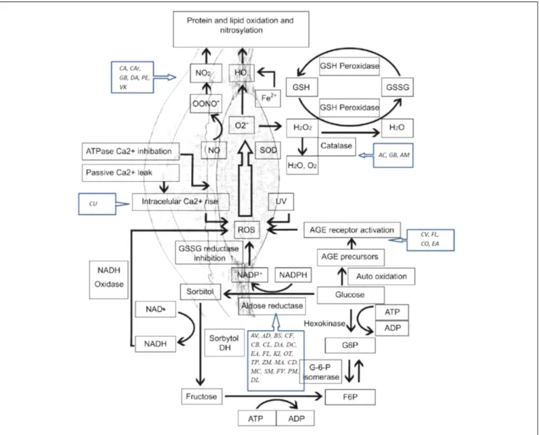
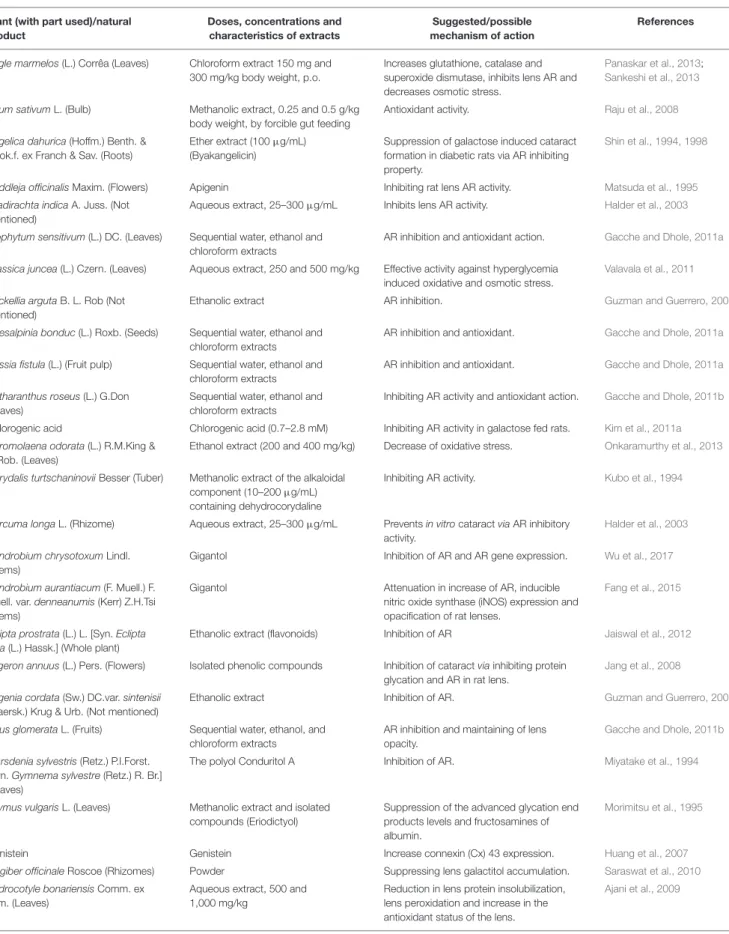
FIGURE 1 | Oxidative stress mechanisms involved in cataract etiology and action mechanisms of several medicinal plants with conducted pharmacological studies for the treatment of cataract. AGE, advanced glycation end-products; ROS, reactive oxygen species; SOD, superoxide dismutase; GSH, glutathione; GSSG, glutathione disulfide; NO, nitric oxide. AC, Allium cepa; CA, Coffea arabica; CU, Curcumin; GB, Ginkgo biloba; AV, Adhatoda vasica; AM, Aegle marmelos; AD, Angelica dahurica; BS, Biophytum sensitivum; CB, Caesalpinia bonduc; CF, Cassia fistula; CV, Cinnamomum verum; CL, Curcuma longa; DA, Dendrobium aurantiacum var. denneanumis; DC, Dendrobium chrysotoxum; EA, Erigeron annuus; FL, Flavonoids; KI, KIOM-79; OT, Ocimum tenuiflorum; VK, Vitamin K; TP, Tephrosia purpurea; ZM, Zea mays; MA, Matteuorienate A; CD, Caesalpinia digyna; CO, Cornus officinalis; MC, Morinda citrifolia; SM, Salvia miltiorrhiza; FV, Foeniculum vulgare; PM, Pueraria montana; DL, Danshenol; C Ar, Citrus aurantium; PE, Phyllanthus emblica.
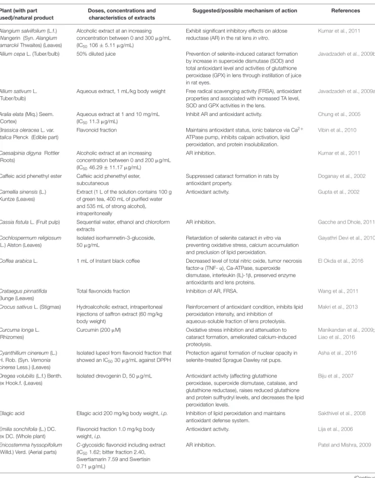
TABLE 1 | Medicinal plants/natural products used against cataract on Selenite/sodium selenite induced cataract models, and suggested/possible mechanisms of action.
Plant (with part used)/natural product
Doses, concentrations and characteristics of extracts
Suggested/possible mechanism of action References
Alangium salviifolium (L.f.) Wangerin (Syn. Alangium lamarckii Thwaites) (Leaves)
Alcoholic extract at an increasing concentration between 0 and 300µg/mL (IC50106 ± 5.11µg/mL)
Exhibit significant inhibitory effects on aldose reductase (AR) in the rat lens in vitro.
Kumar et al., 2011
Allium cepa L. (Tuber/bulb) 50% diluted juice Prevention of selenite-induced cataract formation by increase in superoxide dismutase (SOD) and total antioxidant level and activities of glutathione peroxidase (GPX) in lens through instillation of juice in rat eyes.
Javadzadeh et al., 2009b
Allium sativum L. (Tuber/bulb)
Aqueous extract, 1 mL/kg body weight Free radical scavenging activity (FRSA), antioxidant properties and associated with increased TA level, SOD and GPX activities in the lens.
Javadzadeh et al., 2009a
Aralia elata (Miq.) Seem. (Cortex)
Aqueous extract at 1 and 10 mg/mL (IC5011.3µg/mL)
Inhibit AR and antioxidant activity. Chung et al., 2005
Brassica oleracea L. var. italica Plenck (Edible part)
Flavonoid fraction Maintains antioxidant status, ionic balance via Ca2 +
ATPase pump, inhibits calpain activation, lipid peroxidation, and protein insolubilization.
Vibin et al., 2010
Caesalpinia digyna Rottler (Roots)
Alcoholic extract at an increasing concentration between 0 and 200µg/mL (IC5046.29 ± 11.17µg/mL)
AR inhibition. Kumar et al., 2011
Caffeic acid phenethyl ester Caffeic acid phenethyl ester, subcutaneous
Suppressed cataract formation in rats by antioxidant property.
Doganay et al., 2002
Camellia sinensis (L.) Kuntze (Leaves)
Extract (1 L of the solution contains 100 g of green tea, 400 mL of purified water and 535 mL of strong alcohol), intraperitoneally
Antioxidant activity. Gupta et al., 2002
Cassia fistula L. (Fruit pulp) Sequential water, ethanol and chloroform extracts
AR inhibition. Gacche and Dhole, 2011a
Cochlospermum religiosum (L.) Alston (Leaves)
Isolated isorhamnetin-3-glucoside, 50µg/mL
Retardation of selenite cataract in vitro via preventing oxidative stress, calcium accumulation and preclusion of lipid peroxidation.
Gayathri Devi et al., 2010
Coffea arabica L. 1 mL of Instant black coffee Decreased level of total nitric oxide, tumor necrosis factor-α (TNF- α), Ca-ATPase, superoxide dismutase, interleukin (IL)-1β, preserved enzyme antioxidants and lens proteins.
El Okda et al., 2016
Crataegus pinnatifida Bunge (Leaves)
Total flavonoids fraction Inhibition of AR, FRSA. Wang et al., 2011
Crocus sativus L. (Stigmas) Hydroalcoholic extract, intraperitoneal injections of saffron extract (60 mg/kg body weight)
Reinforcement of antioxidant condition, inhibits lipid peroxidation intensity, and inhibition of
aqueous-soluble fraction of lens proteolysis.
Makri et al., 2013
Curcuma longa L. (Rhizomes)
Curcumin (200µM) Oxidative stress inhibition and attenuation to cataract formation, ameliorated calcium-induced proteolysis.
Manikandan et al., 2009;
Liao et al., 2016
Cyanthillium cinereum (L.) H. Rob. (Syn. Vernonia cinerea Less.) (Leaves)
Isolated lupeol from flavonoid fraction that showed an IC5030µg/mL against DPPH
Protection against formation of nuclear opacity in selenite-treated Sprague Dawley rat pups.
Asha et al., 2016
Dregea volubilis (L.f.) Benth. ex Hook.f. (Leaves)
Isolated drevogenin D, 50µg/mL Antioxidant activity (affecting glutathione peroxidase, superoxide dismutase, catalase, and glutathione reductase), raises reduced glutathione and protein sulfhydryl levels, and decreases the lipid peroxidation levels.
Biju et al., 2007
Ellagic acid Ellagic acid 200 mg/kg body weight, i.p. Inhibition of lipid peroxidation and maintains antioxidant defense system.
Sakthivel et al., 2008
Emilia sonchifolia (L.) DC. ex DC. (Whole plant)
Flavonoid fraction 1.0 mg/kg body weight, i.p.
Antioxidant activity. Lija et al., 2006
Enicostemma hyssopifolium (Willd.) Verd. (Aerial parts)
C-glycosidic flavonoid including extract (IC501.62; bitter fraction 2.40,
Swertiamarin 7.59 and Swertisin 0.71µg/mL)
AR inhibition. Patel and Mishra, 2009
TABLE 1 | Continued
Plant (with part used)/natural product
Doses, concentrations and characteristics of extracts
Suggested/possible mechanism of action References
Eucalyptus deglupta Blume (Not given)
Ethanolic extract AR inhibition. Guzman and Guerrero, 2005
Ginkgo biloba L. (Egb761) Extract 761 (0.35% 100 mg/kg body weight)
Prevents depletion of antioxidant enzymes, reduces oxidative stress, inhibition of lipid peroxidation and suppression of the TGF-β2/Smad pathway activation.
Lu et al., 2014;Cao et al., 2015
Jacobaea maritima (L.) Pelser & Meijden [Syn. Cineraria maritima (L.) L.] (Aerial parts)
Ethanolic extract (300µg/mL) Increase in the activity of antioxidant enzymes and increase in the level of reduced glutathione in lens, reduces free radical generation.
Anitha et al., 2011, 2013
Vitex negundo L. (Leaves) Flavonoids Maintenance of antioxidant status, by inhibition of ROS generation/lipid peroxidation in lens.
Rooban et al., 2012
Moringa oleifera Lam. (Leaves)
Flavonoid fraction 2.5µg/g body weight Improvement of total antioxidant capability in lens, prevention of protein oxidation and lipid peroxidation.
Sasikala et al., 2010
Origanum vulgare L. (Upper crust of beans)
Hydroethanolic extract (70%), 2 g/kg Averts selenite-induced cataract through its antioxidant property.
Dailami et al., 2010
Phyllanthus emblica L. (Syn. Emblica officinalis Gaertn.) (Fruits)
Aqueous extract, 26.19 mg/kg Inhibition of sodium selenite induced cataract in rats though antioxidant property.
Nair et al., 2010
Pleurotus ostreatus (Jacq. ex Fr.) P.Kumm. (Mushroom)
Ethanolic extract 250µg/mL Reduction of lipid peroxidation and increase in antioxidant enzymes.
Isai et al., 2009
Rutin Rutin Alteration in protein profile and insolubilization of soluble protein.
Sasikala et al., 2013
Senna tora (L.) Roxb. (Syn. Cassia tora L.) (Leaves)
Ethyl acetate fraction having anthraquinones and flavonoids, 5µg/g body weight
Prevention of cytoskeletal protein denaturation in the lens, improvement of antioxidant capacity, and reduction in free radical generation.
Sreelakshmi and Abraham, 2016
Spathodea campanulata P.Beauv. (Flowers)
Exudate, 0.1 and 0.2 mg/mL Counteracts cataract by antioxidant activity. Gbemisola et al., 2014
Syzygium malaccense (L.) Merr. & L.M.Perry (Not mentioned)
Ethanolic extract AR inhibition. Guzman and Guerrero, 2005
Tagetes erecta L. (Flowers) Lutein and its ester at doses of 4, 40, and 400 mg/kg body weight
Antioxidant activity. Harikumar et al., 2008
Tephrosia purpurea (L.) Pers. (Whole plant)
Flavonoid rich fraction (40 mg/kg) or alcohol extract (300 mg/kg)
Maintenance of the antioxidant status and prevention of protein oxidation and lipid peroxidation in lens.
Bhadada et al., 2016a
Trigonella foenum-graecum L. (Seeds)
Lyophilized aqueous extract, (25, 50, and 100µg/mL)
Antioxidant. Gupta et al., 2010b
Triphala [An Ayurvedic formulation consisting of Emblica officinalis Gaertn., Terminalia chebula Retz., and Terminalia bellirica (Gaertn.) Roxb.]
Aqueous extract at 25, 50, and 75 mg/kg body weight i.p.
Restoration of GSH and reduced
malondialdehyde levels. Substantial restoration in antioxidant enzymes activities like glutathione peroxidase, superoxide dismutase, catalase, and glutathione-s-transferase.
Gupta et al., 2010a
Vaccinium corymbosum L. (Leaves)
Decoctions (centrifuged, filtered, lyophilized), and dry extract, dissolved in sterilized normal saline, 100 mg/kg
Direct and indirect inhibition of lens calpains, anti-oxidant and chelating properties.
Ferlemi et al., 2016
Vitex negundo L. Flavonoids Enhancement of antioxidant enzymes, maintains ionic balance and reduces the lens oxidative stress, prevention of changes in lens protein, loss of chaperone property, changes in lens structure, protective effect against oxidative damage.
Rooban et al., 2009, 2010, 2011
Vitex negundo L. (Leaves) Luteolin Maintenance of antioxidant status via reducing ROS generation/lipid peroxidation in lens.
Rooban et al., 2012
TABLE 1 | Continued
Plant (with part used)/natural product
Doses, concentrations and characteristics of extracts
Suggested/possible mechanism of action References
Vitis vinifera L. (Seed extract) Proanthocyanidin/procyanidin-rich extract Oxidative stress inhibition, suppression of lipid peroxidation, and free radicals and activation of inducible nitric oxide synthase (iNOS), and calpain II in lenses. Improvement of the antioxidant defense mechanisms of the lens.
Yamakoshi et al., 2002;
Durukan et al., 2006;
Zhang and Hu, 2012;
Mani Satyam et al., 2014
Withania somnifera (L.) Dunal (Procured extract)
Aqueous extract, 25–300µg/mL Inhibits lens AR activity. Halder et al., 2003
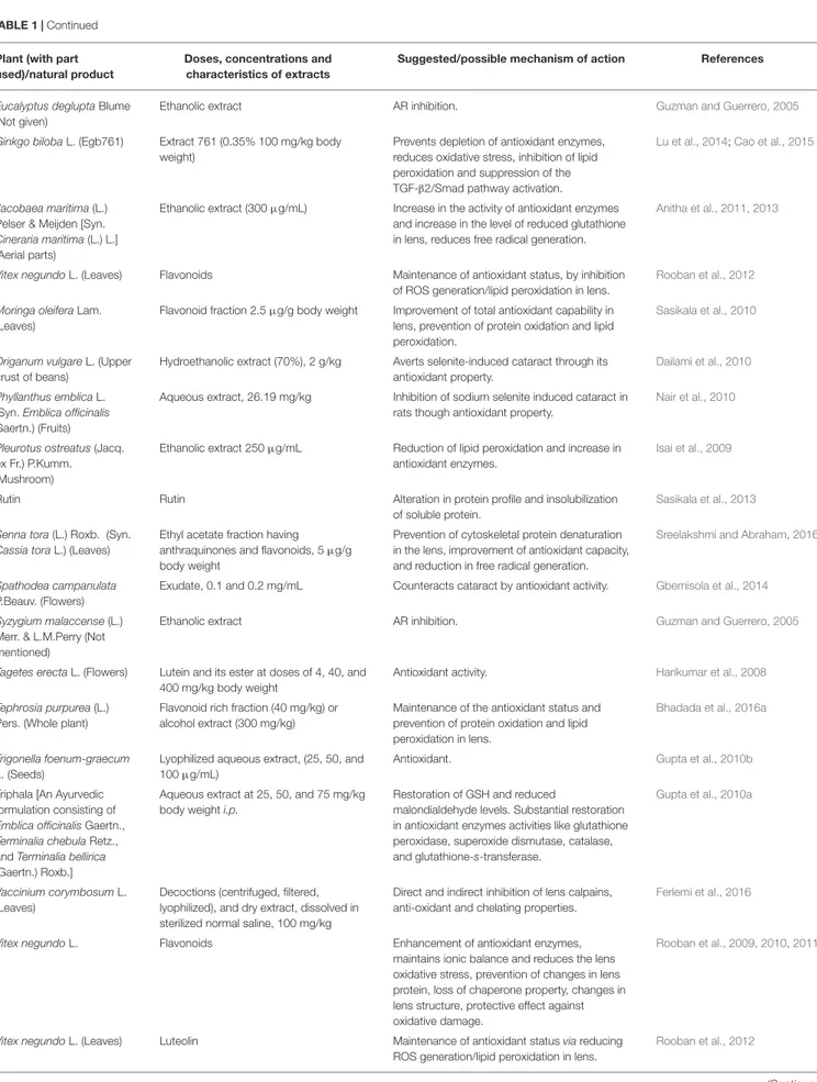
TABLE 2 | Medicinal plants/natural products used against cataract on preventing photo-oxidative damage.
Plant (with part used)/ natural product
Doses, concentrations and characteristics of extracts
Suggested/possible mechanism of action References
Astaxanthin Astaxanthin (0–1 mM) Prevention of cataract through protection of lens from oxidative insults and degradation by calcium-induced calpain.
Wu et al., 2006
Citrus × aurantium L. (Peel) Methanol-water extract, 100 and 200 mg/kg body weight
Delay in onset and maturation of naphthalene induced cataract vis prevention of the photo-oxidative damage produced by naphthalene.
Umamaheswari et al., 2011
Ginkgo biloba L. (Leaves) Standardized EGb761 extract (24% flavonol glycoside and 6% terpene lactones)
Protection from radiation induced cataracts in rat lens via antioxidant property.
Ertekin et al., 2004
Lutein and Zeaxanthin Lutein and Zeaxanthin Protection of eye from oxidative stress and high-energy photons of blue light.
Moeller et al., 2000
induce cell apoptosis in lens epithelial cells and the antioxidant
agents could reduce its effects. Inhibitors of p38 MAPK reduced
ROS levels and apoptosis (
Bai et al., 2015
).
Lipids peroxidation is also a reason of age related cataract. This
process has a negative impact on lipid–lipid and lipid–protein
interactions. Research has shown high levels of hydroperoxides,
oxy derivatives, and diene conjugates of phospholipid fatty acids
in the aqueous humor of cataract patients. Also, studies have
reported high levels of oxidation products of linoleic acid in
patients with early cataract (
Bai et al., 2015
). A schematic
representation of oxidative stress mechanisms involved in
cataract etiology and action mechanisms of several medicinal
plants with conducted pharmacological studies for the treatment
of cataract are presented in Figure 1.
Polyol pathway is associated with diabetic cataract. Enzymes
implicated in the polyol pathway, sorbitol dehydrogenase and AR
are responsible for the conversion of glucose to fructose. Sorbitol,
an intermediate compound, was found to produce cell lesions by
modifying the membrane permeability. Therefore, accumulation
of sorbitol leads to osmotic stress, collapse, and liquefaction of
lens fibers resulting in loss of lens transparency (
Pollreisz and
Schmidt-Erfurth, 2010
;
Hashim and Zarina, 2012
). AR converts
glucose to sorbitol, dependent to NADPH. As a consequence,
the level of NADPH decreases, also having a negative impact
on the glutathione activity and the antioxidant system.
In vivo
and
in vitro studies have shown that by inhibiting the activity
of AR, the progression to cataract in patients with diabetes is
reduced (
Kim et al., 2011a
;
Ramana, 2011
). The glycosylation
pathway has also been linked to diabetic cataract. Excessive
glucose level induces the glycation of proteins, generating
superoxide radicals and AGEs in the process. Recent studies
suggest that there is interdependence between the oxidative stress
and polyol pathway, through AR and iNOS, responsible for the
nitric oxide production during oxidation (
Snow et al., 2015
;
Li et al., 2017
) (Figure 1).
Here, we present details of plants evaluated against cataract
with discussion of their possible mechanism of action (Figure 1
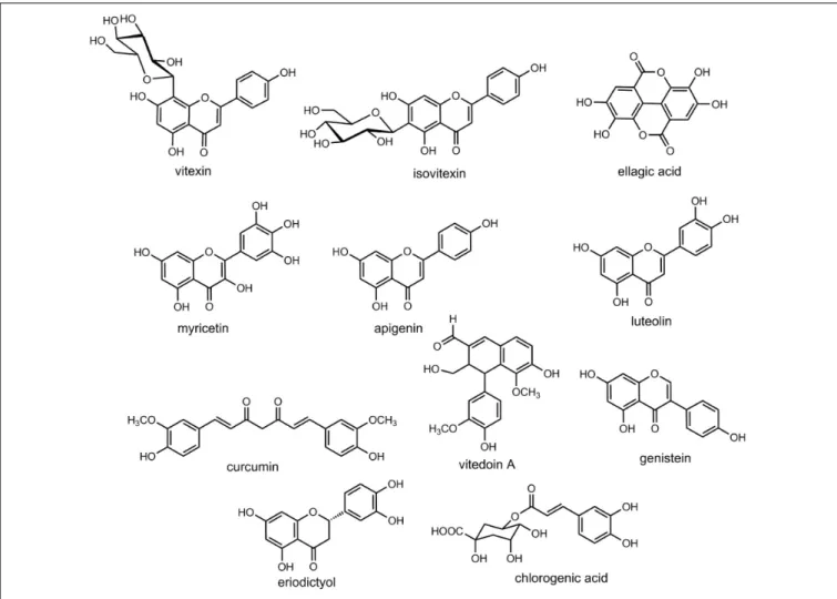
and Tables 1–4). Some important chemical structures of the
natural products that are used against cataract or found in
plants used in the management of cataract are also presented
at Figure 2 (not all chemical structures are presented). We
categorized the plants based upon the models evaluated. Table 1
describes the natural products used against cataract evaluated
on selenite/sodium selenite induced cataracts, in Table 2 natural
products used against cataract on preventive photooxidative
damage is described, Table 3 deals with the natural products used
against cataract on sugar-induced lens opacity/Streptozotocin
induced diabetic cataract/galactose or glucose induced/ZDF
models, in Table 4 AGEs-BSA crosslinking inhibition assay and
lens AR activity models are described, and Table 5 describes the
natural products used against cataract on hydrogen peroxide and
naphthalene induced cataract and other miscellaneous models.
Like in case of any other disease conditions, medicinal
plants are being used in management of various eye ailments
from ancient times. Medicinal plants are used in case of
cataract, eye infections, conjunctivitis, eye dryness, and other
eye disorders in many countries including India (
Sandhu
et al., 2011
;
Das et al., 2013
;
Rothe and Maheshwari, 2016
),
Bangladesh (
Yusuf et al., 2006
;
Das et al., 2007, 2013
), Nepal
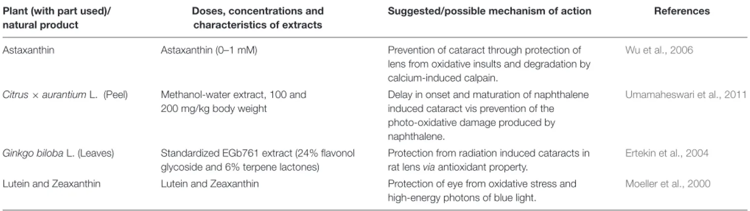
TABLE 3 | Medicinal plants/natural products used against cataract on sugar-induced lens opacity/streptozotocin induced diabetic cataract/galactose, glucose and xylose induced/Zucker diabetic fatty (ZDF) aldose reductase rat models and possible mechanisms of action.
Plant (with part used)/natural product
Doses, concentrations and characteristics of extracts
Suggested/possible mechanism of action
References
Aegle marmelos (L.) Corrêa (Leaves) Chloroform extract 150 mg and 300 mg/kg body weight, p.o.
Increases glutathione, catalase and superoxide dismutase, inhibits lens AR and decreases osmotic stress.
Panaskar et al., 2013;
Sankeshi et al., 2013
Allium sativum L. (Bulb) Methanolic extract, 0.25 and 0.5 g/kg body weight, by forcible gut feeding
Antioxidant activity. Raju et al., 2008
Angelica dahurica (Hoffm.) Benth. & Hook.f. ex Franch & Sav. (Roots)
Ether extract (100µg/mL) (Byakangelicin)
Suppression of galactose induced cataract formation in diabetic rats via AR inhibiting property.
Shin et al., 1994, 1998
Buddleja officinalis Maxim. (Flowers) Apigenin Inhibiting rat lens AR activity. Matsuda et al., 1995
Azadirachta indica A. Juss. (Not mentioned)
Aqueous extract, 25–300µg/mL Inhibits lens AR activity. Halder et al., 2003
Biophytum sensitivum (L.) DC. (Leaves) Sequential water, ethanol and chloroform extracts
AR inhibition and antioxidant action. Gacche and Dhole, 2011a
Brassica juncea (L.) Czern. (Leaves) Aqueous extract, 250 and 500 mg/kg Effective activity against hyperglycemia induced oxidative and osmotic stress.
Valavala et al., 2011
Brickellia arguta B. L. Rob (Not mentioned)
Ethanolic extract AR inhibition. Guzman and Guerrero, 2005
Caesalpinia bonduc (L.) Roxb. (Seeds) Sequential water, ethanol and chloroform extracts
AR inhibition and antioxidant. Gacche and Dhole, 2011a
Cassia fistula (L.) (Fruit pulp) Sequential water, ethanol and chloroform extracts
AR inhibition and antioxidant. Gacche and Dhole, 2011a
Catharanthus roseus (L.) G.Don (Leaves)
Sequential water, ethanol and chloroform extracts
Inhibiting AR activity and antioxidant action. Gacche and Dhole, 2011b
Chlorogenic acid Chlorogenic acid (0.7–2.8 mM) Inhibiting AR activity in galactose fed rats. Kim et al., 2011a
Chromolaena odorata (L.) R.M.King & H.Rob. (Leaves)
Ethanol extract (200 and 400 mg/kg) Decrease of oxidative stress. Onkaramurthy et al., 2013
Corydalis turtschaninovii Besser (Tuber) Methanolic extract of the alkaloidal component (10–200µg/mL) containing dehydrocorydaline
Inhibiting AR activity. Kubo et al., 1994
Curcuma longa L. (Rhizome) Aqueous extract, 25–300µg/mL Prevents in vitro cataract via AR inhibitory activity.
Halder et al., 2003
Dendrobium chrysotoxum Lindl. (Stems)
Gigantol Inhibition of AR and AR gene expression. Wu et al., 2017
Dendrobium aurantiacum (F. Muell.) F. Muell. var. denneanumis (Kerr) Z.H.Tsi (Stems)
Gigantol Attenuation in increase of AR, inducible nitric oxide synthase (iNOS) expression and opacification of rat lenses.
Fang et al., 2015
Eclipta prostrata (L.) L. [Syn. Eclipta alba (L.) Hassk.] (Whole plant)
Ethanolic extract (flavonoids) Inhibition of AR Jaiswal et al., 2012
Erigeron annuus (L.) Pers. (Flowers) Isolated phenolic compounds Inhibition of cataract via inhibiting protein glycation and AR in rat lens.
Jang et al., 2008
Eugenia cordata (Sw.) DC.var. sintenisii (Kiaersk.) Krug & Urb. (Not mentioned)
Ethanolic extract Inhibition of AR. Guzman and Guerrero, 2005
Ficus glomerata L. (Fruits) Sequential water, ethanol, and chloroform extracts
AR inhibition and maintaining of lens opacity.
Gacche and Dhole, 2011b
Marsdenia sylvestris (Retz.) P.I.Forst. [Syn. Gymnema sylvestre (Retz.) R. Br.] (Leaves)
The polyol Conduritol A Inhibition of AR. Miyatake et al., 1994
Thymus vulgaris L. (Leaves) Methanolic extract and isolated compounds (Eriodictyol)
Suppression of the advanced glycation end products levels and fructosamines of albumin.
Morimitsu et al., 1995
Genistein Genistein Increase connexin (Cx) 43 expression. Huang et al., 2007
Zingiber officinale Roscoe (Rhizomes) Powder Suppressing lens galactitol accumulation. Saraswat et al., 2010
Hydrocotyle bonariensis Comm. ex Lam. (Leaves)
Aqueous extract, 500 and 1,000 mg/kg
Reduction in lens protein insolubilization, lens peroxidation and increase in the antioxidant status of the lens.
Ajani et al., 2009
TABLE 3 | Continued
Plant (with part used)/natural product
Doses, concentrations and characteristics of extracts
Suggested/possible mechanism of action
References
Justicia adhatoda L. (Syn. Adhatoda vasica Nees.) (Procured extract)
Sequential water, ethanol and chloroform extracts
AR inhibition and antioxidant action. Gacche and Dhole, 2011a
KIOM-79 (80% ethanol extract of parched Puerariae Radix, gingered). (Magnoliae cortex, Glycyrrhizae Radix and Euphorbiae Radix) (Magnolia officinalis, Pueraria lobata, Glycyrrhiza uralensis, Euphorbia pekinensis) (0–1,000µg/mL)
AR inhibition. KIOM-79, an Inhibitor of AGEs–Protein Cross-linking, Prevents Progression of Nephropathy in Zucker Diabetic Fatty Rats.
Kim et al., 2011b
Magnolia fargesii (Finet & Gagnep.) W. C. Cheng (Flower buds)
Isolated scopoletin and tiliroside Inhibition of rat lens aldose reductase (RLAR) activity; ex vivo cataractogenesis of rat lenses induced by xylose was inhibited by scopoletin.
Lee et al., 2010
Mangifera indica L. Ethanolic extract AR inhibition and antioxidant activity. Guzmán and Guerrero, 2005
Miyamayomena koraiensis (Nakai) Kitam. (Syn. Aster koraiensis Nakai) (Korean starwort) (Aerial part)
Extract of 100 and 200 mg/kg Delay in the progression of lens opacification during the early diabetic cataractogenesis.
Kim et al., 2009
Momordica charantia L. (Fruits) Aqueous and ethanolic extracts, 200 and 400 mg/kg
Prevention of experimental diabetic cataract through reduction of plasma glucose levels.
Rathi et al., 2002
Ocimum tenuiflorum L. (Syn. Ocimum sanctum L.) (Leaves)
Aqueous extract, 25–300µg/mL Prevents in vitro cataract by virtue of its aldose reductase inhibitory activity.
Halder et al., 2003
Peonidin-3-glucoside Peonidin-3-glucoside Inhibits lens AR. Morimitsu et al., 2002
Phyllanthus emblica L. (Syn. Emblica officinalis Gaertn.) (Fruits)
Isolatedβ-glucogallin (0–40 µM) Inhibition of AKR1B1. Puppala et al., 2012
Pterocarpus marsupium Roxb. (Bark) Aqueous extract, 2 g/kg Decreased opacity index. Vats et al., 2004
Pueraria montana (Lour.) Merr. var. lobata (Willd.) Sanjappa & Pradeep. (Roots)
Puerariafuran isolated from methanoilc extract
Inhibition of rat lens AR. Kim et al., 2010
Rutin Rutin (10–100µM) Inhibits advanced glycation end products formation by prevention of dicarbonyls formation.
Muthenna et al., 2012
Silybin Silybin, 231 mg/day for 4 weeks Reductions in the erythrocytic sorbitol level which lead to formation of glycation end products.
Zhang et al., 1995
Silybum marianum (L.) Gaertn. (Seeds) Silymarin 200 mg/kg/d, from extract Antioxidative activity and increase in lens GSH and decrease in lipid peroxides (LPO) levels.
Fallah Huseini et al., 2009
Syzygium cumini (L.) Skeels (Syn. Eugenia jambolana Lam.) (Kernels)
Aqueous and ethanolic extracts, 200 and 400 mg/kg
Significant reduction of plasma glucose. Rathi et al., 2002
Syzygium nervosum A.Cunn. ex DC. [Cleistocalyx operculatus (Roxb.) Merr. & L.M.Perry] (Dried flower buds)
Aqueous extract, 500 mg/kg bw/day Indirect antihyperglycemic effect, decreases the levels of glucose, sorbitol, and fructose in diabetic rat lenses.
Mai et al., 2010
Tephrosia purpurea (L.) Pers. (Whole plant)
Flavonoid rich fraction, 40 mg/kg/day, p.o, whole plant
AR enzyme inhibition and anti-oxidant activity. Bhadada et al., 2016b
Theobroma cacao L. (Cacao liquor) Crude polyphenol fraction (0.5% with diet) (Cyanidin)
Inhibits lens AR. Osakabe et al., 2004
Tinospora sinensis (Lour.) Merr. [Syn. Tinospora cordifolia (Willd.) Miers] (Procured stem extract)
Aqueous and ethanolic extracts, 200 and 400 mg/kg
Prevention of retinal oxidative stress, restoration of antioxidant enzyme levels and reduction in the angiogenic markers, vascular endothelial growth factor (VEGF) and protein kinase C (PKC) that are increased in diabetic retina.
Rathi et al., 2002;
Rajalakshmi et al., 2009;
Agrawal et al., 2012
Triphala Ghrita It’s an Ayurvedic formulation containing gallic acid
Delay in the onset and progression of galactose induced cataract through antioxidant activity.
Mahajan et al., 2011
Vitamin K Vitamin K Lens Ca2 +homeostasis modulation and
inhibition of osmotic and oxidative stress.
Sai Varsha et al., 2014
Zea mays L. (Seed) Hydroalcoholic extract, 2, 10, and 50 mg/mL
Decline in oxidative stress and inhibition of aldose reductase.
Thiraphatthanavong et al., 2014
Zingiber officinale Roscoe (Rhizomes) Powder Reduction in the carbonyl stress, inhibition of osmotic stress by reduction in the activity of the polyol pathway, oxidative stress prevention.
TABLE 4 | Medicinal plants/natural products used against cataract on advanced glycation end products (AGE)- BSA cross-linking inhibition assay and lens aldose reductase activity models and possible mechanisms of action.
Plant (with part used)/natural product
Doses, concentrations and characteristics of extracts Suggested/possible mechanism of action
References
Caesalpinia digyna Rottler (Roots) Alcoholic extract at an increasing concentration between 0 and 200µg/mL (IC5046.29 ± 11.17µg/mL)
AR inhibition and antioxidant action. Kumar et al., 2011
Cinnamomum verum J.Presl (Bark) Ethanolic extract fractions containing Procyanidin-B2, 1–3 mg
AGE inhibition of eye lens proteins under in vitro conditions and inhibition of the formation of glycosylated hemoglobin in human blood in ex vivo conditions.
Muthenna et al., 2013
Cornus officinalis Siebold & Zucc. (Seeds)
EtOAc-soluble fraction (Galloyl glucoses) Inhibition of formation of AGE, AGE-BSA cross-linking, and RLAR.
Lee et al., 2011
Erigeron annuus (L.) Pers. (Leaves and stems)
3,5-Di-O-caffeoyl-epi-quinic acid isolated from methanolic fraction, 5µM
Inhibition of AGEs, AGEs-BSA cross-linking to collagen, RLAR formation, and prevention of lenses opacification.
Jang et al., 2010b
Flavonoids Chrysin, apigenin, and baicalein Inhibition of glycation, glycation induced lens opacity, AGEs, AR and lens protein aggregation.
Patil et al., 2016
Hybanthus enneaspermus (L.) F.Muell. (Whole plant)
Different fractions from ethanolic extract, 0–300µg/mL Not clearly described. Patel et al., 2012
Magnolia biondii Pamp. [Syn. Magnolia fargesii (Finet & Gagnep.) W.C.Cheng] (Flower buds)
Isolated scopoletin and tiliroside RLAR inhibition. Lee et al., 2010
Onoclea orientalis (Hook.) Hook. (Syn. Matteuccia orientalis Trev.) (Rhizomes)
Isolated compound Matteuorienate A, Matteuorienate B from the methanolic extract
AR inhibition. Kadota et al., 1994
Morinda citrifolia L. (Fruits) Sequential water, ethanol, and chloroform extracts AR inhibition and free radical scavenging activity.
Gacche and Dhole, 2011b
Onoclea orientalis (Hook.) Hook. (Syn. Matteuccia orientalis Trev.) (Rhizomes)
Isolated compound from the methanolic extract Matteuorienate C
AR inhibition. Basnet et al., 1995
Platycodon grandiflorus (Jacq.) A.DC. (Flowers)
Isolated compounds from ethyl acetate soluble fractions [apigenin, luteolin, luteolin-7-O-β-D-glucopyranoside, luteolin-7-O-(60 0-O-acetyl)- β-D-glucopyranoside, apigenin-7-O-β-D-glucopyranoside, apigenin-7-O-(60 0 -O-acetyl)-β-D-glucopyranoside, isorhamnetin-3-Oneohesperidoside, 4-O-caffeoylquinic acid, chlorogenic acid methyl ester,
4-O-β-D-glucopyranosyl caffeic acid]
Substantial inhibition of AGEs formation and RLAR.
Jang et al., 2010a
Salvia miltiorrhiza Bunge (Roots) Constituents of methanolic extract Danshenol A Danshenol B, (-)-Danshexinkun A, Dihydrotanshinone I, Tanshinone IIA
AR inhibition. Tezuka et al., 1997
Watanabe et al., 2013
), Sudan (
Khalid et al., 2012
), Tanzania
(
Maregesi et al., 2016
), South Africa (
Pendota et al., 2008
), and
many other regions of the world.
The literature analysis revealed that the sugar-induced or
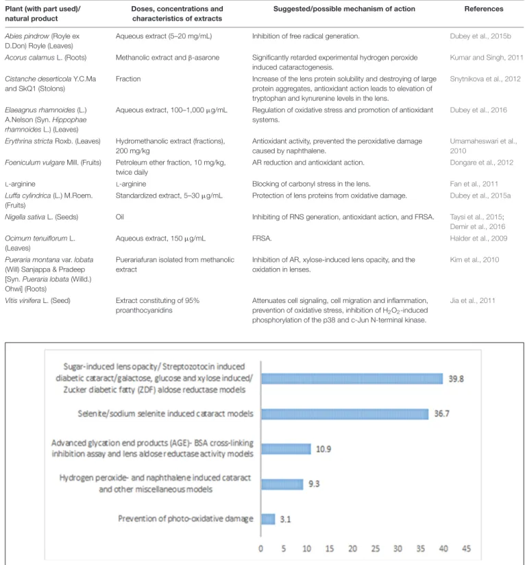
diabetic cataract models were the highest used models which
were applied for the evaluation of around 39.84% of the
plants/natural products. It was followed by selenite/sodium
selenite induced cataract which is another common model of
evaluation of cataract, and it accounts for around 36.71% of the
plants/natural products. AGE-BSA crosslinking inhibition assay
was used for the evaluation of 10.93%, and hydrogen peroxide
and naphthalene induced cataract was account for evaluation
of around 9.38% of the plants (Figure 3). In most of the cases
especially for the diabetic cataract models, it was found that
different antioxidant parameters like soluble protein, reduced
glutathione, superoxide dismutase, lipid peroxidation were used
(
Bhadada et al., 2016b
). Inhibition of AR was found as the most
common hypothesis in these models (
Bhadada et al., 2016b
). Uses
of
in silico studies were also found common in some studies to
explore the binding mode of the phytochemicals with the aldose
reductase enzyme (
Bhadada et al., 2016b
;
Patil et al., 2016
).
In most of the studies, rats or rat pups lens were utilized as
the model (
Bhadada et al., 2016b
;
El Okda et al., 2016
;
Ferlemi
et al., 2016
;
Sreelakshmi and Abraham, 2016
) and in some cases
fresh goat eyeballs were also used (
Patil et al., 2016
).
In vitro
studies were also utilized in large number of experiments. In some
studies lens crystalline turbidity assay was used by estimation of
lens protein turbidity using homogenized decapsulated porcine
lenses which were procured from the local slaughterhouses in
some cases (
Ferlemi et al., 2016
;
Liao et al., 2016
). Some other
important factors in cataractogenesis like UV radiation was
also used by researchers, and it was also proposed that some
compounds can protect
γ-crystallin from UV radiation damage
and can act as potential anticataract agents (
Liao et al., 2016
).
Many of the mentioned plants showed potent anticataract
activity in
in vitro and in vivo models. Vitex negundo and Vitis
FIGURE 2 | Chemical structures of some of the relevant natural products discussed in the context of cataract treatment.
vinifera were the plants in which sufficient preclinical studies
were conducted and they may be of potential clinical use. It is
also interesting that
Vitex negundo was also used in the folk
medicine in India (
Kulkarni et al., 2008
). The genus
Ocimum was
also one such genus which is utilized in folk medicine and was
scientifically validated for its anticataract potential. Some other
interesting findings were the use of
Pleurotus ostreatus extract
that prevented cataract in 75% of the tested rats (
Isai et al.,
2009
). In a clinical study, although not directly against cataract,
silybin improved the peripheral nerve conduction velocity and
was reported as an effective aldose reductase inhibitor that can
improve the disorder of polyol pathway in non-insulin dependent
diabetes patients and prevent chronic complications of diabetes
(
Zhang et al., 1995
) like cataract.
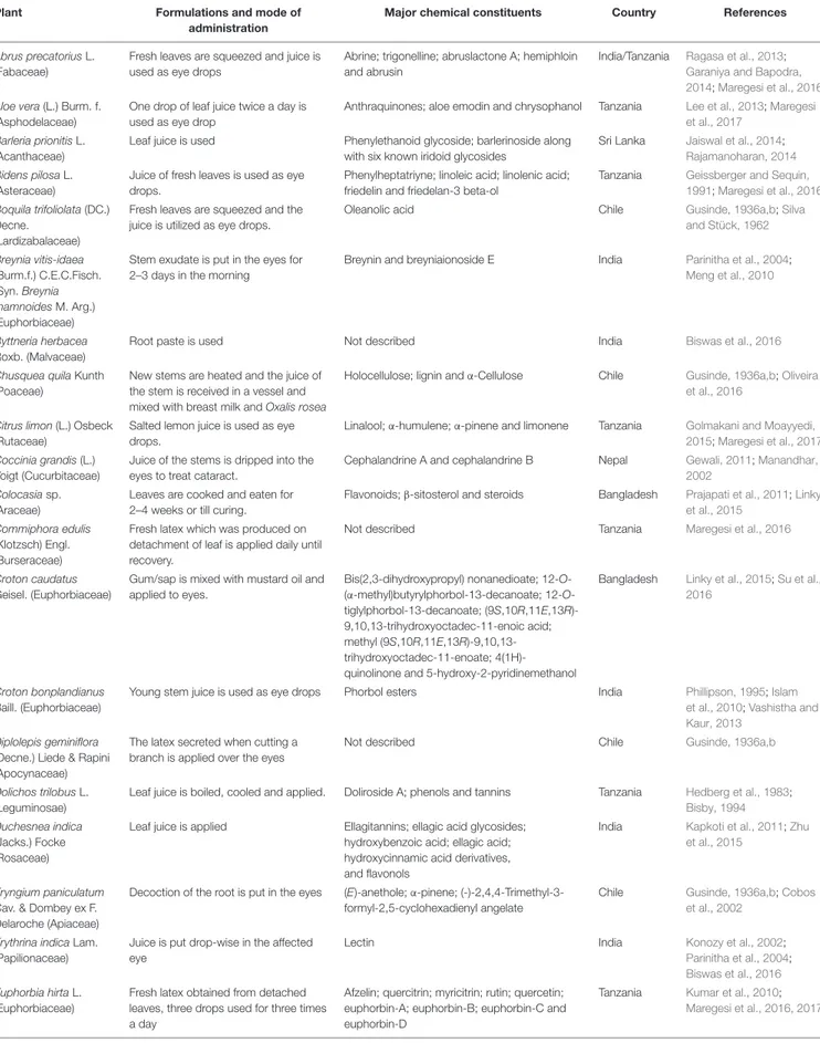
The detailed list of medicinal plants used in the management
of cataract as reported in many ethnopharmacological surveys is
given in Table 6.
Singh et al. (2012)
had also listed the medicinal
plants used in management of cataract, however, the mechanistic
insight was not performed and plants used in the management of
cataract available till 2011 were covered (
Singh et al., 2012
).
It was found that most of the surveys were conducted
in different developing countries like Bangladesh, Chile,
India, Nepal, and Tanzania (see Figure 4). Apart from the
ethnobotanical surveys, several plants used in traditional
medicine systems like Ayurveda were also found beneficial for
cataract. One such good example of use of Ayurvedic formulation
against cataract is the use of
Triphala which showed good effect
against cataract in
in vitro and in vivo (
Gupta et al., 2010a
;
Mahajan et al., 2011
) studies and also was evaluated clinically
and showed promising results (
Bhati and Manjusha, 2015
),
however, more clinical studies are required involving larger
patients for better scientific evidences. Plants used in Ayurveda
like
Momordica charantia, Eugenia jambolana, Pterocarpus
marsupium, and Trigonella foenum-graecum prevented cataract
development when observed in alloxan diabetic cataract model
(
Rathi et al., 2002
;
Vats et al., 2004
).
Although, many plants have been utilized in various folklore
medical practices, most of them are not scientifically validated.
Moreover, some of these traditional practices may be harmful
for the eyes as well. For instance, use of latex/sap of some
Euphorbiaceae plants like
Euphorbia hirta and Croton caudatus
can be dangerous for eyes rather being beneficial. Moreover,
sufficient care is obligatory while using the herbal medication
for any of the eye diseases, as there is a case study that showed
TABLE 5 | Medicinal plants/natural products used against cataract on hydrogen peroxide- and naphthalene induced cataract and other miscellaneous models and possible mechanisms of action.
Plant (with part used)/ natural product
Doses, concentrations and characteristics of extracts
Suggested/possible mechanism of action References
Abies pindrow (Royle ex D.Don) Royle (Leaves)
Aqueous extract (5–20 mg/mL) Inhibition of free radical generation. Dubey et al., 2015b
Acorus calamus L. (Roots) Methanolic extract andβ-asarone Significantly retarded experimental hydrogen peroxide induced cataractogenesis.
Kumar and Singh, 2011
Cistanche deserticola Y.C.Ma and SkQ1 (Stolons)
Fraction Increase of the lens protein solubility and destroying of large protein aggregates, antioxidant action leads to elevation of tryptophan and kynurenine levels in the lens.
Snytnikova et al., 2012
Elaeagnus rhamnoides (L.) A.Nelson (Syn. Hippophae rhamnoides L.) (Leaves)
Aqueous extract, 100–1,000µg/mL Regulation of oxidative stress and promotion of antioxidant systems.
Dubey et al., 2016
Erythrina stricta Roxb. (Leaves) Hydromethanolic extract (fractions), 200 mg/kg
Antioxidant activity, prevented the peroxidative damage caused by naphthalene.
Umamaheswari et al., 2010
Foeniculum vulgare Mill. (Fruits) Petroleum ether fraction, 10 mg/kg, twice daily
AR reduction and antioxidant action. Dongare et al., 2012
L-arginine L-arginine Blocking of carbonyl stress in the lens. Fan et al., 2011
Luffa cylindrica (L.) M.Roem. (Fruits)
Standardized extract, 5–30µg/mL Protection of lens proteins from oxidative damage. Dubey et al., 2015a
Nigella sativa L. (Seeds) Oil Inhibiting of RNS generation, antioxidant action, and FRSA. Taysi et al., 2015;
Demir et al., 2016
Ocimum tenuiflorum L. (Leaves)
Aqueous extract, 150µg/mL FRSA. Halder et al., 2009
Pueraria montana var. lobata (Will) Sanjappa & Pradeep [Syn. Pueraria lobata (Willd.) Ohwi] (Roots)
Puerariafuran isolated from methanolic extract
Inhibition of AR, xylose-induced lens opacity, and the oxidation in lenses.
Kim et al., 2010
Vitis vinifera L. (Seed) Extract constituting of 95% proanthocyanidins
Attenuates cell signaling, cell migration and inflammation, prevention of oxidative stress, inhibition of H2O2-induced
phosphorylation of the p38 and c-Jun N-terminal kinase.
Jia et al., 2011
FIGURE 3 | Percentage of different models used for evaluation of anticataract activity of plants/natural products.
that cataract or development of cataract was aggravated after
treatment with some unrevealed herbal medication in a 11 years
old patient with atopic dermatitis (
Kang et al., 2008
).
This survey reveals that selenite/sodium selenite induced
cataracts was the preferred model in studies with natural
products used against cataract, followed by sugar-induced
TABLE 6 | Medicinal plants reported globally by different ethnopharmacology/ethnobotanical surveys to be used in the treatment of cataract.
Plant Formulations and mode of administration
Major chemical constituents Country References
Abrus precatorius L. (Fabaceae)
Fresh leaves are squeezed and juice is used as eye drops
Abrine; trigonelline; abruslactone A; hemiphloin and abrusin
India/Tanzania Ragasa et al., 2013;
Garaniya and Bapodra, 2014;Maregesi et al., 2016
Aloe vera (L.) Burm. f. (Asphodelaceae)
One drop of leaf juice twice a day is used as eye drop
Anthraquinones; aloe emodin and chrysophanol Tanzania Lee et al., 2013;Maregesi et al., 2017
Barleria prionitis L. (Acanthaceae)
Leaf juice is used Phenylethanoid glycoside; barlerinoside along with six known iridoid glycosides
Sri Lanka Jaiswal et al., 2014;
Rajamanoharan, 2014
Bidens pilosa L. (Asteraceae)
Juice of fresh leaves is used as eye drops.
Phenylheptatriyne; linoleic acid; linolenic acid; friedelin and friedelan-3 beta-ol
Tanzania Geissberger and Sequin, 1991;Maregesi et al., 2016
Boquila trifoliolata (DC.) Decne.
(Lardizabalaceae)
Fresh leaves are squeezed and the juice is utilized as eye drops.
Oleanolic acid Chile Gusinde, 1936a,b;Silva and Stück, 1962 Breynia vitis-idaea (Burm.f.) C.E.C.Fisch. (Syn. Breynia rhamnoides M. Arg.) (Euphorbiaceae)
Stem exudate is put in the eyes for 2–3 days in the morning
Breynin and breyniaionoside E India Parinitha et al., 2004;
Meng et al., 2010
Byttneria herbacea Roxb. (Malvaceae)
Root paste is used Not described India Biswas et al., 2016
Chusquea quila Kunth (Poaceae)
New stems are heated and the juice of the stem is received in a vessel and mixed with breast milk and Oxalis rosea
Holocellulose; lignin andα-Cellulose Chile Gusinde, 1936a,b;Oliveira et al., 2016
Citrus limon (L.) Osbeck (Rutaceae)
Salted lemon juice is used as eye drops.
Linalool;α-humulene; α-pinene and limonene Tanzania Golmakani and Moayyedi, 2015;Maregesi et al., 2017
Coccinia grandis (L.) Voigt (Cucurbitaceae)
Juice of the stems is dripped into the eyes to treat cataract.
Cephalandrine A and cephalandrine B Nepal Gewali, 2011;Manandhar, 2002
Colocasia sp. (Araceae)
Leaves are cooked and eaten for 2–4 weeks or till curing.
Flavonoids;β-sitosterol and steroids Bangladesh Prajapati et al., 2011;Linky et al., 2015
Commiphora edulis (Klotzsch) Engl. (Burseraceae)
Fresh latex which was produced on detachment of leaf is applied daily until recovery.
Not described Tanzania Maregesi et al., 2016
Croton caudatus Geisel. (Euphorbiaceae)
Gum/sap is mixed with mustard oil and applied to eyes.
Bis(2,3-dihydroxypropyl) nonanedioate; (α-methyl)butyrylphorbol-13-decanoate; 12-O-tiglylphorbol-13-decanoate; (9S,10R,11E,13R)-9,10,13-trihydroxyoctadec-11-enoic acid; methyl (9S,10R,11E,13R)-9,10,13-trihydroxyoctadec-11-enoate; 4(1H)-quinolinone and 5-hydroxy-2-pyridinemethanol
Bangladesh Linky et al., 2015;Su et al., 2016
Croton bonplandianus Baill. (Euphorbiaceae)
Young stem juice is used as eye drops Phorbol esters India Phillipson, 1995;Islam et al., 2010;Vashistha and Kaur, 2013
Diplolepis geminiflora (Decne.) Liede & Rapini (Apocynaceae)
The latex secreted when cutting a branch is applied over the eyes
Not described Chile Gusinde, 1936a,b
Dolichos trilobus L. (Leguminosae)
Leaf juice is boiled, cooled and applied. Doliroside A; phenols and tannins Tanzania Hedberg et al., 1983;
Bisby, 1994
Duchesnea indica (Jacks.) Focke (Rosaceae)
Leaf juice is applied Ellagitannins; ellagic acid glycosides; hydroxybenzoic acid; ellagic acid; hydroxycinnamic acid derivatives, and flavonols
India Kapkoti et al., 2011;Zhu et al., 2015
Eryngium paniculatum Cav. & Dombey ex F. Delaroche (Apiaceae)
Decoction of the root is put in the eyes (E)-anethole;α-pinene; (-)-2,4,4-Trimethyl-3-formyl-2,5-cyclohexadienyl angelate
Chile Gusinde, 1936a,b;Cobos et al., 2002
Erythrina indica Lam. (Papilionaceae)
Juice is put drop-wise in the affected eye
Lectin India Konozy et al., 2002;
Parinitha et al., 2004;
Biswas et al., 2016
Euphorbia hirta L. (Euphorbiaceae)
Fresh latex obtained from detached leaves, three drops used for three times a day
Afzelin; quercitrin; myricitrin; rutin; quercetin; euphorbin-A; euphorbin-B; euphorbin-C and euphorbin-D
Tanzania Kumar et al., 2010;
Maregesi et al., 2016, 2017
TABLE 6 | Continued
Plant Formulations and mode of administration
Major chemical constituents Country References
Fascicularia bicolor (Ruiz & Pav.) Mez (Bromeliaceae)
The juice of the young plant parts Not described Chile Gusinde, 1936a,b
Ficus benghalensis L. (Moraceae)
Milky juice is used Alkaloids; glycosides, terpenoids; flavonoids; and tannins
Nepal Acharya and Pokhrel, 2006;
Ogunlowo et al., 2013
Geranium core-core Steud. (Geraniaceae)
Powdered roots are placed on the eyes Hexadecanoic acid; hexahydrofarnesyl acetone and tetracosane
Chile Gusinde, 1936a,b;
Radulovic et al., 2011
Ludwigia hyssopifolia (G.Don) Exell (Onagraceae)
Juice of this plant along with Ocimum americanum L.- Camphor type (2 drops thrice daily for 7–8 days) is given in the eye as a drop.
Piperine Bangladesh Yusuf et al., 2006;Das
et al., 2007
Marchantia polymorpha L. (Marchantiaceae)
Ointment of the crushed plant is prepared, applying it to the eyes
Polymorphatin A; Isorricardin D; 11,10
,130
-trihydroxyisorricardin; 2-[3-(hydroxymethyl)phenoxy]-3-[2-(4-hydroxyphenyl)ethyl]phenol; marchantin J and perrottetin E; 22-hydroxyhopane; 17(21)-hopene; 6α,22-dihydroxyhopane; 20α,22-dihydroxyhopane; 21,22-dihydroxyhopane; 6α, 11α,
22-trihydroxyhopane; 22,28-didroxyhopane; β-sitosterol and daucosterol
Chile Gusinde, 1936a,b;Fang et al., 2007, 2008
Microglossa pyrifolia (Lam.) Kuntze (Asteraceae)
Root juice is used as eye drops α-Humulene and α-pinene, 13-carene, (E)-β-ocimene
and germacrene D
Tanzania Hedberg et al., 1983;Boti et al., 2007
Nepenthes khasiana Hook.f. (Nepenthaceae)
Not described Droserone; 5-O-methyldroserone and naphthoquinones
India Eilenberg et al., 2010;Dhal et al., 2011
Nephrolepis biserrata (Sw.) Schott (Nephrolepidaceae)
Rhizome is scrubbed in the eyes 1β,11α-Diacetoxy-11,12-epoxydrim-7-ene; 1β,6α,11α-triacetoxy-11,12-epoxydrim-7-ene; 1β,3β,11α-triacetoxy-11,12-epoxydrim-7-ene; 9(11)-fernene
Chile Gusinde, 1936a,b;Bottari et al., 1972;Siems et al., 1996
Ocimum americanum L. (Lamiaceae)
Juice of O. americanum with Ludwigia hyssopifolia (two drops thrice daily for 7–8 days) is given in the eye as a drop
1,8-Cineol; camphor;α-pinene and trans-α-bergamotene
Bangladesh Yusuf et al., 2006;Bayala et al., 2014
Oenothera acaulis Cav. (Onagraceae)
Stem juice is given in the eye as a drop. Not described Chile Gusinde, 1936a,b
Oxalis corniculata L. (Oxalidaceae)
Leaf juice is used. Flavonoids; iso-vitexin;
vitexin-2”-O-β–D-glucopyranoside; oleic acid; palmitic acid; linoleic acid; linolenic acid and stearic acid
India Badwaik et al., 2011;
Vashistha and Kaur, 2013
Oxalis rosea Jacq. (Oxalidaceae)
Plant material scrubbed in the eye. Ascorbic acid; oxalic acid; dehydroascorbic acid; pyruvic acid and glyoxalic acid
Chile Gusinde, 1936a,b;Montes and Wilkomirsky, 1985;
Das, 1990
Phyllanthus amarus Schum. &Thonn. (Phyllanthaceae)
Fresh leaves are squeezed and juice is utilized as eyes drops, 2–3 drops thrice daily for 7 days.
Amariin Tanzania Foo, 1993;Maregesi et al.,
2016
Ribes punctatum Ruiz & Pav.
(Grossulariaceae)
Not described Cyanidin-3-glucoside; cyanidin-3-rutinoside; delphinidin-3-rutinoside; delphinidin-3-glucoside; 3-caffeoylquinic acid; (epi)-gallocatechin and (epi)-catechin tetramers
Chile Gusinde, 1936a,b;
Jiménez-Aspee et al., 2016
Rumex usambarensis (Dammer)
(Polygonaceae)
Aerial parts are squeezed and the juice is used as eye drops 2 times daily till recovery.
Chrysophanol, physcion, and emodin Tanzania Midiwo and Rukunga, 1985;Maregesi et al., 2016
Stellaria media (L.) Vill. (Caryophyllaceae)
Aerial parts are scrubbed in the eyes 2,4,5,7-tetramethyloctane; 6-methylheptyl-30
-hydroxy-20
-methylpropanoate; 2, 2,4-trimethyloctan-3-one; apigenin 6-C-β-D -galactopyranosyl-8-C-α-L-arabinopyranoside; apigenin 6-C-α-L- arabinopyranosyl-8-C-α-D-galactopyranoside
Chile Gusinde, 1936a,b;Hodisan and Sancraian, 1989;
Kitanov, 1992;Pande et al., 1995;Hu et al., 2006;
Vanhaecke et al., 2008;
Sharma and Arora, 2012;
Arora and Sharma, 2014
Solanum lycopersicum L. (Solanaceae)
Fresh leaves are squeezed and the juice is used as eye drops.
Adenosine Tanzania Fuentes et al., 2012;
Maregesi et al., 2016
TABLE 6 | Continued
Plant Formulations and mode of administration
Major chemical constituents Country References
Solanum virginianum L. (Solanaceae)
Seed is used Arabinogalactan, glycosides India Pattanayak et al., 2012;
Raja et al., 2014
Swietenia macrophylla King (Meliaceae)
One drop of fresh latex produced from bark is used once daily
Swietemacrophyllanin; catechin and epicatechin Tanzania Falah et al., 2008;
Maregesi et al., 2016
Thunbergia grandiflora (Roxb. ex Rottl.) Roxb. (Acanthaceae)
Bubbles of 1–2 drops of the watery latex from the stem are blown gently into the affected eyes, 3 times a day for 4–5 days
Isounedoside and grandifloric acid India Ismail et al., 1996;
Dipankar, 2012
Typha angustifolia L. (Typhaceae)
New stems are applied on the eye Pentacosanoic acid;β-sitosterol; nonadecanol; naringenin; daucosterol; uracil typhaneoside; nicotinic acid; vanillic acid; succinic acid; thymine; stearic acid propanetriol ester
Chile Gusinde, 1936a,b;Jia et al., 1986;Liao et al., 1990;Chen et al., 2008;
Varghese et al., 2009;
Shukla et al., 2013
Tridax procumbens (L.) L. (Asteraceae)
Leaf juice is dripped into the eyes to treat cataract
Procumbentin Nepal Manandhar, 2002
Vernonia amygdalina Delile (Asteraceae)
Fresh leaves are squeezed and the juice is used as eyes drops, 2–3 drops are used thrice daily for 7 days
Steroidal saponins; tannins; alkaloids; and flavonoids Tanzania Omoregie and Pal, 2016;
Maregesi et al., 2017
Vitex negundo L. (Lamiaceae)
NA Vitedoin A; vitedoamine A; vitexdoin A; flavonoids; lignans; and terpenoids
India Tewar et al., 2013;Shu et al., 2016
FIGURE 4 | Distribution of plants used in folklore medicine.