Abstract. – OBJECTIVE: In this study, we aimed to investigate the relation between the mRNA expression levels of VHL, TIMP-3 and RASSF1A genes, and the histopathological and clinical characteristics of patients with renal tu-mors.
PATIENTS AND METHODS: Radical nephrec-tomy specimens of cases presented without neoadjuvant treatment were confirmed to be cancerous, non-cancerous, benign, and healthy after removal from separate localizations. A to-tal of 69 patients with kidney tumors (138 tissue samples) were included in the study group. RNA isolation, reverse transcriptase PCR (RT-PCR), and quantitative real time PCR (qPCR) were per-formed, and the GAPDH gene was used to nor-malize mRNA levels.
RESULTS: In the RCC cancerous tissue, TIMP-3 levels increased 1.3 times and RASS-F1A levels increased 1.4 times compared to the corresponding levels in non-cancerous tissues, and there was no statistically significant differ-ence in these values. On the other hand, VHL gene expression levels in cancerous tissue were 2.8 times higher than in matched adjacent non‐ cancerous tissues (p < 0.05). In the case of on-cocytomas, TIMP-3 levels were found to be 3.2 times higher, RASSF1A levels 3.8 times high-er, and VHL levels 2.2 times lower than the cor-responding levels in healthy tissues (p < 0.05).
CONCLUSIONS: The roles of VHL, TIMP-3, and RASSF1A mRNA expression in contributing to the development of renal tumors could not be clearly established. Further studies are there-fore required to elucidate the mechanisms un-derlying renal tumors.
Key Words:
Renal cell carcinoma, qPCR, mRNA expression.
Introduction
Renal cell carcinoma (RCC) is a common oncological disorder that accounts for about 3% of all malignancies in adults and 85% of all ma-lignant tumors in the kidney. Over the last thirty years, the incidence rates in Europe and Amer-ica have increased each year1. RCC is usually an asymptomatic type of cancer, and reliable diagnostic and prognostic tumor markers have not yet been identified. Thus, the identification of molecular profiles that are based on global analyses of gene and protein expression could help to identify a new marker specific to RCC2.
The disruption of the function of the Von Hip-pel-Lindau (VHL) gene results in the formation of clear cell carcinoma, the most common renal cancer in adults. The VHL gene product is a component of the ubiquitin-ligase complex and is responsible for the control of multiple gene expression3. The best-defined function of VHL protein (pVHL) is the control of the cellular response to oxygen. More specifically, pVHL activity leads to hypoxia-in-duced factor (HIF) inactivation in the presence of oxygen4. In normal cells, HIF generally helps to coordinate the changes observed in gene expression in response to oxygen5. Generally, the activation of the HIF pathways is low in non-cancerous tissues
in vivo and standard cell culture conditions. When
the amount of oxygen decreases, VHL becomes in-active, and HIF stability increases; the transcription of a large number of target genes is subsequently in-duced following activation of the pathways6. There-fore, alterations in the VHL gene have an important role in the development and progression of RCC7.
I. URE
1, E. KONAC
2, E. ALP
3, H.I. ONEN
2, A.F. BATUR
4, I.I. GONUL
5,
S. MENEVSE
2, S. SOZEN
61Department of Urology, Faculty of Medicine, Osmangazi University, Eskisehir, Turkey
2Department of Medical Biology and Genetics, Faculty of Medicine, Gazi University, Ankara, Turkey
3Department of Medical Biology, Faculty of Medicine, Giresun University, Giresun, Turkey 4Department of Urology, Faculty of Medicine, Selcuk University, Konya, Turkey
5Department of Pathology, Faculty of Medicine, Gazi University, Ankara, Turkey 6Department of Urology, Faculty of Medicine, Gazi University, Ankara, Turkey
Transcriptomic expression levels of the VHL,
TIMP-3, and RASSF1A genes in renal tumors
Matrix metalloproteinases (MMPs) are en-zymes that enable the cleavage of various ex-tracellular matrix (ECM) components and are responsible for remodeling the ECM. However, the irregularity of MMPs has been observed in several disorders, including autoimmune dis-eases and cancers8. In fact, the increase in expression of MMPs is important for tumor invasion and metastasis in many cancers9. Tis-sue metalloproteinase inhibitor (TIMP) family members inhibit the proteolytic activities of active MMPs by forming inhibitor complexes with enzymes. There are four members of the TIMP family: TIMP-1, -2, -3, and -410. TIMP-1 possesses the potential to inhibit the activity of many MMPs other than MMP-2, whereas TIMP-2 is a potential inhibitor of many MMPs other than MMP-9. TIMP-3 binds to MMP-1, -2, -3, -9 and -13, while TIMP-4 binds to MMP-1, -3, -7, and -911.
The Ras-association domain family 1 isoform (RASSF1A) is a promoter in the 3p21.3 region that directly binds to the Ras gene and induces GTP-dependent apoptosis. At the same time, Ras regulates cell proliferation in the molecular path-way and binds to microtubules to induce tumor suppression12. RASSF1A is methylated in lung, breast, ovarian, kidney, prostate, and thyroid can-cers. Re-expression of RASSF1A stops the growth of human cancer cells, and this feature supports the identification of RASSF1A as a tumor-sup-pressing gene13. RASSF1A has seven different isoforms due to its alternative cutting regions and is translated into mRNA from two different pro-moters composed of CpG islets14,15. During RCC development, the relation between the reduction of RASSF1A expression and the early stage of tumor development has been determined16.
The aim of this study was to determine wheth-er thwheth-ere is a relation between clinical, histologi-cal, and pathological differences of patients with renal tumors and the mRNA expression level of VHL, TIMP-3, and RASSF1A genes. If such a relation exists, the role of the gene products with respect to the development of the disease, the general molecular basis of the disease, and the most effective forms of treatment can be deter-mined.
Patients and Methods
Histopathologically, the cancerous and benign tumor tissues in the radical nephrectomy
speci-mens of 79 cases presented without neoadjuvant treatment were confirmed to be benign and can-cerous after they were extracted from separate localizations. A total of 69 pairs of tissues (61 patients with RCC and eight patients with be-nign renal tumors) and their matched adjacent non-cancerous or healthy renal tissues obtained from patients who underwent surgery at the De-partment of Urology, Faculty of Medicine, Gazi University, were histopathologically confirmed.
The Gazi University Medical Faculty Ethics Committee for Medical Research approved the investigation. A consent form was completed by all patients so that patient samples could be used. Tissue specimens were obtained, and transport was secured through nitrogen vapor and storage at –80°C until RNA isolation. In the study, the isolation of RNA from the tissue, reverse tran-scriptase PCR (RT-PCR), and quantitative real time PCR (qPCR) analysis were performed.
Determination of the Expression of VHL, TIMP-3, and RASSF1A Genes in mRNA Levels
With respect to RNA isolation, an approx-imately 0.1 g tissue sample from each kidney tumor was sectioned using a homogenizer (IKA, Wilmington, NC, USA). RNA was extracted us-ing TRIzol reagent (peqGOLD TriFast™, peqlab, Erlangen, Germany) according to the method previously described by Konac et al17.
For the reverse transcriptase PCR (RT-PCR) method, complementary DNA (cDNA) was syn-thesized from 1 µg of total RNA with ran-dom hexamer primers using a Transcriptor First Strand cDNA Synthesis Kit (RocheDiagnostics GmbH, Mannheim, Germany) according to the manufacturer’s instructions.
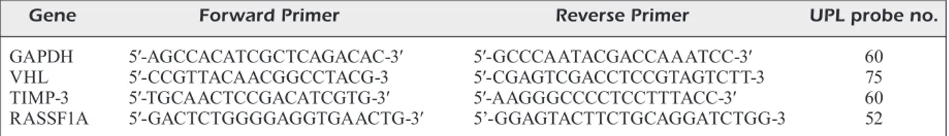
The quantitative real time PCR (qPCR) method was used for the quantitative analysis of expres-sion at the relevant genomic mRNA level. For the identification of VHL, TIMP-3, and RASS-F1A gene expressions, PCR primers [exon–exon junction to allow discrimination between cDNA and genomic DNA (gDNA)] and Universal Probe Library (UPL) probes were used (UPL; Roche Diagnostics, Germany). The primers and UPL probe numbers for this study are provided in Table I.
All PCR reactions were performed with the LightCycler® 480 instrument (Roche Diagnos-tics, GmbH, Mannheim, Germany) using the following program conditions: 50 cycles at 95°C for 15 s and 60°C for 20 s, with the samples
ulti-mately being cooled to 40°C. The mRNA level of the glyceraldehyde-3-phosphate dehydrogenase (GAPDH) gene was used to normalize the mRNA expression levels for the genes of interest.
Statistical Analysis
Pearson’s χ2 and Fisher’s precision tests were
used to compare patients’ demographics. p < 0.05 was considered statistically significant. VHL, TIMP-3, and RASSF1A mRNA expressions in renal tumors and non-cancerous tissues were evaluated by comparison with the REST 2009 V2.0.13 (Qiagen, Hilden, Germany) program.
Results
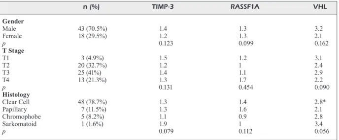
The mean age of the patients was 55.9 (14-78) years. Of the 69 patients, 47 (68.1%) were male and 22 (31.9%) were female. The mean age of patients with RCC was 58.1 (30-78). Of the 61 patients, 43 were male and 18 were female. In these patients, the level of expression of TIMP-3 was found to be 1.3 times higher in the tumor, and the corresponding tumor expression level of RASSF1A was 1.4 times higher than the level in the matched adjacent non-cancerous tissues. Yet, the upregulation of the mRNA levels of TIMP-3 and RASSF1A in RCC cancerous tissue was not statistically different compared with the level in the matched adjacent non-cancerous tis-sues. On the other hand, the level of VHL gene expression in RCC cancerous tissues was found to be 2.8 times higher than that of the matched non-cancerous tissues. This increase was statis-tically significant (p = 0.04). Notably, there was no significant difference in the expression levels of TIMP-3, RASSF1A and VHL genes between male and female patients when RCC cancerous tissues and their non-cancerous tissue counter-parts were compared (p = 0.123, p = 0.099, and p = 0.162, respectively). When RCC patients were grouped according to pathological stages, no sig-nificant differences in TIMP-3, RASSF1A, and VHL gene expressions between RCC cancerous
tissues and its paired non-cancerous tissues were found (p = 0.131, 0.454, and 0.090, respectively). When RCC patients were clustered according to histopathologic type (clear cell, papillary, chro-mophobe, and sarcomatoid renal cell carcinoma), no differences were found in terms of TIMP-3, RASSF1A and VHL gene expression changes (p = 0.079, 0.112, and 0.056, respectively). The expression changes in the genes of RCC patients are summarized in Table II.
The mean age of benign renal tumor patients was 48.7 (14-56). Of the eight patients, four were male and four were female. When these patients were categorized by gender, there were no differ-ences in the expression of TIMP-3, RASSF1A, and VHL genes between benign tumors and their healthy tissue counterparts. Similarly, when patients were classified according to histopatho-logical types, there were no differences in the TIMP-3, RASSF1A, and VHL gene expressions between benign tumors and their paired healthy tissues in the groups with oncocytoma, angiomy-olipoma, and metanephric adenomas (p = 0.642, 0.732, and 0.454, respectively). However, in the case of oncocytomas, TIMP-3 levels increased 3.2 times, RASSF1A levels increased 3.8 times, and VHL levels decreased 2.2 times in the benign tumor tissues in comparison to the corresponding levels in healthy tissues (p < 0.01, p < 0.01, and
p = 0.02, respectively). The clinicopathologic and
statistical data of patients with benign tumors are summarized in Table III.
Discussion
Inactivation of the pVHL is a frequent cause of clear cell renal carcinoma (ccRCC), the most common form of RCC. It has been established that pVHL fulfills a number of functions includ-ing servinclud-ing as a substrate recognition module for a ubiquitin ligase complex, targeting the alpha subunits of the heterodimeric HIF transcription factor for proteasomal degradation18. There have been many studies19-21 conducted on the frequent
Table I. The gene-specific primer sequences and probe numbers.
Gene Forward Primer Reverse Primer UPL probe no.
GAPDH 5′-AGCCACATCGCTCAGACAC-3′ 5′-GCCCAATACGACCAAATCC-3′ 60 VHL 5′-CCGTTACAACGGCCTACG-3 5′-CGAGTCGACCTCCGTAGTCTT-3 75 TIMP-3 5′-TGCAACTCCGACATCGTG-3′ 5′-AAGGGCCCCTCCTTTACC-3′ 60 RASSF1A 5′-GACTCTGGGGAGGTGAACTG-3′ 5’-GGAGTACTTCTGCAGGATCTGG-3 52
occurrence of mutations in the VHL gene in patients with RCC. Gossage et al22 noted that, of the 61 ccRCC patients, inactivation of VHL in cancerous tissues (coding mutation or promoter methylation) was detected in 75% of ccRCCs. It has been shown that somatic non-coding VHL alterations were identified in 29% of these RCCs and may be associated with improved overall survival rates22. In another recent study by Huang et al23, the mRNA and protein expression lev-els of VHL, HIF-1, BCL2 interacting protein 3 (BNIP-3) and vascular endothelial growth factor (VEGF) were measured in 30 ccRCCs and their adjacent non-cancerous tissues23. They found that the expression levels of BNIP3 and VHL were lower in the ccRCC tissues than they were in the
pericarcinous tissues. Also, there was no signifi-cant correlation between the BNIP-3 mRNA and their protein levels and the expressions of VHL, HIF-1alpha, and VEGF. Xiao-Fen et al24 showed thatthe expression levels of VHL and a tumor suppressor gene Jade-1 were analyzed in RCC tissues. Researchers found a downregulation in the expression of the VHL gene in 62.7% of the 75 RCC tissue samples compared to the matched adjacent non-cancerous tissues24 .
However, our findings present a number of dif-ferences from the previously discussed studies. We showed that the RCC cancerous tissues exhib-ited significantly higher expression of VHL, com-pared with the matched non-cancerous tissues. We also detected a diminished expression level
*Difference is statistically significant (p < 0.05).
Table II. Differences (fold) in mRNA expressions of the relevant genes in cancerous tissues relative to non-cancerous tissues of the RCC patients (n = 61). n (%) TIMP-3 RASSF1A VHL Gender Male 43 (70.5%) 1.4 1.3 3.2 Female 18 (29.5%) 1.2 1.3 2.1 p 0.123 0.099 0.162 T Stage T1 3 (4.9%) 1.5 1.2 3.1 T2 20 (32.7%) 1.2 1 2.4 T3 25 (41%) 1.4 1.1 2.9 T4 13 (21.3%) 1.3 1.7 2.2 p 0.131 0.454 0.090 Histology Clear Cell 48 (78.7%) 1.3 1.4 2.8* Papillary 7 (11.5%) 1.3 1.6 2.1 Chromophobe 5 (8.2%) 1.1 0.9 2.8 Sarkomatoid 1 (1.6%) 1.9 1 3.4 p 0.079 0.112 0.056
*Difference is statistically significant (p < 0.05); ↓: downregulation.
Table III. Differences (fold) in mRNA expressions of the genes in benign tumor tissues relative to healthy tissues of the patients (n= 8). n (%) TIMP-3 RASSF1A VHL Gender Male 4 (50%) 3.1 2.7 0.7 Female 4 (50%) 3.3 2.4 0.9 p 0.123 0.099 0.162 Histology Oncocytoma 4 (50%) 3.2* 3.8* ↓2.2* Angiomyolipoma 3 (37.5%) 2.7 2.2 1 Metanephric adenoma 1 (12.5%) 3.9 2.1 0.8 p 0.642 0.732 0.454
of VHL in benign tumors, when compared to the matched healthy tissues. Yet, this decrease was not statistically significant. The increased VHL expression seen in RCC tumors and not in benign tumors suggests that the HIF pathways may in some cases use VHL-independent mechanisms. Another reason why the level of VHL mRNA in RCCs was shown to be significantly higher in cancerous tissues than in benign tumor tissues may be that we have not shown whether there was a mutation in the VHL gene. In other words, although there exists an expression of mRNA, we do not know whether or not there is a functional protein present. This can be regarded as a lim-itation of our study. A report which supports the importance of functional VHL protein was pub-lished in 2011 by Cherkasova et al25. In this study, mutations in the VHL gene were detected in non-RCC tumors even though there was no VHL promoter methylation. Since the VHL protein performs tumor suppressor function, mutations that would prevent active protein formation is more important than promoter methylation status or mRNA expression changes. A study conducted by Banks et al26 emphasizes the importance of mutation in this regard. In this case, the research-ers found promoter methylation in only 19 of 93 (20.4%) sporadic ccRCC samples examined, in one of three transitional cell carcinoma (TCC) samples, in four of eight papillary RCC samples, and in one of two unclassified RCC samples.
Several studies have shown that an increase in the expression of the RASSF1A gene induces apoptosis and cell cycle arrest. Lack of RASSF1A leads to elevated mitotic activity and chromo-somal defect27. Indeed, the reduction in RASS-F1A expression is usually due to promoter hy-permethylation. In many studies28-30, it has been shown that RASSF1A methylation can be used as a prognostic marker in small cell lung cancer, breast cancer, and ccRCC. In many studies, it has been determined that the expression of the RASSF1A gene in RCC is reduced at the mRNA level. As the tumor progresses, this expression appears to decrease31-33. In contrast to some pre-vious studies, Pronina et al34 found an increase in RASSF1A expression levels in renal, breast, ovarian, and colorectal cancers. They noted that RASSF1A expression changes occurred due to tumor specificity and may indicate unstable RASSF1A functions in tumors, that is, RASS-F1A may function as a tumor suppressor gene or a proto-oncogene. In our study, we found no significant differences in the mRNA expression
levels of RASSF1A between the cancerous tis-sues and their non-cancerous tissue counterparts regardless of the histopathological type.
One of the most important factors contributing to the development and especially the progres-sion of RCC is the pro-angiogenic shift of the pro-angiogenic and anti-angiogenic balance. The majority of these activators are kinase receptor ligands such as VEGF. The only known physio-logical antagonist of VEGFR2, one of the VEGF receptors, is TIMP-335. Hagemann et al36 exam-ined the expression patterns of TIMP 1, 2, 3, and 4 in the cancerous tissues of patients with RCC and found that expression of these genes was reduced in all types; however, TIMP-4 increased in the cancerous tissue of the papillary cell type37. This study shows that both the TIMP subtypes and the histopathological type of RCC may differ in their gene expression levels. In our study, there was no significant reduction in the expression of TIMP-3 in the cancerous tissue, which may indicate that it may not be accurate to explain the metastatic potential of RCC cancer cells through only the TIMP-3 mRNA level. It should also be noted that Masson et al37 have found that the expression of TIMP-3 in the cancerous tissue is reduced, especially in Grade 4 tumors, compared to corresponding levels in non-cancerous tissues. We have seen such an increase in our own work, but this increase was statistically insignificant. As in the case of the VHL gene, mutations and mutation-related dysfunctional protein theory may also apply here. Another important finding has shown that as the stage progressed from T1 to T4, the expression of the VHL gene mRNA decreased38. In parallel, we have observed that the expression of mRNA of the VHL gene in cancer-ous tissues was the highest at T1 stage.
There are not many studies in the literature on benign renal cortical tumors that reveal the relation between the expression levels analyzed in our study. Brauch et al38 have investigated the expression of VHL genes in the histopathologi-cal subtypes of renal epithelial tumors and have shown that this gene’s methylation or mutation increases in all subtypes except for chromo-phobe renal cell cancer and oncocytoma. In a study aiming to establish a genomic algorithm to determine the histopathologic type of renal cortical tumors, the absence of the VHL gene mutation appears in the first leg of the algorith-mic stratum advancing to oncocytoma39. In our study, it was observed that there was a significant decrease in the expression of the VHL gene in
oncocytoma cases. Within various human renal cancer subtypes, weak VHL immunohistochemi-cal expression predominates in descending order: Chromophobe RCC > ccRCC > papillary RCC > unclassified RCC > oncocytoma40. Patients with weak VHL expression tended to show a shorter overall survival, indicating loss of VHL protein as a possible negative factor for patient survival. This situation contradicts the limited knowledge in the literature, but it shows that further investi-gations are needed in this regard.
Conclusions
We showed that the absence of any currently valid markers for the diagnosis of RCC generally made it difficult to detect this asymptomatic type of cancer. For this reason, many genomic prod-ucts that can be used as a diagnostic marker for RCC are being investigated today. The expression levels of some of these genes, including RASS-F1A, TIMP-3, and VHL, have been examined in our study, but no evidence has been found to support the use of these gene constructs as mark-ers for RCC. If RCC pathogenesis is thought to be based on multifactorial genetic factors, it is evident that more in-depth studies are needed to clarify this issue.
Conflict of Interest
The Authors declare that they have no conflict of interests.
Ethical Approval
24.06.2009 – No: 392 Gazi University Faculty of Medicine Clinical Research Ethics Committee.
Acknowledgements
This study was conducted with financial support from the Gazi University Research Fund with the project code num-ber 01/2010-25.
References
1) Banumathy G, Cairns P. Signaling pathways in renal
cell carcinoma. Cancer Biol Ther 2010; 10: 658-664.
2) KovaCs G, aKhtar m, BeCKwith BJ, BuGert P, Coo -Per Cs, Delahunt B, eBle Jn, FleminG s, lJunGBerG
B, meDeiros lJ, moCh h, reuter ve, ritz e, roos G,
sChmiDt D, sriGley Jr, störKel s, vanDen BerG e, zBar
B. The Heidelberg classification of renal cell tu-mours. J Pathol 1997; 183: 131-133.
3) steinBaCh F, stöCKle m, hohenFellner r. Clinical
ex-perience with nephron-sparing surgery in the presence of a normal contralateral kidney. Semin Urol Oncol 1995; 13: 288-291.
4) KirKali z, tuzel e, munGan mu. Recent advances
in kidney cancer and metastatic disease. BJU Int 2001; 88: 818-824.
5) lanDis sh, murray t, BolDen s, winGo Pa.
Can-cer statistics, 1999. CA CanCan-cer J Clin 1999; 49: 8-31.
6) Jemal a, murray t, warD e, samuels a, tiwari rC,
GhaFoor a, Feuer eJ, thun mJ. Cancer statistics,
2005. CA Cancer J Clin 2005; 55: 10-30. 7) Kim BJ, Kim Jh, Kim hs, zanG Dy. Prognostic and
predictive value of VHL gene alteration in renal cell carcinoma: a meta-analysis and review. On-cotarget 2017; 8: 13979-13985.
8) JabłońsKa-tryPuć a, mateJCzyK m, rosoChaCKi s.
Matrix metalloproteinases (MMPs), the main extracellular matrix (ECM) enzymes in colla-gen degradation, as a target for anticancer drugs. J Enzyme Inhib Med Chem 2006; 31: 177-183.
9) leon sa, shaPiro B, sKl aroFF Dm, yaros mJ. Free
DNA in the serum of cancer patients and the effect of therapy. Cancer Res 1977; 37: 646-650.
10) ChanG hw, lee sm, GooDman sn, sinGer G, Cho sK,
soKoll lJ, montz FJ, roDen r, zhanG z, Chan Dw,
Kurman rJ, shih iem. Assessment of plasma DNA
levels, allelic imbalance, and CA 125 as diagnos-tic tests for cancer. J Natl Cancer Inst 2002; 94: 1697-1703.
11) wanG BG, huanG h-y, Chen yC, Bristow re, Kas -sauei K, ChenG CC, roDen r, soKoll lJ, Chan Dw,
shih iem. Increased plasma DNA integrity in
can-cer patients. Cancan-cer Res 2003; 63: 3966-3968. 12) linehan wm, walther mm, zBar B. The genetic
basis of cancer of the kidney. J Urol 2003; 170: 2163-2172.
13) ilioPoulos o. Molecular biology of renal cell
can-cer and the identification of therapeutic targets. J Clin Oncol 2006; 24: 5593-5600.
14) airola K, ahonen m, Johansson n, heiKKilä P, Kere
J, Kähäri vm, saarialho-Kere uK. Human TIMP-3 is
expressed during fetal development, hair growth cycle, and cancer progression. J Histochem Cy-tochem 1998; 46: 437-447.
15) ahonen m, BaKer ah, Kähäri vm.
Adenovirus-me-diated gene delivery of tissue inhibitor of metallo-proteinases-3 inhibits invasion and induces apop-tosis in melanoma cells. Cancer Res 1998; 58: 2310-2315.
16) Bian J, wanG y, smith mr, Kim h, JaCoBs C, JaCKman
J, KunG hF, ColBurn nh, sun y. Suppression of in
vivo tumor growth and induction of suspension cell death by tissue inhibitor of metalloprotein-ases (TIMP)-3. Carcinogenesis 1996; 17: 1805-1811.
17) KonaC e, alP e, onen hi, KoruCuoGlu u, Biri aa,
ma-trix metalloproteinases, their tissue inhibitors and cell adhesion molecules in unexplained infertility and implantation failure patients. Reprod Biomed Online 2009; 19: 391-397.
18) shen C, Kaelin wG Jr. The VHL/HIF axis in clear
cell renal carcinoma. Semin Cancer Biol 2013; 23: 18-25.
19) lessi F, mazzanti Cm, tomei s, Di CristoFano C, min -ervini a, meniCaGli m, aPollo a, masieri l, ColleC -Chi P, minervini r, Carini m, BevilaCqua G. VHL and
HIF-1α: gene variations and prognosis in ear-ly-stage clear cell renal cell carcinoma. Med On-col 2014; 31: 840.
20) arJumanD w, sultana s. Role of VHL gene mutation
in human renal cell carcinoma. Tumour Biol 2012; 33: 9-16.
21) neal Cs, miChael mz, rawlinGs lh, van Der hoeK
mB, GleaDle Jm. TheVHL-dependent regulation
of microRNAs in renal cancer. BMC Med 2010; 8: 64.
22) GossaGe l, murtaza m, slatter aF, liChtenstein CP,
warren a, haynes B, marass F, roBerts i, shanahan
sJ, Claas a, Dunham a, may aP, rosenFelD n, For -shew t, eisen t. Clinical and pathological impact
of VHL, PBRM1, BAP1, SETD2, KDM6A, and JARID1c in clear cell renal cell carcinoma. Genes Chromosomes Cancer 2014; 53: 38-51.
23) huanG l, zhao t, tian y, wanG h, Chen X, Xu m, li
X. Expression and significance of BNIP3 in clear cell renal cell carcinoma. Sichuan Da Xue Xue Bao Yi Xue Ban 2014; 45: 396-399.
24) Xiao-Fen w, tinG C, Jie l, DenG-yanG m, qinG-FenG
z, Xin l. Correlation analysis of VHL and Jade-1
gene expression in human renal cell carcinoma. Open Med (Wars) 2016; 11: 226-230.
25) CherKasova e, malinzaK e, rao s, taKahashi y,
senChenKo vn, KuDryavtseva av, niCKerson ml, me -rino m, honG Ja, sChrumP Ds, srinivasan r, linehan
wm, tian X, lerman mi, ChilDs rw. Inactivation of
the von Hippel-Lindau tumor suppressor leads to selective expression of a human endogenous retrovirus in kidney cancer. Oncogene 201; 30: 4697-4706.
26) BanKs re, tiruKonDa P, taylor C, horniGolD n, as -tuti D, Cohen D, maher er, stanley aJ, harnDen P,
JoyCe a, Knowles m, selBy PJ. Genetic and
epigen-etic analysis of von Hippel-Lindau (VHL) gene al-terations and relationship with clinical variables in sporadic renal cancer. Cancer Res 2006; 66: 2000-2011.
27. Kawai y, saKano s, oKayama n, suehiro y, matsuyama
h, hinoDa y. Association of RASSF1A genotype
and haplotype with the progression of clear cell renal cell carcinoma in Japanese patients. BJU Int 2012; 110: 1070-1075.
28) yanaGawa n, tamura G, oizumi h, KanauChi n, en -Doh m, saDahiro m, motoyama t. Promoter
hyper-methylation of RASSF1A and RUNX3 genes as an independent prognostic prediction marker in surgically resected non-small cell lung cancers. Lung Cancer 2007; 58: 131-138.
29) müller hm, wiDsChwenDter a, FieGl h, ivarsson l,
GoeBel G, PerKmann e, marth C, wiDsChwenDter m.
DNA methylation in serum of breast cancer pa-tients: an independent prognostic marker. Cancer Res 2003; 63: 7641-7645.
30) Kawai y, saK ano s, suehiro y, oK aDa t, Korena -Ga y, hara t, naito K, matsuyama h, hinoDa y.
Methylation level of the RASSF1A promoter is an independent prognostic factor for clear-cell renal clear-cell carcinoma. Ann Oncol 2010; 21: 1612-1617.
31) KlaCz J, wierzBiCKi Pm, wronsKa a, ryBarCzyK a,
stanislawowsKi m, sleBioDa t, oleJniCzaK a, matusze -wsKi m, KmieC z. Decreased expression of
RASS-F1A tumor suppressor gene is associated with worse prognosis in clear cell renal cell carcino-ma. Int J Oncol 2016; 48: 55-66.
32) liu y, sun l, FonG P, yanG J, zhanG z, yin s, JianG s,
liu X, Ju h, huanG l, Bai J, GonG K, yan s, zhanG
C, shao G. An association between
overexpres-sion of DNA methyltransferase 3B4 and clear cell renal cell carcinoma. Oncotarget 2017; 8: 19712-19722.
33) DreiJerinK K, BraGa e, Kuzmin i, Geil l, Duh Fm, an -Geloni D, zBar B, lerman mi, stanBriDGe eJ, min -na JD, ProtoPoPov a, li J, KashuBa v, Klein G,
zaBarovsKy er. The candidate tumor
suppres-sor gene, RASSF1A, from human chromosome 3p21.3 is involved in kidney tumorigenesis. Proc Natl Acad Sci USA 2001; 98: 7504-7509.
34) Pronina iv, loGinov vi, KholDyrev Ds, KazuBsKaia
tP, BraGa Éa. Alterations of expression level of
RASSFIA gene in primary epithelial tumors of various locations. Mol Biol (Mosk) 2012; 46: 260– 268.
35) BalDewiJns mm, thiJssen vl, van Den eynDen
GG, van laere sJ, BlueKens am, rosK ams t, van
PoPPel h, De Bruïne aP, GriFFioen aw, vermeulen
PB. High-grade clear cell renal cell carcino-ma has a higher angiogenic activity than low-grade renal cell carcinoma based on histo-morphological quantification and qRT-PCR mRNA expression profile. Br J Cancer 2007; 96: 1888-1895.
36) haGemann t, Gunawan B, sChulz m, Füzesi l, BinD -er C. MRNA expression of matrix
metalloprote-ases and their inhibitors differs in subtypes of renal cell carcinomas. Eur J Cancer 2001; 37: 1839-1846.
37) masson D, riouX-leClerCq n, FerGelot P, Jouan F,
mottier s, thÉoleyre s, BaCh-nGohou K, PatarD JJ,
Denis mG. Loss of expression of TIMP3 in clear
cell renal cell carcinoma. Eur J Cancer 2010; 46: 1430-1437.
38) BrauCh h, weiriCh G, BrieGer J, GlavaC D, röDl h,
eiChinGer m, Feurer m, weiDt e, PuranaKanitstha
C, neuhaus C, Pomer s, Brenner w, sChirmaCher P,
störKel s, rotter m, masera a, GuGeler n, DeCK -er hJ. VHL alterations in human clear cell renal
cell carcinoma: association with advanced tumor stage and a novel hot spot mutation. Cancer Res 2000; 60: 1942-1948.
39) GowrishanKar B, PrzyByCin CG, ma C, nanDula sv,
rini B, CamPBell s, Klein e, ChaGanti rs, maGi-Gallu -zzi C, houlDsworth J. A genomic algorithm for the
molecular classification of common renal cortical neoplasms: development and validation. J Urol 2015; 193: 1479-1485.
40) höGner a, Krause h, JanDriG B, Kasim m, Fuller
tF, sChostaK m, erBersDoBler a, PatzaK a, KiliC e.
PBRM1 and VHL expression correlate in human clear cell renal cell carcinoma with differential as-sociation with patient’s overall survival. Urol On-col 2018; 36: 94.e1-94.e14.