Arch Neuropsychiatr 2015; 52: 200-1 • DOI: 10.5152/npa.2015.7316
Neuro-Behçet’s Disease with Chorea
Selen GÜR ÖZMEN
1,2, Haşmet HANAĞASI
3, Hakan GÜRVİT
3, Murat EMRE
3, Gülşen AKMAN DEMİR
4Correspondence Address: Dr. Selen Gür Özmen, Department of Neuroscience, Institute for Experimental and Medical Research (DE-TAE), Istanbul University, İstanbul, Turkey Phone: +90 532 394 10 11 E-mail: [email protected]
Received: 08.07.2013 Accepted: 11.04.2014
1Department of Neuroscience, Institute for Experimental and Medical Research (DETAE), Istanbul University, İstanbul, Turkey 2Clinic of Neurology, Iğdır State Hospital, Iğdır, Turkey
3Department of Neurology, İstanbul University İstanbul Faculty of Medicine, İstanbul, Turkey 4Department of Neurology, İstanbul Bilim University, İstanbul, Turkey
Behçet’s disease (BD) was described as a three-symptom complex comprising uveitis, oral aphthae, and genital ulcerations. It is a multisystemic, recurrent, inflammatory disorder and it is of unknown cause. Neuro-Behçet (NB) is present in 5%-7% of BD. Movement disorders have rarely
been reported in NB. Here, we report a case of chronic parenchymal NB presenting with chorea.
Keywords: Neuro-Behçet’s disease, chorea, movement disorders ABSTRACT
200
INTRODUCTION
Behçet’s disease (BD) was first described by the Turkish dermatologist Hulusi Behçet as a three- symptom complex comprising uveitis, oral aphthae, and genital ulcerations (1). Today, it is known that the disease is a multisystemic, recurrent, inflammatory disorder, although it is of unknown cause (2). Neuro-Behçet (NB) is present in 5%–7% of BD. Movement disorders have rarely been reported in NB. Here, we report a case of chronic parenchymal NB presenting with chorea and apathy.
CASE
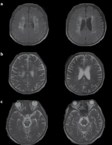
A forty-seven-year-old man had a history of recurrent oral aphthae and genital ulceration, anterior uveitis, and erythema nodosum that led to a diagnosis of BD 15 years ago. For a while, he was under cyclosporine and azathioprine treatment; however, he discontinued this treatment 5 years ago. Three years ago, he had an attack with dysarthria and truncal ataxia, and 2 years ago, he developed apathy. One year ago, abnormal involuntary movements started from the left side of his body and gradually involved both sides. Neurological examination revealed that he was apathic and had severe dysarthria and truncal ataxia. He had brief, abrupt, and irregular movements on both sides of arms and legs, which were compatible with chorea. He did not have any vascular risk factors and had not received any neuroleptic treatment before. Molecular analysis for Huntington’s disease was negative. Serum ceruloplasmin and urinary copper levels were normal. Anti-cardiolipin antibodies and lupus anticoagulant were negative. HIV test was negative. Peripheral blood smear that was repeated on three occasions revealed no acanthocytes. Cerebrospinal fluid protein and glucose levels were normal and acellular. Oligoclonal bands were pattern two positive. Cranial magnetic resonance imaging (MRI) revealed atrophy of the brain stem, pontine punctate hyperintensities on T2 weighted images, also bilateral T2 hyperintensities in centrum semiovale, and corpus callosum. These signs were even more evident on fluid-attenuated inversion-recovery (FLAIR) sequence (Figure 1). He was diagnosed of chronic pa-renchymal NB. Abdominal ultrasonography revealed an infrarenal abdominal aortic dilatation with a diameter of 4 cm that was thought to be a complication and bad prognostic factor of BD. Intravenous (IV) methylprednisolone was administered 1000 mg/day for 5 days, followed by once a week administration of 1000 mg/day IV methylprednisolone for 4 weeks in addition to monthly 1000 mg IV cyclo-phosphamide. Between IV steroid pulses, 32 mg/day oral methylprednisolone was also administered. After following the patient at the 32 mg/day dose for a few months, methylprednisolone was very slowly reduced to 4–8 mg/day, and the patient was maintained at this basal dose. Monthly cyclophosphamide infusions were planned for 12 months; afterwards switch to azathioprine was planned for long term immunosuppressive treatment. He was administered haloperidol 2 × 5 mg for chorea and then switched to olanzapine 2 × 5 mg because of ineffectiveness and extrapyramidal side effects (parkinsonism). A modest improvement in chorea was observed after treat-ment. He was followed up for a year at the outpatient clinic without any complications. After 1 year, he was lost to follow-up. Later on, we obtained the information regarding his exitus due to unknown reasons. The patient’s written consent for the publication of material related to him in a journal had been initially obtained.
Case Report
Arch Neuropsychiatr 2015; 52: 200-1 Gür Özmen et al. Neuro-Behcet’s Disease with Chorea
DISCUSSION
Movement disorders have rarely been reported in NB (3,4,5,6,7,8). In a study of Benamour et. al. (8), 925 patients with BD were analyzed, and 154 of them had NB in which only one patient had chorea. A literature review revealed that the onset of chorea in cases of BD varied from the time of onset of BD to 31 years after the onset of the disease (3). In our case, onset of chorea is observed 14 years after the onset of BD. Brain stem atrophy and small scattered lesions that do not involve an arterial territory on the brain MRI of patients with chronic parenchymal NB are well known (9). The symptoms and findings of this patient are compatible with chronic parenchymal involvement of NB disease, which seemingly had not been appropriately treated at the time of the acute attack. The clinical picture, pathology, and brain MRI may not be concordant in cas-es with NB (10). Autopsy studicas-es reveal that in NB disease, low-grade chronic lymphocytic meningo-encephalitis with widespread perivenular neutrophilic or lymphocytic and plasmocytic cuffing, multifocal necrotic foci are observed. These tend to mostly accumulate in the brain stem and
basal ganglion region and to a lesser extent in the spinal cord, which is ir-relevant to the presenting clinical picture (11,12). Although basal ganglion is frequently involved in neuro-Behçet’s disease, chorea is rare. Usually, the few cases of NB with chorea in the literature reveal high intensity on T2-weighted images and on FLAIR sequence in the periventricular white matter and basal ganglia. FLAIR sequence demonstrating lesions are clearer than the T2-weighted MRI (3,4). In our case, also on FLAIR image bilateral white matter lesions in centrum semiovale and in corpus callosum were better observed than the T2-weighted image (Figure 1). In the case of Kurikawa et al. (3), treatment with prednisolone resolved the chorea, suggesting that chorea was caused by an autoimmune mechanism. In our case, a modest improvement in chorea was observed after treat-ment with both the immunosuppresants and olanzapin, and it is hard to differentiate which one made the difference.
In conclusion, in the differential diagnosis of chorea, it is important to con-sider NB as well because the disease progression may be prevented by early initiation of immunosuppresants.
Conflict of Interest: No conflict of interest was declared by the authors. Financial Disclosure: The authors declared that this study has received no fi-nancial support.
REFERENCES
1. Behçet H. Über residivierende aphtöse durch ein Virus verursachtes Geschwüre am Mund, am Auge und an der Genitalien. Dermatol Wochen-schr 1937; 105:1152-1157.
2. Inaba G. Behçet’s disease. In: Vinken PJ, Bruyn GW, Klawans HL, editors. Hand-book of clinical neurology, vol 56 Amsterdam: Elsevier, 1989. p 593-610. 3. Kuriwaka R, Kunishige M, Nakahira R, Inoue H, Higashi T, Tokumoto Y, Mitsui
T. Neuro-Behçet’s disease with chorea after remission of intestinal Behçet’s disease. Clin Rheumatol 2004; 23:364-367. [CrossRef]
4. Kimura N, Sugihara R, Kimura A, Kumamoto T, Tsuda T. A case of neuro-Behçet’s disease presenting with chorea. Rinsho Shinkeigaku. 2001; 41:45-49.
5. Revilla FJ, Racette BA, Perlmutter JS. Chorea and jaw-opening dystonia as a manifestation of NeuroBehcet’s syndrome. Mov Disord 2000; 15:741-744.
[CrossRef]
6. Bussone G, La Mantia L, Boiardi A, Giovannini P. Chorea in Behcet’s Syndrome. J Neurol 1982; 227:89-92. [CrossRef]
7. Joseph FG, Scolding NJ. Neuro-Behçet’s disease in Caucasians: a study of 22 patients. Eur J Neurol 2007; 14:174-180. [CrossRef]
8. Benamour S, Naji T, Alaoui FZ, El-Kabli H, El-Aidouni S. Neurological involve-ment in Behçet’s disease. 154 cases from a cohort of 925 patients and review of the literature. Rev Neurol (Paris) 2006; 162:1084-1090. [CrossRef]
9. Akman-Demir G, Bahar S, Coban O, Tasci B, Serdaroglu P. Cranial MRI in Behçet’s disease: 134 examinations of 98 patients. Neuroradiology 2003; 45:851-859. [CrossRef]
10. Akman-Demir G, Serdaroğlu P, Tasci B. Clinical patterns of neurological in-volvement in Behçet’s disease: evaluation of 200 patients. The Neuro-Behçet Study Group. Brain 1999; 122:2171-2182. [CrossRef]
11. Sugihara H, Muto Y, Tsuchiyama H. Neuro-Behçet’s syndrome: report of two autopsy cases. Acta Pathol Jpn 1969; 19:95-101. [CrossRef]
12. Hadfield MG, Aydin F, Lippman HR, Sanders KM. Neuro-Behçet’s disease. Clin Neuropathol 1997; 16:55-60.
201
Figure 1. a-c. Axial FLAIR and T2 images of a patient diagnosed with NB disease. (a) Bilateral white matter lesions in centrum semiovale and in corpus callosum are shown and lateral ventricular atrophic dilatation can be seen on FLAIR image. (b) The same signs are seen in T2 weighted image. (c) Brain stem atrophy and pontine punctate hyperintensities can be seen on T2 weighted images