Introduction
Idiopathic granulomatous mastitis (IGM) is a rare, benign, chronic inflammatory lesion of the breast that was first described in 1972 (1). Although benign, it is of clinical significance because it can imitate breast cancer clinically and radiologically, requires long-term follow-up, and is associated with high rates of recurrence after treatment (2, 3).
To diagnose IGM, the inflammation-causing granuloma should be demonstrated histologically, and the inflammation should not be related to a specific factor known to be associated with a granulomatous reaction (4, 5). The pathogenesis of IGM remains unclear. Currently, the most widely supported theory for this condition is autoimmunity, although other possible etiological causes include hormone imbalance, microbiological agents, hyperprolactinemia, smoking, and alpha-1 anti-trypsin deficiency (6-8).
The possible role of various bacterial agents in the etiology has long been discussed. Especially, Corynebacterium species, a component of the normal dermal flora, are suspected of being involved in the etiology and have been isolated in some cases (7, 9). By the isolation of this bacteria, some authors preferred to use “Cystic Neutrophilic Granulomatous Mastitis” term for the IGM cases associated with gram-positive bacilli (10, 11).
This study investigated the presence of bacteriological agents considered to be involved in the etiology of IGM, using a universal primer after isolating deoxyribonucleic acid (DNA) from pathological specimens.
Role of Bacteriological Agents in Idiopathic
Granulomatous Mastitis: Real or Not?
Taner Kıvılcım
1,2, Fatih Altıntoprak
3,4, Bahar Memiş
5, Murat Ferhat Ferhatoğlu
1, Abdulcabbar Kartal
1, Enis
Dikicier
2, İhsan Hakkı Ciftçi
6, Fatma Hüsniye Dilek
71Department of General Surgery, Okan University School of Medicine, İstanbul, Turkey
2Department of General Surgery, Sakarya University Training and Research Hospital, Sakarya, Turkey 3Department of General Surgery, Sakarya University School of Medicine, Sakarya, Turkey
4Department of General Surgery, İstinye University School of Medicine, İstanbul, Turkey 5Department of Pathology, Sakarya University Training and Research Hospital, Sakarya, Turkey 6Department of Microbiology, Sakarya University School of Medicine, Sakarya, Turkey 7Department of Pathology, İzmir Katip Çelebi University School of Medicine, İzmir, Turkey
Corresponding Author:
Taner Kıvılcım, e-mail: [email protected]
Submitted: 26.06.2018 Accepted: 29.08.2018 Available Online Date: 05.11.2018
DOI: 10.5152/ejbh.2018.4249
32
ABSTRACT
Objective: Granulomatous mastitis is a rare, benign, chronic inflammatory disease of the breast of unknown etiology. This study evaluated
bac-teriologic agents that might play a role in the etiology of granulomatous mastitis using a molecular method with a universal primer after isolating deoxyribonucleic acid (DNA) from pathology specimens from patients diagnosed with granulomatous mastitis.
Materials and Methods: Breast biopsy material in the pathology department obtained between July 2008 and June 2013 was analyzed. The
history of the granulomatous mastitis patients was examined in detail and paraffin block sections of the biopsy material were used to determine the presence of bacteria with a universal DNA primer.
Results: This study examined 45 granulomatous mastitis patients who had been diagnosed using excisional, incisional, or core biopsies. We
evalu-ated multiple bacterial taxa, but obtained no positive result using a nucleic-acid-based assay with a universal primer.
Conclusion: The etiology of idiopathic granulomatous mastitis remains unclear. Further studies with a large number of patients should aim to
identify the causative agent.
Keywords: Mastitis, granulomatous mastitis, idiopathic granulomatous mastitis, bacteria, etiology
Cite this article as: Kıvılcım T, Altıntoprak F, Memiş B, Ferhatoğlu MF, Kartal A, Dikicier E, Çiftçi İH, Dilek FH. Role of Bacteriological Agents in Idiopathic Granulomatous Mastitis: Real or Not? Eur J Breast Health 2018; 15(1): 32-36.
Materials and Methods
The current study was conducted with department of General Surgery, Pathology and Microbiology. The breast biopsy materials, available in the Pathology Clinic between July 2008 and June 2013, examined ret-rospectively.
The records of patients who were diagnosed with granulomatous mas-titis based on a histopathological examination were evaluated in detail. Core biopsy, or incisional or excisional biopsy was performed depending on the clinical findings at the time of admission. For all of the biopsy specimens, Gram, periodic acid–Schiff (PAS) and Ziehl–Neelsen (ZN) staining procedures were performed for the analysis of microbiologi-cal agents, and culture methods were used for tuberculosis and fungal analyses. No antibiotic treatment was started before the biopsy process. However, patients admitted with inflammation in the breast were started on antibiotic treatment after taking the biopsy samples. For abscesses, surgical or imaging-guided percutaneous drainage was performed. For patients who did not recover after the initial treatment, options for surgi-cal or steroid treatment were evaluated.
Patients biopsied using fine-needle aspiration biopsy (FNAB) were excluded because of insufficient material. Patients who also identified an etiological factor were excluded from the study. The study included patients for whom no etiological factor could be established to explain the granulomatous reaction in the breast after clinical, radiological and biochemical examinations and were consequently diagnosed with IGM.
Study protocol
Consecutive formalin-fixed paraffin-embedded (FFPE) tissue sections from each breast biopsy were used for molecular analyses. DNA was isolated using a FFPE kit (RNeasy FFPE Kit, QIAGEN), according to the manufacturer’s protocol. The quality (OD260/OD280) and quan-tity (OD260) of DNA obtained was measured spectrophotometrically using the standard method (12).
Primers were designed to amplify DNA from all bacteria considered po-tential causes. The primer designed by Nadkarni et al. (13) to amplify 39 specific agents was used. In addition, the compatibility of the
Entero-bacter spp., Klebsiella spp., Ureaplasma spp., and CoryneEntero-bacterium spp.
genomes with this primer in a region of the 16S ribosomal RNA gene was checked through a sequence alignment. The investigated agents are listed in Table 1. The human glyceraldehyde-3-phosphate dehydroge-nase (GAPDH) gene, which is a heterologous target, was used as an internal control (Table 2). Escherichia coli ATCC 25922 was used as a positive control.
Polymerase chain reaction (PCR) was performed as described by Nad-karni et al. (13) distilled water was used as a negative control. Electro-phoresis was carried out with 10 µl of PCR product on 1% agarose gels in a gel imaging system (EDAS 290, Eastman Kodak Company). The PCR products were compared with a 1-kb ladder (Fermentas, Thermo-Fisher Scientific). The presence of an approximately 245 base pair (bp) band was considered to indicate the presence of bacteria, while a 120-bp band was deemed positive for the internal control.
No specific statistical test was used because only positivity/negativity was assessed in this study. The study was approved by the Ethics Commit-tee for Sakarya University School of Medicine Ethics CommitCommit-tee for Non-Invasive Clinical Trials (71522473.050.01.04/101). This was a ret-rospective study. No new biopsy materials were obtained from patients for diagnostic or investigational purposes in this study. Therefore, no informed consent was taken from the patients.
Results
The records of 51 female patients who clinically and pathologically diagnosed with granulomatous mastitis were evaluated in detail. After excluding two patients (3.9%) for whom FNAB was used as the biopsy method and four patients (7.8%) for whom an etiological factor was established (tuberculosis and sarcoidosis in two cases each), 45 patients diagnosed with IGM, were included in the study.
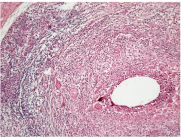
The mean age of the patients included in the study was 35.4 (range 19– 65) years. Twenty-one patients had right breast involvement; 22 patients had left breast involvement; and 2 patients had bilateral involvement. Histopathological examination revealed features of non-caseous granu-lomatous inflammation around breast lobules (Figure 1).
Although the nucleic acid quality of samples no. 12, 20, and 40 was low, there was sufficient DNA for the study. The nucleic acid quality and quantity required for PCR are summarized in Table 3.
During standard PCR reactions, the most intense bands for GAPDH and E. coli ATCC 25922 were obtained at 55°C. Therefore, 55°C was used as the annealing temperature in all PCR reactions. No non-spe-cific product or primer dimer was observed upon increasing the primer concentration or decreasing the annealing temperature. PCR was re-
33
Figure 1 Histopathological examination showed replacement of
breast tissue by granulomatous type inflammation that composed of histiocytes and lymphocytes. In the center of inflammation, epithelial histiocytes, scattered Langhans type giant cells and granular debris without caseous necrosis was seen (Hematoxylin and eosin, x100)
Figure 2 Gel showing the internal control (GAPDH, 120 bp) and
peated three times for each sample. All samples were positive for the internal control (GAPDH, 120 bp) in all three runs, but not positive with the designed universal primer, with the exception of the E. coli ATCC 25922 positive control (Figure 2).
Discussion
Although numerous studies, the etiology of IGM remains unclear. Various factors, including hormonal imbalance, autoimmunity, un-known microbiological agents, etc. have been mentioned (14-21).
In this study, the microbiological agents, which mentioned in the literature investigated and concluded that any bacteriological agent is not primary cause of IGM etiology.
Several studies have investigated the role of microbiological agents in the etiology of IGM. These studies have focused on Corynebacterium (9-11, 22, 23). The normal endogenous bacterial flora of the breast is similar to the dermal flora. The predominant organisms are coagulase-negative staphylococci, Propionibacterium spp., and Corynebacterium spp. It is thought that these bacteria can penetrate deeper into the breast tissue from the skin through the ductal system (7).
34
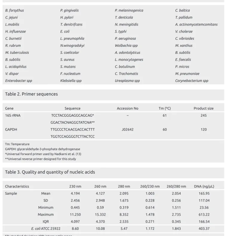
Table 1. The investigated specific agents by using the primer designed by Nadkarni et al. (13)
B. forsythus P. gingivalis P. melaninogenica C. baltica
C. jejuni H. pylori T. denticola T. pallidum
L.mobilis T. denitrifcans N. meningitidis A. actinomycetemcomitans
H. influenzae E. coli S. typhi V. cholerae
C. burnetii L. pneumophila P. aeruginosa C. vibrioides
R. rubrum N.winogradskyi Wolbachia spp M. xanthus
M. tuberculosis S. coelicolor A. odontolyticus B. subtilis
B. subtilis S. aureus L. monocytogenes E. faecalis
L. acidophilus S. mutans C. botulinum P. micros
V. dispar F. nucleatum C. Trachomatis M. pneumoniae
Enterobacter spp Klebsiella spp Ureaplasma spp Corynebacterium spp
Table 2. Primer sequences
Gene Sequence Accession No Tm (ºC) Product size
16S rRNA TCCTACGGGAGGCAGCAG* – 61 245
GGACTACNAGGGTATCNA**
GAPDH TTGCCCTCAACGACCACTTT J02642 60 120
TGGTCCAGGGGTCTTACTCC
Tm: Temperature
GAPDH: glyceraldehyde-3-phosphate dehydrogenase *Universal forward primer used by Nadkarni et al. (13) **Universal reverse primer designed for this study
Table 3. Quality and quantity of nucleic acids
Characteristics 230 nm 260 nm 280 nm 260/230 nm 260/280 nm DNA (ng/μL) Sample Mean 4.194 4.127 2.095 1.003 2.054 165.95 SD 2.456 2.948 1.675 0.228 0.256 117.04 Minimum 0.445 0.59 0.319 0.614 1.511 23.56 Maximum 11.250 15.332 8.352 1.478 2.735 613.22 IQR 4.097 4.370 2.535 0.271 0.345 166.54 E. coli ATCC 25922 8.60 10.08 5.47 1.172 1.843 403.37
In 2003, Taylor et al. (9) identified Corynebacterium in 34 of 62 pa-tients who were diagnosed histologically with granulomatous mastitis; in comparison with the remaining 28 cases, fever, neutrophilia, and fistula formation were more common in bacteria-positive cases. In that study, C. kroppenstedtii was the species identified most commonly, in 14 cases. Paviour et al. (24) isolated Corynebacterium spp. from breast tissue or abscess or deep wound in 24 patients and in 12 cases gram-positive bacilli seen histopathologically. Nine cases diagnosed as IGM. In their study, the species isolated most commonly was also C. kroppenstedtii (15/19 cases) and C. amycolatum and C. tuberculo-stearicum were also identified. In addition, some case reports identi-fied Corynebacterium species (25-27): two of these studies did not specify the species, while Ang et al. (25) isolated C. accolens. All three studies reported that antibiotic therapy was an effective treatment. However, Corynebacterium species are members of the dermal flora and it is difficult to distinguish infection, colonization, and contamina-tion by these organisms (24). Despite this difficulty, it was noteworthy that Corynebacterium spp. were identified in pus or were predominant (>104 CFU/mL) within abscesses (25). Funke et al. (26) reported that the presence of Gram-positive bacilli and polymorphonuclear leukocytes in a specimen or the presence of Corynebacterium in tissue that should normally be sterile, indicate that such bacteria are a potential cause. We conducted a molecular based assay of many flora bacteria including Corynebacterium spp., as well as the most common infective agents. However, no bacterial DNA was detected. In addition, there was no growth in culture samples taken before initiating treatment (surgical or steroid), the antibiotic therapy used conferred no benefit to any of the patients, and the treatment did not lead to clinical recovery. Combined, our findings make it unlikely that bacteriological agents are involved in the etiology of IGM.
Moreover, some authors have suspected that IGM might be the result of undiagnosed tuberculosis and mention the requirement for more detailed examinations for tuberculosis when IGM is diagnosed in de-veloping countries (8, 28-30). Due to tuberculosis is still an endemic disease in our country, we added M. tuberculosis primer to our study. Nevertheless, it was also negative. This weakens the postulate that “IGM patients concentrated in certain geographical regions might be undiagnosed tuberculosis cases”.
Several limitations of our study should be acknowledged. First, the primary limitation of the study design is that not all related cases were examined because of the exclusion criteria. A second study limitation was that fungi and viruses were not evaluated. Third, pro-inflammato-ry mediators and cytokines, which are produced in abundance during the inflammatory process, were not assayed.
Conclusion
We believe that known bacteriological agents, including Corynebac-terium spp., which are members of the dermal flora and are most commonly discussed in the etiology of IGM, may not be the prima-ry cause of IGM. A prospective study, including more cases, which also considers viruses, might be more informative in this matter.
Ethics Committee Approval: Ethics committee approval was received for this study from the ethics committee of Sakarya University School of Medicine Eth-ics Committee for Non-Invasive Clinical Trials (71522473.050.01.04/101).
Informed Consent: Informed consent was not received due to the retrospective nature of the study.
Peer-review: Externally peer-reviewed.
Author Contributions: Concept - T.K., I.H.C.; Design - T.K., F.A.; Super-vision - F.A., I.H.C.; Resources - T.K., I.H.C.; Materials - T.K., B.M.; Data Collection and/or Processing - E.D., T.K. Analysis and/or Interpretation - T.K., I.H.C.; Literature Search - T.K., F.F., A.K.; Writing Manuscript - T.K., F.F., A.K.; Critical Review - F.A.
Conflict of Interest: The authors have no conflicts of interest to declare.
Financial Disclosure: The authors declared that this study has received no fi-nancial support.
References
1. Kessler E, Wolloch Y. Granulomatous mastitis: a lesion clinically simulating car-cinoma. Am J Clin Pathol 1972; 58: 642-646. (PMID: 4674439) [CrossRef]
2. Kiyak G, Dumlu EG, Kilinc I, Tokaç M, Akbaba S, Gurer A, Ozkardes AB, Kilic M. Management of idiopathic granulomatous mastitis: di-lemmas in diagnosis and treatment. BMC Surg 2014; 14: 66. (PMID: 25189179) [CrossRef]
3. Sakurai K, Fuhsaki S, Enomoto K, Amano S, Sugitani M. Evaluation of follow-up strategies for corticosteroid therapy of idiopathic granulomatous mastitis. Surg Today 2011; 41: 333-337. (PMID: 21365412) [CrossRef]
4. Mizrakli T, Velidedeoglu M, Yemisen M, Mete B, Kilic F, Yilmaz H, Oz-turk T, Ozaras R, Aydogan F, Perek A. Corticosteroid treatment in the management of idiopathic granulomatous mastitis to avoid unnecessary surgery. Surg Today 2015; 45: 457-465. (PMID: 24993812) [CrossRef]
5. Yau FM, Macadam SA, Kuusk U, Nimmo M, Van Laeken N. The surgical management of granulomatous mastitis. Ann Plast Surg 2010; 64: 9-16. (PMID: 20023450) [CrossRef]
6. Akbulut S, Yilmaz D, Bakir S. Methotrexate in the management of idio-pathic granulomatous mastitis: review of 108 published cases and report of four cases. Breast J 2011; 17: 661-668. (PMID: 21951547) [CrossRef]
7. Pereira FA, Mudgil AV, Macias ES, Karsif K. Idıopathic Granulomatous lobular mastitis. Int J Dermatol 2012; 51: 142-151. (PMID: 22250621) [CrossRef]
8. Gautier N, Lanolde L, Tran-Tranh D, Khoury ME, David J, Labelle M, Patocskai E, Trop I. Chronic Granulomatous Mastitis: Imaging, Pa-thology and Management. Eur J Radiol 2013; 82: 165-175. (PMID: 23200627) [CrossRef]
9. Taylor GB, Paviour SD, Musaad S, Jones WO, Holland DJ. A clinico-pathological review of 34 cases of inflammatory breast disease showing an association between corynebacteria infection and granulomatous mastitis. Pathology 2003; 35: 109-119. (PMID: 12745457) [CrossRef]
10. Renshav AA, Derhagopian RP, Gould EW. Cystic Neutrophilic Granulo-matous Mastitis: An Underappreciated Pattern Strongly Associated With Gram- Positive Bacilli. Am J Clin Pathol 2011; 136: 424-427. (PMID: 21846918) [CrossRef]
11. D'Alfonso TM, Moo TA, Arleo EK, Cheng E, Antonio LB, Hoda SA. Cystic Neutrophilic Granulomatous Mastitis: Further Characterization of a Distinctive Histopathologic Entity Not Always Demonstrably Attribut-able to Corynebacterium Infection. Am J Surg Pathol 2015; 39: 1440-1447. (PMID: 26200100) [CrossRef]
12. Sambrook J, Fritsch EF, Maniatis T. Molecular Cloning: A Laboratory Manual. 2nd Ed. Vol. 9. New York: Cold Spring Harbor, 1989. 13. Nadkarni MA, Martin FE, Jacques NA, Hunter N. Determination of
bacte-rial load by real-time PCR using a broad-range (universal) probe and prim-ers set. Microbiology 2002; 148: 257-266. (PMID: 11782518) [CrossRef]
14. Asoglu O, Ozmen V, Karanlik H, Tunaci M, Cabioglu N, Igci A, Selcuk UE, Kecer M. Feasibility of surgical management in patients with granu-lomatous mastitis. Breast J 2005; 11: 108-114. [CrossRef]
15. Al-Khaffaf B, Knox F, Bundred NJ. İdiopathic Granulomatous Masti-tis: A 25-year Experience. J Am Coll Surg 2008; 206: 269-273. (PMID:
36
16. Cserni G, Szajki K. Granulomatous Lobular Mastitis Following Drug-Inducing Galactorrhea and Blunt Trauma. Breast J 1999; 5: 398-403. (PMID: 11348321) [CrossRef]
17. Baslaim MM, Khayat HA, Al-Amoudi SA. Idiopathic granulomatous mastitis: A heterogeneous disease with variable clinical presentation. World J Surg 2007; 31: 1677-1681. (PMID: 17541683) [CrossRef]
18. Bani-Hani KE, Yaghan RJ, Matalka II, Shatnawi NJ. Idiopathic granulo-matous mastitis: Time to avoid unnecessary mastectomies. Breast J 2004; 10: 318-322. (PMID: 15239790) [CrossRef]
19. Oran EŞ, Gürdal SÖ, Yankol Y, Öznur M, Calay Z, Tunacı M, Soybir GR. Management of Idıopathic Granulomatous Mastitis Diagnosed by Core Biopsy: A Retrospective Multicenter Study. Breast J 2013; 19: 411-418. (PMID: 23663101) [CrossRef]
20. Ozel L, Unal A, Kara M, Erdogdu E, Krand O, Gunes P, Karagol M, Demiral S, Titiz MI. Granulomatous mastitis: is it an autoimmune dis-ease? Diagnostic and therapeutic dilemmas. Surg Today 2012; 42: 729-733. (PMID: 22068681) [CrossRef]
21. Altintoprak F, Karakece E, Kivilcim T, Dikicier E, Cakmak G, Celebi F, Ciftci İH. Idiopathic Granulomatous Mastitis: An Autoimmune Disease?. Scientific World Journal 2013; 2013: 148727. (PMID: 24082849) 22. Tauch A, Fernandez-Natal I,Soriano F. A microbiological and clinical
re-view on Corynebacterium kroppenstedtiiInt J Infect Dis 2016; 48: 33-39. (PMID: 27155209)
23. Yu HJ, Deng H, Ma J, Huang SJ, Yang JM, Huang YF, Mu XP, Zhang L, Wang Q. Clinical metagenomic analysis of bacterial communities in
breast abscesses of granulomatous mastitis. Int J Infect Dis 2016; 53: 30-33. (PMID: 27777091) [CrossRef]
24. Paviour S, Musaad S, Roberts S, Taylor G, Taylor S, Shore K, Lang S, Hol-land D. Corynebacterium Species Isolated from Patients with Mastitis. Clin Infect Dis 2002; 35: 1434-1440. (PMID: 12439810) [CrossRef]
25. Ang LM, Brown H. Corynebacterium accolens isolated from breast ab-scess: possible association with granulomatous mastitis. J Clin Microbiol 2007; 45: 1666-1668. (PMID: 17344355) [CrossRef]
26. Funke G, Graevenitz A, Clarridge JE 3rd, Bernard KA. Clinical micro-biology of coryneform bacteria. Clin Microbiol Rev 1997; 10: 125-159. (PMID: 8993861) [CrossRef]
27. Stary CM, Lee YS, Balfour J. Idiopathic granulomatous mastitis associ-ated with corynebacterium sp. Infection, Hawaii Med J 2011; 70: 99-101. (PMID: 21857740)
28. Ocal K, Dag A, Turkmenoglu O, Kara T, Seyit H, Konca K. Granuloma-tous mastitis: clinical, pathological features, and management. Breast J 2010; 16: 176-182. (PMID: 20030652) [CrossRef]
29. Akcan A, Akyildiz H, Ali Deneme M, Akgun H, Aritas Y. Granulomatous lobu-lar mastitis: a complex diagnostic and therapeutic problem. World J Surg 2006; 30: 1403-1409. (PMID: 16847715) [CrossRef]
30. Al-Khawari HAT, Al-Manfouhi HA, Madda JP, Kovacs A, Sheikh M, Roberts O. Radiologic features of granulomatous mastitis. Breast J 2011; 17: 645-650. (PMID: 21929558) [CrossRef]