Cellular and Molecular Biology
E-ISSN : 1165-158X / P-ISSN : 0145-5680www.cellmolbiol.org Original Research
Gene variants of TCF7L2 are histopathologically important in colorectal cancers but do
not have direct association with MYC expression
Nazlı Ezgi Ozkan1, Cem Horozoglu2, Ozlem Timirci Kahraman1, Soykan Arikan3, Ammad Ahmad Farooqi4,
Saime Sürmen1, Gurbet Korkmaz1, Esra Canan Kelten Talu5, Uteuliyev Yerzhan Sabitaliyevich6, Ilhan Yaylim1*
1 Molecular Medicine Department, Aziz Sancar Institute of Experimental Medicine / Istanbul Medical Faculty at Istanbul University, Istanbul,
Turkey
2 Department of Medical Services and Techniques, İstanbul Gelisim University, İstanbul-Turkey 3 General Surgery Clinics, Istanbul Training and Research Hospital, Istanbul, Turkey
4 Laboratory for Translational Oncology and Personalized Medicine, Rashid Latif Medical College (RLMC), Lahore, Pakistan. 5 Pathology Clinics, Istanbul Training and Research Hospital, Istanbul, Turkey
6 Kazakhstan Medical University "KSPH", Almaty 050060, Kazakhstan
*Correspondence to: [email protected]
Received October 14, 2018; Accepted June 1, 2019; Published ADecember 31, 2019
Doi: http://dx.doi.org/10.14715/cmb/2019.65.8.1
Copyright: © 2019 by the C.M.B. Association. All rights reserved.
Abstract: Rapidly accumulating preclinical and clinical studies have helped us to unveil underlying mechanisms of colorectal cancer development and
progres-sion. Deregulated signaling pathways play instrumental role in carcinogenesis, drug resistance and metastasis. Wnt signaling cascade has attracted considerable attention in colorectal cancer as many ground-breaking researches have highlighted central role of Wnt pathway in pathogenesis of colorectal cancer. T-Cell Transcription Factors (TCFs) have been shown to work synchronously with β-catenin to fuel colorectal cancer development and progression. Chromatin immuno-precipitation coupled with high-throughput sequencing (ChIP-Seq) data sets has deepened our knowledge about critical role of risk-associated SNPs. Increasingly it is being reported that many risk-associated SNPs are located within binding sites for transcription factors and consequently risk status of these SNPs may modify binding pattern of transcriptional factors and thus rewire the transcriptional regulation. DNA was extracted from peripheral blood samples of 117 colorectal cancer patients and 127 healthy subjects. TCF7L2 variants (rs6983267, rs7903146) were examined by the PCR-RFLP method. Tumor and the surrounding tissues were dissected from 37 CRC patients and RNA isolation was performed. The gene expression of c-myc was determined by RT-PCR. T allele carriage of rs6983267 variant was found to be associated with CRC (p=0.042). TT genotype of rs7903146 was associated with late tumor stage (T3+T4) (p=0.037) and presence of mucinous component (p=0.031). TTCT haplotype was found to be statistically higher in CRC compared to the control group (p=0.007). There was no statistically significant difference in c-myc gene expression. TCF7L2 gene variants may play an important role in histopathologic aspects associated with CRC and it is inde-pendent of c-myc gene expression.
Key words: TCF7L2; Colorectal Cancers; Tumor stage; Gene expression; Signaling.
Introduction
It is becoming progressively more understandable that colorectal cancer is therapeutically challenging and recent advancements in high-throughput technologies have helped us to uncover extra-ordinary complexity and suggest multiple cancer-related functions for many genes, which are often cancer stage-dependent or tis-sue-dependent (1,2). Large-scale sequencing of human cancers has unveiled a myriad of genomic alterations. Furthermore, the past few years have witnessed tremen-dous breakthroughs in the biology of colorectal can-cer and development of multiplexed and quantitative approaches has revolutionized the field of molecular oncology (3,4,5).
Wnt-β-catenin signaling is involved in modulation of different molecular mechanisms (6,7). β-catenin is shipped to the nucleus where it interacts with the TCF family of transcription factors (TCF1, LEF, TCF3, and TCF4). T-Cell Transcription Factors (TCFs) have
gai-ned considerable attention because of their ability to transcriptionally modulate wide ranging target genes. TCF proteins have been shown to physically interact with transcriptional repressors, such as Groucho/Grg/ TLE (transducin-like enhancer of split) CtBP, histone deacetylases (HDACs) and other factors and induce transcriptional inactivation of different genes (6,7). However, intriguingly, interaction of TCF proteins with β-catenin converted TCFs into transcriptional activators (6,7).
It has recently been convincingly revealed that TC-F7L2 with rs138649767-A allele harbored the ability to activate the MYC enhancer with rs6983267-G allele and enhanced proliferation of colorectal cancer cells (8). Functional silencing of TCF7L2 provided evidence of its pro-metastatic role in proliferation, adhesion and migration of colorectal cancer cells (9). TCF7L2 has previously been reported to be frequently overexpressed in therapy-resistant colorectal cancer cells (10, 11). TC-F7L2 inhibition re-sensitized colorectal cancer cells to
therapeutic strategies (10, 11).
Accumulating evidence has started to shed light on involvement of long non-coding RNAs in facilitating Wnt-catenin and TCF7L2 driven pathway.
CCAT2, a long non-coding RNA has been shown to promote TCF7L2 binding to the MYC promoter. Chromatin-immunoprecipitation analysis demonstrated higher binding of TCF7L2 to promoter region of MYC in CCAT2-overexpressing colorectal cancer cells. Data clearly suggested that TCF7L2 worked synchronously with versatile regulators to promote genomic instability and colorectal cancer 12). In accordance with this ap-proach, another long non-coding RNA, colorectal neo-plasia differentially expressed (CRNDE) sequestered TCF7L2 away from miRNA-217. CRNDE promoted Wnt signaling mainly through interfering with miR-217 mediated targeting of TCF7L2 in colorectal cancer cells (13). Stable knockdown of TCF7L2 exerted inhibitory effects on anchorage-independent growth of colorectal cancer cells (14).
Previously published high-impact research has pro-vided evidence of central role of Wnt signaling in regu-lation of MYC-335 (15). MYC-335 elements contai-ning the T and G alleles of rs6983267 were cloned into a reporter vector. It was observed that MYC-335 had Wnt-responsive enhancer activity and interestingly, G allele demonstrated 1.5-fold stronger Wnt responses as compared to T allele (15). It has been experimentally verified that rs6983267-containing region showed no-table binding affinity with TCF7L2 and the transcriptio-nal activator β-catenin (15).
Furthermore, circumstantial evidence indicated that rs6983267 risk region physically interacted with MYC in colorectal cancer cell lines (16).
Certain hints have emerged which highlighted that some splicing forms of TCF7L2 might play functionally important role in transcriptional regulation of MYC in colon tissue but this regulation was not dependent di-rectly on rs6983267 (17). MYC expression was studied in colorectal cancer tissue samples and data clearly sug-gested that although MYC expression was associated with some of the TCF7L2 splicing forms but not with genotypes of rs6983267, or relationship of rs6983267 with TCF7L2 expression (17). These exciting findings suggested some splicing forms of TCF7L2 might play functionally important role in transcriptional regulation of MYC in colon tissue but this regulation was not de-pendent directly on rs6983267.
We aimed to investigate the distribution of variants of TCF7L2 (rs6983267, rs7903146) between Turkish colorectal cancer patients and healthy individuals. We also aimed to study any possible effect of these variants on c-myc gene expression, and the contribution of both of these conditions to tumor histopathology.
Materials and Methods Subjects
In our study, peripheral blood samples of 117 pa-tients, 42 female and 75 male, who were diagnosed as Colorectal Cancer by the departmant of General Surgery, Cerrahpasa Faculty of Medicine and Clinic of General Surgery, Istanbul Training and Research Hospital were taken .The tumor tissues and tumor surrounding tissue
samples of 36 patients were resected at these clinics. Our work was carried out by the permission of the Eth-ics Committee of Istanbul University Cerrahpaşa Medi-cal School with the permission number 1671-1265.
Histopathological examinations
Patients with colorectal cancer, who were diagnosed with the radiological, and routine pathological and his-topathological examination of the tissue, were enrolled into the study. Parameters, such as the results of the mediastinoscopy were performed in order to determine the lymph involvement, the performed operation tech-niques, the stage of the tumor which is determined ac-cording to the last edition of 7th staging system (pTNM) was determined as a result of the pathological examina-tion of the resected tissue, and the grade of the tumor detected in the pathological examination.
The specimens were evenly planed within a cryo-stat. Sections (3 to 4 mm) were sliced and followed by hemotoxylin and eosin staining. Special attention was given to tumor invasions at the lymphatic and vascular tissues. The surrounding tissue of the tumor without the findings of the macroscopic invasion were included in the study as the control group.
Genotyping
DNA isolation was performed by salting out the pe-ripheral blood samples from Colorectal Cancer patients and healthy subjects. For the rs6983267 of TCF7L2 polymorphism, the forward primer 5'- ATGAAGGC-GTCGTCCAAATGA -3' and reverse primer 5'- TTG-GCTGGCACTGTCTGTAT -3' were used at a con-centration of 10 pmol/μl for each primer. Also for the rs7903146 of TCF7L2 polymorphism, the forward primer 5'- AATTAGAGAGCTAAGCACTTTTTAGG-TA-3' and reverse primer 5'- CAAGCTTCTCAGTCA-CACAGG-3' were used at a concentration of 5 pmol/μl for each primer.
The PCR reaction mixture contained 150 ng DNA template, 1.75 mM MgCl2, 50 mM KCl, 10 mMTris-HCl (pH 8.4), 600 μM dNTP (iNtRON Biotechnology Co., Korea) and 0.06 unit Taq DNA polymerase (iN-tRON Biotechnology Co., Korea). The PCR conditions were determined as first denaturation at 95 °C for 5 min-utes, 35 cycles at 94 °C for 45 seconds, 56 °C for 45 seconds, 72 °C for 45 seconds, and a final extension at 72 °C for 5 minutes. PCR product of rs7903146 was digested by RsaI (MBI Fermentas, CA) at 37°C for 2.5 hours. Also, PCR product of rs6983267 was digested by Tsp45I (MBI Fermentas, CA) at 37°C for 2.5 hours. After enzymatic restriction; For rs6983267, two fragments having 334 and 198 bp (G allele) or one fragment having 498 bp (T allele) were identified. For rs7903146, one fragment having 176 bp (T allele) or two fragments having 149 and 27 bp (C allele) were identified with agarose gel electrophoresis.
Gene expression
Tumor tissue and the tumor surrounding tissue sam-ples were surgically dissected. Samsam-ples were stored in liquid nitrogen until use. Total RNA was isolated from tissues using the TRIzol method. cDNA synthesis from total RNA was performed by using the High-Capacity cDNA Reverse Transcription Kit (LifeTech, Applied
The distribution of rs6983267 and rs7903146 vari-ants according to tumor histopathology is shown in Ta-ble 3. When rs6983267 variant is examined histopatho-logically, there are some differences which are close to statistical significance. It is observed in our study that in the advanced tumor stage (T3-T4) the G allele car-rier is higher than the early tumor stage (T1-T2) (p = 0.054, OR = 1.407, Cl: 0.936-2.113). Similarly, when patients were examined according to node metastasis, the presence of G allele (GG-GT) in patients with node metastasis was found to be higher (74.1%) than in pa-tients without node metastasis (57.6%) (p = 0.060, OR = 1.287, Cl: 0.986-1.679).
We also studied relationship between smoking and rs6983267 variance. It was observed that both genotype and environment synergistically promoted colorectal cancer development and progression. Phenotypic ef-fects of GT genotype appeared to be more pronounced in smokers.
The TT genotype carriage of rs7903146 was not de-tected in all early-onset (T1-T2) patients. It was seen in 15.4% of patients with advanced tumor (T3-T4) (p=0.037; OR=0.846; CI:0.775-0.924). CT genotype carriage of rs7903146 was 69.2% in early tumor stage (T1-T2) and 39.6% in advanced tumor stage (T3-T4). The frequency of the patients with advances tumor stage who have CT genotype was lower than those with early tumor stage (p=0.007; OR=0.571; CI: 0.398-0.820).
The CT genotype for the rs7903146 variant was higher in patients without node metastasis (N0) (55%) than patients with node metastasis (N1,N2,N3) (36.8%) and statistical significance were found to be close (p = 0.049, OR = 0.670, Cl: 0.445-1.009). The patients with rs7903146 TT genotype were found higher in tumors with mucinous component than in those without mu-cinous component (p = 0.031, OR = 3.268, CI: 1.245-8.576). Similarly, C allele carriers were found to be higher in smokers compared to non-smokers (p=0,022; OR=0,555; CI:0,249-1,238).
The TTCT haplotype of both variants (rs6983267TT ve rs7903146CT) was significantly higher in CRC pa-tients than in the control group (p=0.007; OR=2.653; CI:1.274-5.527). The same haplotype (TTCT) was found to be statistically significant in the low tumor Biosystem, USA). As a result of our literature researches
and pilot studies, it was determined that β-Actin gene is suitable as housekeeping gene. PCR primers and probes for the c-MYC and β-Actin gene were selected using the Single Tube TaqMan Gene Expression Assay (LifeTech, Applied Biosystem, USA).
The real-time PCR reaction mixture contained 100 ng cDNA, 2X PZR Master Mix (LifeTech, Applied Biosystem, USA) and 20X Primer and Probe ready mix (LifeTech, Applied Biosystem, USA) for both primers. Real-time PCR conditions were determined as UDG in-cubation at 50° C for 2 minutes, AmpliTaq Gold UP en-zyme activation at 95 ° C for 10 minutes, and 40 cycles at 95° C for 15 seconds and at 60° C for 1 minute. Re-al-time PCR analysis was performed using Stratagene Mx3005p instrument (Agilent Technologies, CA, USA) with TaqMan Gene Expression Master Mix (LifeTech, Applied Biosystem, USA).
Statistical analysis
In our study, Chi square and further tests were ap-plied by using SPSS 7 package program for statistical analysis of TCF7L2 variants, c-myc gene expression values, demographic data and histopathological para-meters. Mann-Whitney U test, which is one of the non-parametric tests, was applied by using GraphPad Prism 5 program for the statistical analysis of gene expres-sion levels of tumors and control tissues. The data were verified with both statistical programs. P values that are lower than 0.05 were regarded as statistically signifi-cant.
Results
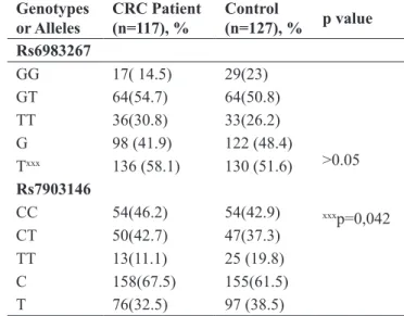
The demographic data of the patients are shown in Table 1 and the allele and genotype distribution of
va-riants Rs6983267 and rs7903146 are shown in Table 2. There was no statistical difference between patient and control groups in terms of genotype and allele distribu-tions in both variants (rs6983267 and rs7903146).
However, the T allele carrier (TT-GT) for the rs6983267 variant was found to be higher in CRC patients compared to the control group (p=0.042; OR=1.130; CI:1.005-1.270).
Genotypes
or Alleles CRC Patient (n=117), % Control (n=127), % p value
Rs6983267 GG 17( 14.5) 29(23) >0.05 xxxp=0,042 GT 64(54.7) 64(50.8) TT 36(30.8) 33(26.2) G 98 (41.9) 122 (48.4) Txxx 136 (58.1) 130 (51.6) Rs7903146 CC 54(46.2) 54(42.9) CT 50(42.7) 47(37.3) TT 13(11.1) 25 (19.8) C 158(67.5) 155(61.5) T 76(32.5) 97 (38.5)
Table 2. Allele and genotype distribution of Rs6983267 and
rs7903146 variants in CRC patient and control group.
Groups Control (n=127) % CRC Patient (n=117) p value
Gender
Woman, n(%) 59 (46.9) 42(35.9)
>0.05
xxxp=0.029
Men, n(%) 68(53,5) 75(64.1)
Average age (year) 58.0 61.2
Family cancer story
Positive, n(%) 8(6.3) 8(6.8) Negative, n(%) 119(93.7) 109(93.2) Alcohol Use Positive, n(%) 4 (3.1) 4(3.4) Negative, n(%) 123 (96.9) 113(96.6) Smoking xxx Positive, n(%) 4 (3.1) 6(5.1) Negative, n(%) 123(96.6) 111(94.9)
stage (p = 0.001, OR = 0.286, CI = 0.140-0.583) and without node metastasis (p = 0.007; OR = 0.310, CI = 0.122-0.784) (Table 4).
The c-myc gene expression levels of tumor and surrounding tissue are shown in figure 1. The fold changes of different genotypes of variants rs6983267 and rs7903146 on c-myc gene expression are shown in
Table 5. When c-myc gene expression levels between
tumor and surrounding tissue were compared, it was observed that c-myc expression in tumor tissue was in-creased 2-fold compared to surrounding tissues, but no statistically significant difference was found (p> 0,05).
There was no statistically significant difference between rs6983267 and rs7903146 variants and c-myc expression levels in tumor tissue (p>0.05). When the
GG (wild type) genotype was referenced for rs6983267, the expression of c-myc was increased 1.36-fold in TT (homozygous mutant type) genotypes and 1.53-fold with GT (heterozygous) genotypes (p> 0,05). When the CC genotype was referenced for the rs7903146 variant, the c-myc gene expression level decreased by 1.14 fold in the CT (heterozygous) genotype and 2.14 fold in the TT genotype (p> 0,05). There was no statistically si-gnificant correlation between the level of c-myc gene expression and histopathologic factors. Similarly, c-myc gene expression increased 1.2-fold in patients with lymphatic involvement and 1.07-fold in patients with N2+N3 nod phase. When c-myc gene expression is as-sessed for metastasis, it is seen that c-myc expression in metastatic patients is 1.1 times lower (p>0,05).
TCF7L2 rs7903146 8q24 rs6983267 CC n(%) CT n(%) TT n(%) GG n(%) GT n(%) TT n(%) Gender women 13 (31,0) 26(61,9) 3(7,1) 7(16,7) 19(45,2) 16(38,1) men 36(48,0) 28(37,3) 11(14,7) 8(10,7) 43(57,3) 24(32,0) T stage T1 4(50,0) 4(50,0) 0 (0,0) 0(0,0) 3(37,5) 5(62,5) T2 4(22,2) 14(77,8) 0(0,0) 1(5,6) 9(50,0) 8(44,4) T3 34(47,2) 29(40,3) 9(12,5) 11(15,3) 37(51,4) 24(33,3) T4 7(36,8) 7(36,8) 5(26,3) 3(15,8) 13(68,4) 3(15,8)
Lymph Node Involvement
N0 22(36,7) 33 (55,0) 5(8,3) 6(10,0) 29(48,3) 25(41,7) N1 16(50,0) 12(37,5) 4(12,5) 7(21,9) 17(53,1) 8(25,0) N2 6(33,3) 7(38,9) 5(27,8) 1(5,6) 12(66,7) 5(27,8) N3 5(71,4) 2(28,6) 0 (0,0) 1(14,3) 4(57,1) 2(28,6) Distant metastasis Yes 11 (42,3) 11 (42,3) 4(15,4) 4(15,4) 14(53,8) 8(30,8) No 38 (41,8) 43(47,3) 10(11,0) 11 (12,1) 48 (52,7) 32(35,2) Angiolymphatic Invasion Positive 14(38,9) 16(44,4) 6(16,7) 4(11,1) 23(63,9) 9(25,0) Negative 35(43,2) 38(46,9) 8(9,9) 11(13,6) 39(48,1) 31(38,3) Perineural Invasion Positive 16 (42,1) 18(47,4) 4(10,5) 5(13,2) 23(60,5) 10(26,3) Negative 33 (41,8) 36(45,6) 10(12,7) 10(12,7) 39(49,4) 30(38,0)
Table 3. The Genotype Distribution of c-myc and TCF7L2 gene variants according to the CRC patient’s histopathological data.
Groups/ c-myc and TCF7L2
genotype combinations n(%) CCGG CCGT CCTT CTGG CTGT CTTT TTGG TTGT TTTT
CRC patients 7 (6%) 27 (23.1%) 15 (12.8%) 6 (5/1%) (22.2%)26 22 (18.8%) 2 (1.7%) 9 (7.7%) 3 (2.6%)
Controls 10 (7.9%) 24 (18.9%) 20 (15.7%) 14 (11%) 25 (19.7%) 9 (7.1%) 5 (3.9%) 16 (12.6% 4 (3.1%)
Table 4. The distribution of c-myc and TCF7L2 genotype combinations in our study groups.
Variant/Genotip Fold Change %95 CI Fold Regulation p value
rs6983267 TT 1,3624 0.00001-2.88 1,3624 >0.05 rs6983267 GT 1,5268 0.00001-3.17 1,5268 rs6983267 GT-TT 1,4458 0.00001-2.89 1,4458 Rs7903146 CT 1,1482 0.18-2.12 1,1482 Rs7903146 TT 2,1443 0.55- 3.74 2,1443 Rs7903146 CT-TT 1,3192 0.31-2.33 1,3192
Discussion
Advent of next generation sequencing technologies has leveraged our understanding related to risk-associa-ted SNPs to a next level. Substantial fraction of infor-mation is continuously being added into pre-existing pool of cancer risk associated SNPs and we now know that risk allele (G) of SNP rs6983267 has been noted to demonstrate preferential binding affinity for the TC-F7L2 as compared to (T) allele.
In our study, rs6983267, T allele carrier was found to be higher in the patient group compared to the control group and it was found to be related to cigarette use in GT genotype carriers (p> 0.05). There was no corre-lation between c-myc gene expression and rs6983267 variant (p> 0.05).
According to our study, no correlation was found between c-myc gene expression and rs6983267 variant. In this respect, this data does not support the work of Poerantz et al. Although we did not find it statistically significant, we observed that the expression of c-myc gene in T-allele carriers of the Rs6983267 variant was slightly lower in tumor tissue (p>0.05). Accordingly, our opinion is that the related genotype does not have a great effect on c-myc gene expression. Prokunina et al. (17) obtained data that c-myc expression was inde-pendent from rs6983267 variant and that co-expression was not associated with colorectal cancers. Our results are in accordance with the findings reported by Proku-nina et al.
In our study, T allele carriage of Rs6983267 variant was statistically high in patients with colorectal cancer. The results of Folsom et al. are parallel to our results (18). Nan et al's study examined the association of aspi-rin use and rs6983267 variant and found that T allele carriage is associated with a lower risk of colorectal cancer (19). It is noteworthy that the T allele carriage of rs6983267 variant is important in both studies, although it is not appropriate to compare the results with our results due to the regulatory effects of aspirin on Wnt /β-catenin pathway.
Another consequence of our study is that GT geno-type of Rs6983267 is associated with cigarette use in CRC (p<0.05). These results are of original value due to the fact that this is the first data in the literature. In the rs7903146 variant, CT genotype and C allel carriage was correlated histologically with early tumor stage (T1-T2), mucinous component carrier and cigarette smokers in colorectal cancer cases. In the rs7903146 variant, TT genotype was correlated histologically with late tumor stage (T3+T4).
This data is of original value because there is no similar data in the related literature. According to the meta-analysis results of Zhang et al. (20), The variants of TCF7L2 are associated with different cancers. Part of the evidence of our work supports this belief. In another study, similarly, the study of Mexican colorectal car-cinoma cases shows that TT genotype of rs7903146 is associated with rectal localization of the tumor, and at the same time, there are indications that multiple gene variants of TCF7L2 are important for TNM staging (21). In a study of colorectal cancer patients with type 2 diabetes, it was determined that the T allele carriage of rs7903146 variant was higher in the patient group than
in the control group (22).
Similarly to these studies, for rs7903146 in our study, genotypes bearing the TT genotype were found higher in tumors with mucinous component than in those with-out mucinous component. We think that the difference between the studies is due to different ethnicity and case selection criteria.
Mucinous component presence in colorectal cancers is used as an important clinical parameter in the progno-sis and histopathologic condition. One of the genes en-coding the mucinous component, MUC1-C, was found in breast cancer cases where the TCF7L2 gene could be regulated by the β-catenin pathway (23,24). In our study, the relationship between the TT genotype carrier of the rs7903146 variant and the presence of the mucin component in the tumor tissue was a supporting factor in terms of the common carcinogenesis mechanisms re-ported in the literature.
There was no statistical difference between c-myc gene expression level and rs7903146 (p> 0.05). Al-though there was no statistically significant difference in our study, there was a decrease in the gene expression of c-myc in CC-CT genotypes of rs7903146 (CC-CT) and an increase in gene expression in T-allele carriers (p> 0.05). This is the first study in the literature exami-ning the relationship of rs7903146 variant of TCF7L2 to c-myc gene expression. The TTCT haplotype of two polymorphisms (rs7903146 and rs6983267) that we stu-died in cases with colorectal cancer in our study was found to be statistically high in early tumor stage (T1-T2) and colorectal cancer patients (p<0.05). It has been observed that these variants are associated with Turkish colorectal cancer cases, as the cases of other literature. However, in our study no statistically significant effect of TCF7L2 variants on c-myc expression was detec-ted. It is noted that different alleles and genotypes of TCF7L2 variants may cause slight alterations in gene expression of c-myc (p>0.05). Overall our findings provided clues of association between rs7903146 and c-myc expression however, we did not find any correla-tion between rs6983267 and c-myc in colorectal cancer patients.
Immunohistochemical studies are planned to be carried out by increasing the number of cases with the support of statistically significant data obtained in our study. This will provide a more detailed look at the cli-nicopathological and histopathological effects of TC-F7L2 and other TCF gene family members in colorectal cancers. Thus, the role of the TCF gene family in the tumor stage will be demonstrated tissue-specific. The relationship of TCF7L2 protein level with T cell clus-tering will be determined. At the same time, the effect of TCF7L2 level change on both epithelial tissue and T-lymphocyte proliferation will be observed separately.
Our study was the first study to examine the histo-pathological relationship of gene variants of TCF7L2 in Turkish cases and this study will support the researcher who is studying Wnt and Wnt/β-catenin pathways.
References
1. Wang H, Liang L, Fang JY, Xu J. Somatic gene copy number alterations in colorectal cancer: new quest for cancer drivers and
biomarkers. Oncogene. 2016 Apr 21;35(16):2011-9. doi: 10.1038/ onc.2015.304.
2. Davies RJ, Miller R, Coleman N. Colorectal cancer screening: prospects for molecular stool analysis. Nat Rev Cancer. 2005 Mar;5(3):199-209.
3. Brenner H, Chen C. The colorectal cancer epidemic: challenges and opportunities for primary, secondary and tertiary prevention. Br J Cancer. 2018 Oct;119(7):785-792. doi: 10.1038/s41416-018-0264-x.
4. Lièvre A, Blons H, Laurent-Puig P. Oncogenic mutations as predictive factors in colorectal cancer. Oncogene. 2010 May 27;29(21):3033-43. doi: 10.1038/onc.2010.89.
5. Tenesa A, Dunlop MG. New insights into the aetiology of colorec-tal cancer from genome-wide association studies. Nat Rev Genet. 2009 Jun;10(6):353-8. doi: 10.1038/nrg2574.
6. Barker N, Clevers H. Mining the Wnt pathway for cancer thera-peutics. Nat Rev Drug Discov. 2006 Dec;5(12):997-1014.
7. Segditsas S, Tomlinson I. Colorectal cancer and genetic altera-tions in the Wnt pathway. Oncogene. 2006 Dec 4;25(57):7531-7. 8. Chang J, Tian J, Yang Y, Zhong R, Li J, Zhai K, Ke J, Lou J, Chen W, Zhu B, Shen N, Zhang Y, Gong Y, Zhu Y, Zou D, Peng X, Huang K, Miao X. A Rare Missense Variant in TCF7L2 Associates with Colorectal Cancer Risk by Interacting with a GWAS-Identified Regulatory Variant in the MYC Enhancer. Cancer Res. 2018 Sep 1;78(17):5164-5172. doi: 10.1158/0008-5472.CAN-18-0910. 9. Torres S, García-Palmero I, Marín-Vicente C, Bartolomé RA, Calviño E, Fernández-Aceñero MJ, Casal JI. Proteomic Charac-terization of Transcription and Splicing Factors Associated with a Metastatic Phenotype in Colorectal Cancer. J Proteome Res. 2018 Jan 5;17(1):252-264. doi: 10.1021/acs.jproteome.7b00548. 10.Ghadimi BM, Grade M, Difilippantonio MJ, Varma S, Simon R, Montagna C, et al. Effectiveness of gene expression profiling for response prediction of rectal adenocarcinomas to preoperative chemoradiotherapy. J Clin Oncol 2005;23(9):1826-38 doi 10.1200/ JCO.2005.00.406.
11.Kendziorra E, Ahlborn K, Spitzner M, Rave-Frank M, Emons G, Gaedcke J, et al. Silencing of the Wnt transcription factor TCF4 sen-sitizes colorectal cancer cells to (chemo-) radiotherapy. Carcinoge-nesis 2011;32(12):1824-31 doi 10.1093/carcin/bgr222.
12.Ling H, Spizzo R, Atlasi Y, Nicoloso M, Shimizu M, Redis RS, Nishida N, Gafà R, Song J, Guo Z, Ivan C, Barbarotto E, De Vries I, Zhang X, Ferracin M, Churchman M, van Galen JF, Beverloo BH, Shariati M, Haderk F, Estecio MR, Garcia-Manero G, Patijn GA, Gotley DC, Bhardwaj V, Shureiqi I, Sen S, Multani AS, Welsh J, Ya-mamoto K, Taniguchi I, Song MA, Gallinger S, Casey G, Thibodeau SN, Le Marchand L, Tiirikainen M, Mani SA, Zhang W, Davuluri RV, Mimori K, Mori M, Sieuwerts AM, Martens JW, Tomlinson I, Negrini M, Berindan-Neagoe I, Foekens JA, Hamilton SR, Lanza G, Kopetz S, Fodde R, Calin GA. CCAT2, a novel noncoding RNA mapping to 8q24, underlies metastatic progression and
chromoso-mal instability in colon cancer. Genome Res. 2013 Sep;23(9):1446-61. doi: 10.1101/gr.152942.112.
13.Yu B, Ye X, Du Q, Zhu B, Zhai Q, Li XX. The Long Non-Co-ding RNA CRNDE Promotes Colorectal Carcinoma Progression by Competitively Binding miR-217 with TCF7L2 and Enhancing the Wnt/β-Catenin Signaling Pathway. Cell Physiol Biochem. 2017;41(6):2489-2502. doi: 10.1159/000475941.
14.Kundu S, Ali MA, Handin N, Padhan N, Larsson J, Karoutsou M, et al. Linking FOXO3, NCOA3, and TCF7L2 to Ras pathway phe-notypes through a genome-wide forward genetic screen in human colorectal cancer cells. Genome Med. 2018;10(1):2.
15.Tuupanen S, Turunen M, Lehtonen R, Hallikas O, Vanharanta S, Kivioja T, et al. The common colorectal cancer predisposition SNP rs6983267 at chromosome 8q24 confers potential to enhanced Wnt signaling. Nat Genet. 2009;41(8):885-90.
16.Pomerantz MM, Ahmadiyeh N, Jia L, Herman P, Verzi MP, Dod-dapaneni H, et al. The 8q24 cancer risk variant rs6983267 shows long-range interaction with MYC in colorectal cancer. Nat Genet. 2009;41(8):882-4.
17.Prokunina-Olsson L, Hall JL. No effect of cancer-associated SNP rs6983267 in the 8q24 region on co-expression of MYC and TC-F7L2 in normal colon tissue. Mol Cancer. 2009;8:96.
18.Folsom AR, Pankow JS, Peacock JM, Bielinski SJ, Heiss G, Boe-rwinkle E. Variation in TCF7L2 and increased risk of colon cancer: the Atherosclerosis Risk in Communities (ARIC) Study. Diabetes Care. 2008;31(5):905-9.
19.Nan H, Morikawa T, Suuriniemi M, Imamura Y, Werner L, Ku-chiba A, et al. Aspirin use, 8q24 single nucleotide polymorphism rs6983267, and colorectal cancer according to CTNNB1 alterations. J Natl Cancer Inst. 2013;105(24):1852-61.
20.Zhang M, Tang M, Fang Y, Cui H, Chen S, Li J, et al. Cumu-lative evidence for relationships between multiple variants in the VTI1A and TCF7L2 genes and cancer incidence. Int J Cancer. 2018;142(3):498-513.
21.Rosales-Reynoso MA, Arredondo-Valdez AR, Juárez-Vázquez CI et al. TCF7L2 and CCND1 polymorphisms and its association with colorectal cancer in Mexican patients. Cell Mol Biol (Noisy-le-grand). 2016;62(11):13-20.
22.Sainz J, Rudolph A, Hoffmeister M, Frank B, Brenner H, Chang-Claude J et al. Effect of type 2 diabetes predisposing gene-tic variants on colorectal cancer risk. J Clin Endocrinol Metab. 2012;97(5):E845-51.
23.Betge J, Schneider NI, Harbaum L, Pollheimer MJ, Lindtner RA, Kornprat P, et al. MUC1, MUC2, MUC5AC, and MUC6 in colorec-tal cancer: expression profiles and clinical significance. Virchows Arch. 2016;469(3):255-65.
24.Rajabi H, Ahmad R, Jin C, Kosugi M, Alam M, Joshi MD, et al. MUC1-C oncoprotein induces TCF7L2 transcription factor acti-vation and promotes cyclin D1 expression in human breast cancer cells. J Biol Chem. 2012 Mar 23;287(13):10703-13.