Giant Pericardial Tube Roll Aneurysm after the
Treatment of Aortic Interruption
Mustafa O. Ulukan, MD
1Yahya Yildiz, MD
2Emin C. Ata, MD
1Didem M. Oztas, MD
3Korhan Erkanli, MD
1Murat Ugurlucan, MD
1Halil Turkoglu, MD
11Department of Cardiovascular Surgery, Istanbul Medipol University Medical Faculty, Istanbul, Turkey
2Department of Anesthesiology and Reanimation, Istanbul Medipol University Medical Faculty, Istanbul, Turkey
3Department of Cardiovascular Surgery, Bagcilar Training and Research Hospital, Istanbul, Turkey
AORTA 2020;8:152–154.
Address for correspondence Murat Ugurlucan, MD, TEM Avrupa Otoyolu, Goztepe Cikisi, No: 1, Bagcilar 34214, Istanbul, Turkey (e-mail: [email protected]).
Interrupted aortic arch is a rare congenital cardiac pathology that requires emergency repair during infancy. Different tech-niques, such as aortic mobilization and ascending aorta to descending aorta anastomosis, patch augmented anastomosis, or biologic or synthetic graft interposition, have been de-scribed in the literature for the treatment of this pathology.1
The patient is a 3-year-old boy who underwent autolo-gous pericardial tube graft interposition between the as-cending and desas-cending aorta for the treatment of Type B interrupted aortic arch during infancy. He had two insigni fi-cant apical muscular ventricular septal defects that did not require closure or pulmonary artery banding.
He has been followed periodically in the outpatient clinic, and the ventricular septal defects closed spontaneously. However, the pericardial roll dilated gradually. Originally it was 7 mm in diameter, and the size enlarged to 1.5 to 2.4 to 3.1 and finally 4.2 cm in diameter at 12, 18, 28, and 36 months, respectively.
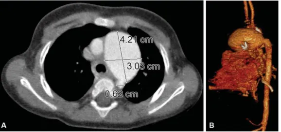
Aneurysm formation was confirmed with computed to-mography (CT) angiography that revealed a 4.2-cm dilatation of the pericardial tube (►Fig. 1A,B,►Video 1). There was a stenosis visualized at the proximal anastomosis region.
However, cardiac catheterization did not indicate a signi fi-cant gradient (►Fig. 2A,B,►Video 2).
Video 1
Three-dimensional computerized tomography angiography video. Online content including video sequences viewable at: https://www.thieme-connect. com/products/ejournals/html/10.1055/s-0040-1714122.
Video 2
Cardiac catheterization video. Online content including video sequences viewable at: https://www. thieme-connect.com/products/ejournals/html/ 10.1055/s-0040-1714122. Keywords
►
interrupted aortic
arch
►
congenital cardiac
surgery
►
pericardial roll
Abstract
Various techniques have been described for the treatment of interrupted aortic arch
pathology. Graft interposition, either autologous or synthetic, is included among these
methods. In this article, we present the images of giant pericardial roll aneurysm that
was used for the treatment of aortic interruption during the newborn period.
received April 21, 2019 accepted after revision May 30, 2020
DOI https://doi.org/ 10.1055/s-0040-1714122. ISSN 2325-4637.
Copyright © 2020 by Thieme Medical Publishers, Inc., 333 Seventh Avenue, New York, NY 10001, USA.
Tel: +1(212) 760-0888. Images in Aortic Disease
THIEME
152
We performed surgical reconstruction of the aortic arch. The anterior wall of the aneurysm was resected leaving the posterior wall to enable growth. Reconstruction was done with a xenograft pericardial patch (Edwards Lifesciences, Irvine, CA). Operative and postoperative courses were un-eventful. This patient was discharged on postoperative day 5. He remains asymptomatic, without evidence of further aortic dilatation for more than 12 months.
The reason for occurrence of the pericardial roll autograft aneurysm was attributed to the fresh use of the pericardial material and stenosis at the proximal anastomosis zone, that is, poststenotic dilatation. The advantages of pericardial roll bypass technique includes protecting the patient from nega-tive effects of cardiopulmonary bypass, providing relanega-tively shorter operation time and growth potential due to use of native tissue. However, while fresh materials may cause graft aneurysm, in the condition of longer gluteraldehydefixation, calcification may occur and the growth potential may be
compromised. Also, xenografts may be calcified. The appro-priate fixation of the pericardium as a native tissue is beneficial to prevent complications. Complications including aneurysm, infection, or calcification have been reported in the literature by different authors following pericardial tube into position.2 In complicated and life-threatening cases, resection of the pericardial materials and reconstruction of the remaining anatomy has been performed.3Autologous pericardial tube aneurysm after aortic interruption repair is possible but very rare. Treatment is required when symp-toms of compression of the adjacent structures and progres-sive enlargement of the aneurysm occur.
As a limitation, we did not have a postoperative control CT, the patient was followed-up with echocardiography to prevent negative effects of CT such as radiation and contrast. If we had any doubts about gradient or dilatation, we were going to perform CT. But the patient was followed-up uneventfully.
Fig. 1 Axial (A) and 3-dimentional (B) computerized tomography angiography reveals dilatation of the pericardial tube.
Fig. 2 Preoperative cardiac catheterization: saggital (A) and frontal (B) view.
AORTA Vol. 8 No. 5/2020
Funding
None.
Conflict of Interest
The authors declare no conflict of interest related to this article.
Acknowledgments
None.
References
1 LaPar DJ, Baird CW. Surgical considerations in interrupted aortic arch. Semin Cardiothorac Vasc Anesth 2018;22(03):278–284 2 Eloakley RM, Grotte GJ. Severe calcification of a parietal
pericar-dial patch used in an aortic root enlargement: case report. J Thorac Cardiovasc Surg 1994;107(02):634–635
3 Seybold-Epting W, Chiariello L, Hallman GL, Cooley DA. Aneurysm of pericardial right ventricular outflow tract patches. Ann Thorac Surg 1977;24(03):237–240
AORTA Vol. 8 No. 5/2020
Pericardial Roll Aneurysm Ulukan et al. 154