Pathology–ResearchandPractice209 (2013) 429–432
ContentslistsavailableatSciVerseScienceDirect
Pathology
–
Research
and
Practice
jo u r n al ho me p a g e :w w w . e l s e v i e r . c o m / l o c a t e / p r p
Original
article
Presence
and
extent
of
estrogen
receptor-alpha
expression
in
patients
with
simple
steatosis
and
NASH
Gulbanu
Erkan
a,∗,
Guldal
Yilmaz
b,
Ceyla
Konca
Degertekin
c,
Gulen
Akyol
b,
Seren
Ozenirler
daUfukUniversityHospital,DepartmentofGastroenterology,FacultyofMedicine,06520Balgat,Ankara,Turkey
bGaziUniversityHospital,DepartmentofPathology,FacultyofMedicine,06500Besevler,Ankara,Turkey
cGaziUniversityHospital,DepartmentofEndocrinology,FacultyofMedicine,06500Besevler,Ankara,Turkey
dGaziUniversityHospital,DepartmentofGastroenterology,FacultyofMedicine,06500Besevler,Ankara,Turkey
a
r
t
i
c
l
e
i
n
f
o
Articlehistory:
Received4January2013
Receivedinrevisedform15March2013
Accepted16April2013 Keywords: Estrogenreceptor-alpha Nonalcoholicsteatohepatitis Simplesteatosis
a
b
s
t
r
a
c
t
Lossofestrogenreceptor-alpha(ER-␣)intheliverisassociatedwithhepaticsteatosisandinflammation. WeconductedastudyinordertoinvestigatethepresenceandextentofER-␣expressioninNASH,and itsrelationshipwithhistologicalfindings.Fifty-fourpatientswithhistologicallyconfirmedNASH,12 patientswithsimplesteatosis(SS),and6patientswithnormallivertissue(NLT)wereincluded.NASH activityscoreandfibrosisscorewerecalculatedaccordingtobiopsyfindings.Liverbiopsyspecimens wereimmunohistochemicallystainedforER-␣expression.NuclearER-␣expressionpercentage(staining index)wascalculated.MeanstainingindexwassignificantlydifferentacrosstheNASH,SS,andNLTgroups (6.3±9.9vs.22.1±26.4vs.44.2±24.8,respectively,p<0.001forallcomparisons).Stainingindexwas significantlyhigherinwomenthaninmen(19.4±22.2vs.7.9±15.3,respectively,p=0.003).Staining indexnegativelycorrelatedwithserumALT(r=−0.240;p=0.04),fastingplasmaglucose(r=−0.261; p=0.027),andfibrosisscore(r=−0.312;p=0.011).Asaconclusion,hepaticnuclearER-␣expression percentage(stainingindex)islowerinpatientswithNASHwhencomparedtoSSandNLTgroups.Staining indexisnegativelycorrelatedwithserumALTlevels,plasmaglucose,andfibrosisscore.Furtherstudies arerequiredtoclarifythesignificanceofER-␣expressioninNASH.
© 2013 Elsevier GmbH. All rights reserved.
Introduction
Theterm“nonalcoholicsteatohepatitis”(NASH)hasbeenused
todescribeadistinctdiseaseprocesscharacterizedbyliverbiopsy
findingsidenticaltoalcoholichepatitis,withoutahistoryofnotable
alcoholintake[7].Itisapartofthenon-alcoholicfattyliver
dis-eases(NAFLD)spectrum,inwhichhepatocyteinjury,inflammation,
andfibrosisareobservedinadditiontomacrovesicularhepaticfat
accumulation(simplehepaticsteatosis)[8].
RecentstudieshavedemonstratedthatNAFLDismorecommon
inmenthaninwomen[15].Yatsujietal.reportedthatwomenolder
than55yearsofagehaveahigherincidenceofNAFLDthanmenin
thesameagegroup[17],suggestingthatmenopausemayhavean
impactontheinitiationofNAFLD.Breastcancerpatientstreated
withestrogenreceptorantagonisttamoxifendevelopprominent
hepaticsteatosis,andinsomecases,full-blownNASH[13].These
observations suggest that estrogens retard thedevelopment of
NAFLDandNASH.
∗ Correspondingauthorat:UfukUniversitesiTıpFakultesiMevlanaBulvarı
No:86-88,06520Balgat,Ankara,Turkey.Tel.:+903122044172;fax:+903122044055.
E-mailaddress:[email protected](G.Erkan).
Theeffectsofestrogenaremediatedviaitsspecificreceptor,the
estrogenreceptor(ER).ERisanuclearreceptorthatfunctionsas
atranscriptionfactorandmodifiestheexpressionofsomecertain
genes.Estrogenactionsarebasicallycarriedoutby2distinct
recep-tors:estrogenreceptor-alpha(ER-␣)and estrogenreceptor-beta
(ER-)[10].
Chowetal.demonstratedthatonlyER-␣isexpressedinthe
mouseliverandthatestrogensactviaER-␣toregulatetriglyceride
homeostasis.AselectiveER-␣agonistalleviateshepaticsteatosisin
themalearomataseknockoutmice[2].Ribasetal.reportedthatloss
ofER-␣bringsaboutlipiddeposition,inflammation,andinsulin
resistance,andimpairedglucosetoleranceinfemalemice[12].
WhileestrogenispresumedtoretardthedevelopmentofNAFLD
and NASH, thereis nopublisheddata concerninghepatic ER-␣
expressioninhumansubjectswithNASH.Inthisstudy,weaimed
toinvestigatetherelationshipbetweenthepresenceandextentof
ER-␣expressionanditsrelationshiptohistopathologicfindingsin
patientswithnormallivertissue(NLT),simplesteatosis(SS),and
NASH.
Materialandmethods
Fifty-four patients with histologically confirmed NASH, 12
patients with histologically confirmed SS, and 6 patients with
0344-0338/$–seefrontmatter © 2013 Elsevier GmbH. All rights reserved.
430 G.Erkanetal./Pathology–ResearchandPractice209 (2013) 429–432 Table1
Demographicandbiochemicalfindingsofthestudygroup.
NASH(n:54) SS(n:12) NLT(n:6) p-value Age(years) 45.5±9.6 42.8±12.2 42.8±15.1 0.654 Gender(female/male) 18/36 5/7 3/3 0.663 AST(IU/L) 50.0±33.1 43.1±40.6 31±6.6 0.033 ALT(IU/L) 86.9±43.0 63.4±46.8 36.2±12.2 0.001 ALP(IU/L) 94.9±34.2 81.3±29.4 106.7±18.1 0.213 GGT(IU/L) 83.5±77.3 89.2±50.2 70.2±32.3 0.279 Totalbilirubin(mg/dl) 0.73±0.27 0.7±0.45 0.91±0.64 0.343 Directbilirubin(mg/dl) 0.26±0.11 0.24±0.07 0.35±0.14 0.241
Fastingplasmaglucose(mg/dl) 107.4±19.8 89.8±15.5 91.5±12 0.003
ERpositivity 28(51.8%) 9(75%) 6(100%) 0.012
ERexpressionpercentage(%) 6.3±9.9 22.1±26.4 44.2±24.8 <0.001
Resultsexpressedasmean±SD.Numbersinparenthesesdenotepercentages.
histologicallyconfirmed NLTwere includedinthe study.NASH
and SS groups consisted of individuals with persistently
ele-vated liver enzymes (>6 months) and ultrasound-proven fatty
liver. NLT group consisted of patients with unexplained,
per-sistent liverenzyme elevation (>6 months) whose liver biopsy
resultsrevealednormalliverhistology.Allpatientsunderwenta
completelaboratoryexaminationinordertoexcludeother
pos-sible etiologies of liver disease (viral hepatitis, primary biliary
cirrhosis,sclerosingcholangitis,autoimmunehepatitis,
hemochro-matosis, ␣1-antitrypsin deficiency, Wilson’s disease). Exclusion
criteriaalsoincludedhistoryofexposuretodrugsknowntocause
hepaticsteatosis(corticosteroids,methotrexate,tetracycline,
cal-ciumchannelblockers,oramiodarone)intheprevious6months,
clinicalevidenceofhepaticdecompensation,andcurrentorpast
consumptionofmorethan20gofalcoholperday.Afterthe
exclu-sion of other causes, the patients underwent liver biopsy, the
techniqueofwhichisdescribedbelow.
Patients were diagnosed with diabetes mellitus in case of
documentedantidiabeticmedicationuseorafastingplasma
glu-cose≥126mg/dL or a random plasma glucose≥200mg/dL on
2separatemeasurements.The remainingpatientsunderwent a
standard75-goral glucosetolerancetestandwereclassifiedas
“normal”, “impaired glucose tolerance”, or “diabetes mellitus”
basedontheAmericanDiabetesAssociationguidelines[1].
Histopathologicalanalysis
Percutaneousliverbiopsywasperformedinallpatientsusing
adisposableMenghinitypeneedle(Hepafix1.6mm;Braun
Mel-sungenAG,Melsungen,Germany).Thebiopsyspecimensofliver
tissuewerestainedwithhematoxylin–eosinandMassontrichrome
stain,andallbiopsyspecimenswereanalyzedbytwoexperienced
pathologistsblindedtoclinicaldata.
Presenceofmacrovesicularsteatosis,lobularinflammation,and
acinar zone 3 hepatocellular injury or ballooning degeneration
wererequiredfordiagnosisofsteatohepatitisonliverbiopsy[5].
Thedegreeofsteatosiswasgradedasgrade0if<5%hepatocytes
containmacrovesicularfatdroplets;grade1if5–33%hepatocytes
containmacrovesicularfatdroplets;grade2if33–66%ofthe
hepa-tocytescontainmacrovesicularfatdroplets;andgrade3if>66%
ofthehepatocytescontainmacrovesicularfatdroplets.Thestages
offibrosiswerescoredasstage0,ifnofibrosiswaspresent;stage
1,ifperivenularand/orperisinusoidalfibrosiswasfoundinzone
3;stage2, ifcombinedpericellular portal fibrosiswasevident;
stage3,ifseptal/bridgingfibrosiswerepresent;andstage4,ifthe
patienthadcirrhosis.TheNASHactivityscore(NAS)wascalculated
asproposedbyKleineretal.[5].
Liverbiopsyspecimenswereimmunohistochemicallystained
forthepresenceofER-␣expression.Allspecimenswerefixated
ina10%formalinsolution.Cross-sectionswereobtainedfromthe
paraffinblocksinordertoassesstheestrogenreceptorexpression.
Immunohistochemical staining was performed using an
auto-matedsystemandapplyingthestreptavidin–biotintripleindirect
immunoperoxidasemethod.TheantibodyemployedforER-␣was
IgGtypeandofmonoclonalcharacter(SantaCruzBiotechnology,
CA,USA).ER-positivebreastcancertissuewasusedasapositive
control.NuclearERexpressionpercentagewascalculatedinall
ER-positivecases.NuclearERexpressionpercentagewascomputedby
countingatleast1000cellsatrandomlyselected10fieldsunder
highmagnification,andthus“stainingindex”wascalculated.
Thestudywasapprovedbythelocalethicscommittee,and
writ-teninformedconsentwasobtainedfromallpatientspriortostudy
entry.Thestudywasconductedinaccordancewiththedeclaration
ofHelsinki.
Statisticalanalysis
DatawasanalyzedusingtheSPSS15.0softwarepackage(SPSS
Inc.,Chicago,IL,USA).Frequenciesandpercentagedistributionsof
thevariablesweregiven.Normaldistributionwasverified
accord-ingtotheKolmogorov–Smirnovtest.Forvariableswhichdidnot
displaynormaldistribution,comparisonsbetween2groupswere
madeusingtheMann–WhitneyUtest,andcomparisonsbetween
multiplegroupsweremadeusingtheKruskal–WallisHtestwith
Bonferronicorrection.
Level of significance was assumed as 0.05 for comparisons
betweengroups.Apvaluelessthan0.05denotesthatthedifference
betweengroupsissignificant.
Correlationbetweencategoricalvariableswasanalyzedusing
theChi-squaretest.Relationshipbetweennumericalvariableswas
assessedusingtheSpearmancorrelationanalysis.Dependencyof
thevariableswasconsideredassignificantifthepvaluewasless
than0.05.
Results
Fifty-four patients with histologically confirmed NASH, 12
patientswithhistologicallyconfirmedSS,and6patientswith
nor-malliverhistologywereincludedinthestudy.
Demographic and biochemical findings are summarized in
Table1.Therewasnosignificantdifferencewithregardtoageand
genderbetweenthethreegroups(p>0.05).
AllofthepatientsintheNLTgroup(n:6),75%ofthepatients
intheSSgroup(n:9),and51.8%ofthepatientsintheNASHgroup
(n:28)werepositiveforER-␣expression.Therewasasignificant
differencebetweenthegroupswithregardtopresenceof ER-␣
expression(100%vs.75%vs.51.8%,p=0.012).Therewasno
statis-ticallysignificantdifferencebetweentheER-␣positiveandER-␣
negativepatientsintermsofage,diabeticstatus,NASscore,degree
G.Erkanetal./Pathology–ResearchandPractice209 (2013) 429–432 431
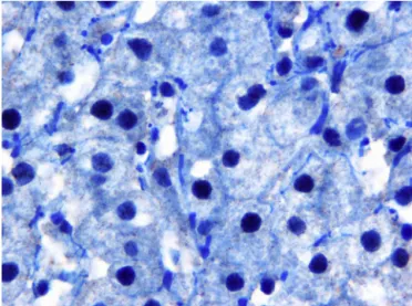
Fig.1. Estrogenreceptorexpressioninpatientswithnonalcoholicsteatohepatitis.
andbilirubin(p>0.05).Nevertheless,therewasasignificant
dif-ferencebetweentheER-␣positiveandnegativegroupsintermsof
gender:ER-␣positivitywassignificantlymoreprevalentinfemale
subjects(76.9%vs.50%,infemaleandmalesubjects,respectively,
p=0.04).FibrosisscorewassignificantlylowerinER-␣positive
sub-jectswhencomparedtotheER-␣negativesubjects(1.0±0.8vs.
1.4±0.9,respectively,p=0.047).
MeanER-␣expressionpercentage(stainingindex)was
signif-icantlydifferentacross theNASH,SS and NLT groups (6.3±9.9
vs.22.1±26.4vs.44.2±24.8,respectively,p<0.001forall
com-parisons)(Figs.1 and 2).Meanstaining indexwassignificantly
higherinwomenwhencomparedtomen(19.4±22.2vs.7.9±15.3,
respectively,p=0.003).Genderspecificanalysisrevealedthatthere
wasasignificantdifferenceintermsofER-␣expression
percent-agebetweenNASH,SS,andNLTgroupsinmen.Nevertheless,in
women,ER-␣expressionpercentagewasnotsignificantly
differ-entacrossNASH,SS,andNLTgroups(Table2).Inourstudygroup,
36patientswerenon-diabetic,19patientshadimpairedglucose
tolerance,and17patientswerediabetic.Therewasnostatistical
differenceintermsofmeanstainingindexaccordingtothe
dia-beticstatus(p>0.05).Meanstainingindexnegativelycorrelated
withserumALTlevel(r=−0.240;p=0.04),fastingplasmaglucose
(r=−0.261;p=0.027),andfibrosisscore(r=−0.312;p=0.011).No
statisticallysignificantrelationshipwasfoundbetweenthemean
Fig.2. Estrogenreceptorexpressioninpatientswithsimplesteatosis.
stainingindexandtheNASscore,steatosis,inflammation,or
bal-looning.
Discussion
NAFLDrepresentsaspectrumofhistopathologicalfindings
vary-ingfromsimplesteatosis(SS)tosteatosisplusnecroinflammation
(non-alcoholic steatohepatitis, NASH), with or without fibrosis.
Liverbiopsyshouldbeperformedtodiscriminatetheseentities.
SSisexpectedtohaveafavorableprognosis;nevertheless,NASH
mayproceedtocirrhosisin20–25%ofthepatientsinaperiodof10
years[11].
Findingsof previousstudieshaveconsistently demonstrated
thatNAFLDismorecommoninmenthaninwomen;andismore
likelytobeencounteredinpostmenopausalwomenthanitisin
premenopausalwomen,suggestingthatestrogensmayhavea
pro-tectiveeffectagainstthedevelopmentNAFLD[3].
Inananimalmodel,itwasdemonstratedthathepatic
steato-sisspontaneouslydevelopsinaromatase-deficientmice,whichare
notcapableofproducingestrogenandhaveimpaired
hepatocellu-larfattyacidbeta-oxidation. Replacementofestradiolalleviates
hepatic steatosis and improvesmitochondrial and peroxisomal
fattyacidbetaoxidationtothelevelsobservedinwild-typemice
[9].
Chowetal.demonstratedthatonlyER-␣isexpressedinthe
mouseliver.TheirfindingssuggestthatestrogensactviaER-␣to
regulatetriglyceridehomeostasisinthemouseliver.Aselective
ER-␣agonistalleviates hepaticsteatosisin themale aromatase
knockoutmice[2].
Ribasetal.reportedthatlossofER-␣bringsaboutdiminished
oxygenutilization,lipid deposition,inflammation,insulin
resis-tance,andimpairedglucosetolerance.Moreover,itwasshownthat
ER-␣playsacrucialroleagainstthedeleteriouseffectsofhigh-fat
dietinfemalemice[12].
ThesetwostudiesdemonstratedthatlossofER-␣intheliveris
associatedwithhepaticsteatosisandinflammation[2,12].
Estradiolisapotentendogenousantioxidantthatcounteracts
liverfibrosisinanimalmodels[14].Moreover,neutralizing
anti-bodiesagainstestradiol inmaleratsandovariectomyinfemale
ratsresultsinincreasedfibrogenesis[16].
Inourstudy,therewasasignificantdifferencewithregardto
presenceofER-␣expressionbetweenNASH,SS,andNLTgroups.
MeanhepaticER-␣expressionpercentage(stainingindex)was
sig-nificantlylowerinNASHwhencomparedtotheSSandNLTgroups
(6.3±9.9 vs. 22.1±26.4 vs. 44.2±24.8, respectively, p<0.001)
Stainingindexwassignificantlyhigherinwomenwhencompared
tomen(19.4±22.2vs.7.9±15.3,respectively,p=0.003).As
previ-ouslyreportedintheliterature,estrogenshaveaprotectiveeffect
againsthepatic steatosisandfibrosis.Moreover,NAFLDismore
prevalentinmen.Therefore,hepaticER-␣expressionpercentage
(stainingindex)mightbeexpectedtobelowerinmencomparedto
womenandintheNASHpatientscomparedtoSSandNLTgroups.
Indeed,thefindingsofourstudysupportthishypothesis.However,
gender-specificanalysisrevealedthattherewasasignificant
differ-encewithregardtoER-␣expressionpercentageacrosstheNASH,
SS,andNLTgroupsinmenbutnotinwomen.Thisfindingmay
beattributedtosmallsamplesizeandshouldbeverifiedinlarge
scalestudies.Theoretically,steatosisandfibrosisareexpectedto
occurlessintheER-positivegroupcomparedtotheER-negative
group,nevertheless,therewasnosignificantdifferencebetween
theER-positiveandER-negativegroupswithregardtoNASscore,
steatosis,andinflammation.However,therewasasignificant
dif-ferencebetweenERpositiveandnegativegroupintermsoffibrosis
432 G.Erkanetal./Pathology–ResearchandPractice209 (2013) 429–432 Table2
ER-␣expressionpercentageacrosstheNASH,SSandNLTgroupswithregardtogender.
Female(n:26) Male(n:46)
NASH(n:18) SS(n:5) NLT(n:3) p NASH(n:36) SS(n:7) NLT(n:3) p
ERexpressionpercentage(%) 11.9±13.6 37±34.9 35±22.9 0.063 3.5±5.7 11.4±12.2 53.3±27.5 0.004
Resultsexpressedasmean±SD.
Loombaetal.havereportedthatdiabetesisstronglycorrelated
withtheriskofNASHandfibrosis[6].Inourstudy,therewasno
significantdifferencebetweendiabetic,non-diabetic,andimpaired
glucosetolerancegroupsintermsoffibrosis,NASscore,steatosis,
inflammation,ER-␣presenceandpercentage.Nevertheless,ER-␣
expressionpercentage(stainingindex)wasnegativelycorrelated
withplasmaglucoselevel.Further,large-scalestudiesareneeded
toelucidatethisissue.
Kamada et al. studied the effects of estrogen deficiency in
ovariectomized mice which were fed on a high-fat and
high-cholesterol diet for 6 weeks, which resulted in enhanced liver
injurywithincreasedlivermacrophageinfiltrationandelevated
serumcholesterollevels.Hepatocytemonocytechemoattractant
protein-1 (MCP-1) expression, and hepatic inflammatory gene
expressionsweresignificantlyelevatedinovariectomizedmiceon
ahigh-fatdiet.Estrogentreatmentreducedserumcholesterol
lev-els,liverinjury, macrophageinfiltration,MCP-1 expression,and
inflammatorygeneexpressionsinovariectomizedmice.Thisstudy
demonstratedthatestrogendeficiencyacceleratedNASH
progres-sioninovariectomizedmicefedonahigh-fatdiet,andthatthis
effectwasreversedbyestrogentherapy[4].
ThefindingsofKamadaandRibas[4,12]suggestthatestrogen
deficiencyisassociatedwithhepaticinflammation.Nevertheless,
wedidnotobserveasignificantcorrelationbetweenER-␣
expres-sionpercentage(stainingindex)andthedegreeofinflammationin
liverbiopsyspecimens.Thismaybeduetotherelativelysmall
sam-plesize.Itmayalsobeassertedthattheanti-inflammatoryeffects
andtheprotectionfromsteatosisconferred byestrogenmaybe
mediatedbyotherpathwaysratherthanER-␣.Thisassertionalso
warrantsfurtherresearch.
Themainlimitationofourstudyistherelativelysmallsample
size.
Asa conclusion, in ourstudy, hepatic ER-␣expression
per-centage(stainingindex)isdecreasedinpatientswithNASH,and
this expression percentage is negatively correlated with serum
ALT, plasma glucose levels, and fibrosis score. On the other
hand, ER-␣ expression percentage is not correlated with NAS
score, steatosis, inflammation, and ballooning. Further studies
are required to clarify the significance of ER-␣ expression in
NASH.
Authors’contributions
G.Erkan,S.Ozenirler,G.Yilmaz,G.Akyoldesignedthestudy;
G.ErkanandC.KoncaDegertekinrecruitedthepatients,G.Yilmaz
andG.Akyolperformedthehistopathologicexaminations,andG.
Erkanwrotethemanuscript.
Funding
None.
Conflictofinterest
None.
References
[1]AmericanDiabetesAssociation,Diagnosisandclassificationofdiabetes melli-tus,DiabetesCare32(Suppl.1)(2009)62–67.
[2]J.D.Chow,M.E.Jones,K.Prelle,E.R.Simpson,W.C.Boon,Aselective estro-genreceptor␣agonistameliorateshepaticsteatosisinthemalearomatase knockoutmouse,J.Endocrinol.210(3)(2011)323–334.
[3]Y.Gutierrez-Grobe,G.Ponciano-Rodríguez,M.H.Ramos,M.Uribe,N. Méndez-Sánchez,Prevalenceofnonalcoholicfattyliverdiseaseinpremenopausal, posmenopausalandpolycysticovarysyndromewomen.Theroleofestrogens, Ann.Hepatol.9(4)(2010)402–409.
[4]Y.Kamada,S.Kiso,Y.Yoshida,N.Chatani,T.Kizu,M.Hamano,M.Tsubakio, T.Takemura,H.Ezaki,N.Hayashi,T.Takehara,Estrogendeficiencyworsens steatohepatitisinmicefedhigh-fatandhigh-cholesteroldiet,Am.J.Physiol. Gastrointest.LiverPhysiol.301(6)(2011)1031–1043.
[5]D.E.Kleiner,E.M.Brunt,M.VanNatta,C.Behling,M.J.Contos,O.W.Cummings, L.D.Ferrell,Y.C.Liu,M.S.Torbenson,A.Unalp-Arida,M.Yeh,A.J.McCullough, A.J.Sanyal,Nonalcoholicsteatohepatitisclinicalresearchnetwork,designand validationofahistologicalscoringsystemfornonalcoholicfattyliverdisease, Hepatology41(6)(2005)1313–1321.
[6]R.Loomba,M.Abraham,A.Unalp,L.Wilson,J.Lavine,E.Doo,N.M.Bass,NASH ClinicalResearchNetwork,Associationbetweendiabetes,familyhistoryof dia-betesandriskofnonalcoholicsteatohepatitisandfibrosis,Hepatology56(3) (2012)943–951.
[7]J.Ludwig,T.R.Viggiano,D.B.McGill,B.J.Oh,Nonalcoholicsteatohepatitis:Mayo clinicexperienceswithahithertounnameddisease,MayoClin.Proc.55(1980) 434–438.
[8]G. Marchesini,E. Bugianesi,G. Forlani,F. Cerrelli,M.Lenzi, R. Manini,S. Natale,E.Vanni,N.Villanova,N.Melchionda,M.Rizzetto,Nonalcoholicfatty liver,steatohepatitis,andthemetabolicsyndrome,Hepatology37(4)(2003) 917–923.
[9]Y.Nemoto,K.Toda,M.Ono,K.Fujikawa-Adachi,T.Saibara,S.Onishi,H.Enzan, T.Okada,Y.Shizuta,Alteredexpressionoffattyacid-metabolizingenzymesin aromatase-deficientmice,J.Clin.Invest.105(12)(2000)1819–1825.
[10]S.Nilsson,S.Mäkelä,E.Treuter,M.Tujague,J.Thomsen,G.Andersson,E. Enmark,K.Pettersson,M.Warner,J.A.Gustafsson,Mechanismsofestrogen action,Physiol.Rev.81(4)(2001)1535–1565.
[11]V.Ratziu,S.Bellentani,H.Cortez-Pinto,C.Day,G.Marchesini,Aposition state-mentonNAFLD/NASHbasedontheEASL2009specialconference,J.Hepatol. 53(2010)372–384.
[12]V.Ribas,M.T.Nguyen,D.C.Henstridge,A.K.Nguyen,S.W.Beaven,M.J.Watt, A.L.Hevener,Impairedoxidativemetabolismandinflammationareassociated withinsulinresistanceinERalpha-deficientmice,Am.J.Physiol.Endocrinol. Metab.298(2)(2010)304–319.
[13]T.Saphner,S.Triest-Robertson,H.Li,P.Holzman,Theassociationof nonalco-holicsteatohepatitisandtamoxifeninpatientswithbreastcancer,Cancer115 (14)(2009)3189–3195.
[14]I.Shimizu,N.Kohno,K.Tamaki,M.Shono,H.W.Huang,J.H.He,D.F.Yao,Female hepatology:favorableroleofestrogeninchronicliverdiseasewithhepatitisB virusinfection,WorldJ.Gastroenterol.13(32)(2007)4295–4305.
[15]S.R.Weston,W.Leyden,R.Murphy,N.M.Bass,P.B.Bell,M.M.Manos,N.A. Terrault, Racialandethnicdistributionofnonalcoholicfattyliverin per-sons with newly diagnosed chronic liverdisease, Hepatology41 (2005) 372–379.
[16]M.Yasuda,I.Shimizu,M.Shiba,S.Ito,Suppressiveeffectsofestradiolon dimethylnitrosamine-inducedfibrosisoftheliverinrats,Hepatology29(3) (1999)719–727.
[17]S.Yatsuji,E.Hashimoto,M.Tobari,K.Tokushige,K.Shiratori,Influenceofage andgenderinJapanesepatientswithnonalcoholicsteatohepatitis,Hepatol. Res.37(2007)1034–1043.