Original Paper
Urol Int 2013;91:182–186 DOI: 10.1159/000350512Time between First and Second Transurethral
Resection of Bladder Tumors in Patients with
High-Grade T1 Tumors: Is It a Risk Factor for
Residual Tumor Detection?
Evren Süer
a
Cihat Özcan
a
Sümer Baltacı
a
Ömer Gülpınar
a
Berk Burgu
a
Ahmet Haliloğlu
b
Yaşar Bedük
a
a Department of Urology, University of Ankara, and b Department of Urology, Ufuk University, Ankara , Turkey
± 9.1 vs. 39.3 ± 10.9 days, respectively, p = 0.001). Multivari-ate analysis demonstrMultivari-ated that time elapsed between first and second TURBT is the most important parameter for re-sidual tumor detection. Conclusion: Our study revealed that multiple tumors, tumors >3 cm in size, absence of detrusor muscle in the initial TURBT specimen, TURBT performed by trainees and finally, as a new finding, prolonged interval be-tween first and second TURBT are independent predictors for residual tumor detection in patients with high-grade T1
tumors. © 2013 S. Karger AG, Basel
Introduction
Transurethral resection of bladder tumors (TURBT) is the first stage in the diagnosis and treatment of bladder tumors. The major objectives of TURBT are to remove all visible tumor and provide the necessary information to accurately stage the patients. However, there are signifi-cant problems in the initial transurethral resection of non-muscle-invasive bladder cancer (NMIBC) concern-ing correct stagconcern-ing and complete resection of the tumor [1] . A high number of recurrences and the requirement of adjuvant therapy indicate incomplete tumor resection in most of the patients [2–4] . Moreover understaging due
Key Words
Bladder tumor · Transurethral resection · Second transurethral resection · Residual tumor
Abstract
Purpose: We evaluated the risk factors for residual tumor detection after transurethral resection of bladder tumors (TURBT) in patients with newly diagnosed high-grade T1 transitional cell carcinoma of the bladder. Patients and Methods: Overall 132 patients underwent TURBT for primary
bladder tumors and were diagnosed as high-grade T1 blad-der cancer. Patients with incomplete resections were exclud-ed from the study. Clinical and pathologic characteristics of the patients were compared and multivariate analysis was performed to determine independent prognostic factors. Results: Residual tumor was demonstrated in 57 (43.1%) of the patients. The residual tumor rate was significantly lower in patients with solitary tumors, tumors <3 cm in diameter, muscle presence in the initial TURBT pathologic sample and treated by an expert surgeon. In patients with solitary blad-der tumors, tumors at the dome and posterior wall of the bladder exhibited higher rates of residual tumor (p < 0.0001). The time elapsed between first and second TURBT was sig-nificantly shorter in patients without residual tumor com-pared to patients with residual tumor at second TURBT (32.6
Received: October 31, 2012
Accepted after revision: February 21, 2013 Published online: June 6, 2013
Internationalis
to inadequate resection may influence the progression rate in NMIBC.
Especially in high-grade tumors, good and complete TURBT is the essential part of initial management. Early recurrence is an unfavorable prognostic factor for these tumors and is mostly attributed to incomplete resection at the initial TURBT [5] . Although the quality of TURBT is accepted as the mainstay feature in the management of NMIBC, experienced surgeons reported considerable re-sidual tumor rates and understaging even after resection of all visible tumors [6, 7] . Furthermore there are increas-ing data which demonstrate the favorable effects of sec-ond TURBT on the recurrence-free survival of these pa-tients [8] . Due to these findings, a second transurethral resection is recommended when a high-grade or T1 tu-mor has been detected at initial transurethral resection [9] . There is no established timing for the second TURBT, however 2–6 weeks after the initial resection is mostly ac-cepted.
Many clinical and pathologic factors may alter the out-comes of second TURBT [10, 11] . In this study our aim was to investigate the risk factors and their impact on re-sidual tumor detection by second TURBT in high-grade T1 bladder cancer.
Patients and Methods
We reviewed our bladder tumor database and selected patients with bladder tumor who were diagnosed between 2005 and 2011. The resections were performed by 4 experienced urooncologists and trainees in years 3–5 (urology training is 5 years in our coun-try). The trainees performed these operations under the supervi-sion of senior urologists. White light cystoscopy and conventional resection equipments were utilized during TURBT. Fluorescence cystoscopy was not performed in the initial TURBT. All visible tu-mors, the tumor bed and the margins were resected separately. Results of these resections were reported in the pathology report. We implemented intravesical 40 mg mitomycin C within 24 h of resection except for patients with serious hematuria and suspicion of bladder perforation. We performed second TURBT in patients with incomplete initial resection, multiple and/or large tumors, high-grade or T1 tumors and tumor specimens without detrusor muscle samples. Tumors were classified according to the TNM staging system [12] and were graded according to the WHO grad-ing system [13] . The inclusion criteria were (1) primarily diag-nosed urothelial bladder cancer, (2) macroscopically complete ini-tial TURBT without any visible residual tumor, (3) T1 or high-grade tumors, and (4) no extravesical urothelial cancer.
The recorded clinicopathologic parameters in these patients were age, gender, medical history, tumor size, tumor grade, tumor stage, multifocality, tumor localization for solitary tumors, operat-ing surgeon, timoperat-ing of the second TURBT and presence of detru-sor muscle in the pathologic specimen.
The aforementioned parameters were compared between pa-tients with and without residual tumors detected by second TURBT after the initial resection. We investigated the impact of localization in solitary bladder tumors by dividing tumor localiza-tion according to the accessibility of the tumors. We excluded mul-tifocal tumors from this analysis as these tumors are generally lo-cated in different sites of the bladder. Solitary tumors at the dome or posterior wall and tumors at the lateral or inferior bladder wall were accepted as tumors difficult and easy to access with the resec-toscope, respectively. The interval between initial and second TURBT was recorded using days as the time unit. All statistical evaluations were performed using SPSS. Pearson χ 2 and
Mann-Whitney U tests were used for standard statistical procedures. To determine independent prognostic factors, multivariate survival analysis was performed by a Cox regression model with respect to potential influencing factors. Statistical significance in this study was defined as p < 0.05.
Results
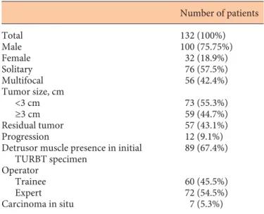
Overall 132 patients underwent TURBT for primary bladder cancer and were diagnosed as T1 or high-grade urothelial cancer. Mean age was 63.35 ± 10.51 years. Sec-ond TURBT was performed at a mean of 35.7 ± 8.03 years (20–69). Residual cancer and progression was demon-strated in 57 (43.1%) and 12 (9.09%) of all patients, re-spectively. Associated carcinoma in situ was reported in 7 patients at initial TURBT pathologic report. The clinical and pathologic characteristics of the study population are demonstrated in table 1 .
Table 1. Clinical and pathologic characteristics of patients who underwent initial TURBT for primary bladder tumors
Number of patients Total 132 (100%) Male Female 100 (75.75%) 32 (18.9%) Solitary Multifocal 76 (57.5%) 56 (42.4%) Tumor size, cm <3 cm ≥3 cm 73 (55.3%) 59 (44.7%) Residual tumor 57 (43.1%) Progression 12 (9.1%) Detrusor muscle presence in initial
TURBT specimen 89 (67.4%) Operator Trainee Expert 60 (45.5%) 72 (54.5%) Carcinoma in situ 7 (5.3%)
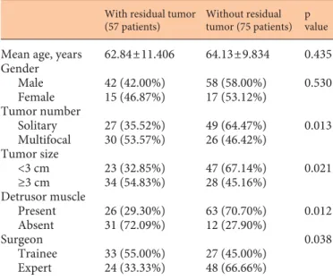
The comparison of patients with and without residual tumor after the initial TURBT is shown in table 2 . Resid-ual tumor rate did not display any significant difference according to age and gender (p > 0.05). Residual tumor rate was significantly lower in patients with solitary tu-mors, tumors <3 cm, muscle presence in the initial TURBT pathologic sample and treated by an expert surgeon (all p < 0.05) ( table 2 ). In accordance with the demonstrated
difference of residual tumor rates between TURBT per-formed by trainees and experts, detrusor muscle presence was reported to be significantly higher in TURBT per-formed by the experts (61.1 vs. 80.7%, p = 0.032). The time elapsed between first and second TURBT was significant-ly shorter in patients without residual tumor compared to patients with residual tumor at second TURBT (32.6 ± 9.1 vs. 39.3 ± 10.9 days, respectively, p = 0.001).
The impact of bladder cancer localization on the re-sidual tumor rate was evaluated in solitary bladder cancer patients. Of the 76 patients with solitary bladder cancer, residual tumor was found in 27 (35.5%). A total of 24 (31.57%) had tumors at the dome or posterior wall of the bladder (group I) and 52 (68.42%) had tumors at the lat-eral or inferior wall of the bladder (group II). The residu-al tumor rate was 58.3 and 25.0% for patients in group I and II, respectively. This difference was demonstrated to be significant (p < 0.0001).
Cox proportional model was formed according to re-sidual tumor rate ( table 3 ). According to this analysis, multiple tumors, tumor size >3 cm, prolonged interval between first and second TURBT, absence of detrusor muscle and trainees were independent predictors of re-sidual tumor detection. Upon these parameters timing for the second TURBT was the most important factor (odds ratio 8.1, 95% confidence interval 2.7–23.6, p < 0.0001).
Discussion
Complete resection is the irrefutable section for the treatment of NMIBC. To achieve high success rates fur-ther resections are performed in selected patients. The European Association of Urology guidelines recommend a second TURBT when a high-grade NMIBC or a T1 tu-mor has been detected at the initial transurethral resec-tion [1] . Herr [7] demonstrated a 76% residual tumor rate even in experienced hands and recommended routine re-peat TURBT. Divrik et al. [14] assessed second TURBT for primary bladder cancer and demonstrated a residual tumor rate of 33.3%. They showed a high correlation be-tween grade and residual tumor rate. In their prospective study, Grimm et al. [6] reported a residual tumor rate of 51% in grade 2–3 T1 bladder cancer patients. In our study, the residual tumor rate was 43.1%. As all of our patients had high-grade T1 cancers, this rate is acceptable.
Many clinical and pathologic factors may affect the outcomes of second TURBT. However, to our knowl-edge, no study evaluated the risk factors for residual
tu-Table 2. Comparison of patients with and without residual tumor after initial TURBT
With residual tumor (57 patients)
Without residual tumor (75 patients)
p value
Mean age, years 62.84±11.406 64.13±9.834 0.435 Gender Male Female 42 (42.00%) 15 (46.87%) 58 (58.00%) 17 (53.12%) 0.530 Tumor number Solitary Multifocal 27 (35.52%) 30 (53.57%) 49 (64.47%) 26 (46.42%) 0.013 Tumor size <3 cm ≥3 cm 23 (32.85%) 34 (54.83%) 47 (67.14%) 28 (45.16%) 0.021 Detrusor muscle Present Absent 26 (29.30%) 31 (72.09%) 63 (70.70%) 12 (27.90%) 0.012 Surgeon Trainee Expert 33 (55.00%) 24 (33.33%) 27 (45.00%) 48 (66.66%) 0.038
Table 3. Cox proportional model according to residual tumor de-tection HR 95% CI p value Age 0.943 0.896–0.993 0.56 Tumor size <3 cm ≥3 cm 3.737 1.280–10.878 0.016 Tumor number Solitary Multifocal 5.283 1.657–16.841 0.005 Detrusor muscle Present Absent 2.981 1.007–10.046 0.049 Surgeon Expert Trainee 3.128 1.317–10.931 0.014
Time elapsed between first and second TURBT
mors after initial TURBT in a specific group of patients, namely patients with high-grade T1 tumors. Our study revealed that multiple tumors, tumors >3 cm in size, ab-sence of detrusor muscle in the initial TURBT specimen, TURBT performed by trainees and finally prolonged in-terval between first and second TURBT are independent predictors for residual tumor detection in patients with high-grade T1 tumors.
Divrik et al. [8] classified further resections after initial TURBT according to their indication. They defined sec-ond TURBT as transurethral resection after complete re-section and pathologic specimen with detrusor muscle. It is not possible to make an accurate staging without detru-sor resection. In our study only 71.2% of the patients had detrusor muscle in their initial TURBT specimen. The rates of absence of detrusor muscle in previous series are also not negligible and were reported in a range between 15 and 50% [7, 15, 16] . In a recent study Mariappan et al. [11] demonstrated the presence of detrusor muscle in the pathologic sample of initial TURBT as a favorable predic-tive factor for recurrence after first cystoscopy. They specified detrusor presence as a surrogate marker for the quality of initial TURBT. Surgeon experience was also linked with obtaining detrusor muscle. In their study, de-trusor muscle presence was 56 and 72% for junior and senior surgeons, respectively. This outcome was in accor-dance with our results (61.1 and 80.7% for trainees and expert surgeons, respectively).
High-grade T1 patients stand at the border of muscle-invasive disease status, and second TURBT is strongly ad-vocated for better staging and long-term outcomes in these patients. Brausi et al. [10] in many cases attributed early recurrences to incomplete resection. Many sur-geons are aware of incomplete resection because of large, multiple tumors and tumors which are difficult to access by resectoscope. However residual tumors may be detect-ed even after the impression of an adequate resection. Tu-mor grade, tuTu-mor size and multifocality of tuTu-mors are the most recognized risk factors for residual tumors [8, 10, 17] . In our study tumor size >3 cm and multifocal tumors were also independent risk factors for residual tumor de-tection.
Learning curve and experience are other crucial fac-tors which affect the residual tumor rate. Initially Zurkirchen et al. [18] demonstrated the residual tumor rate as 37% for beginners and 26% for experts. Brausi et al. [10] emphasized the importance of surgeon experience and attributed the heterogeneity of various centers’ recur-rence rate following first cystoscopy to the quality of the surgery. Although all TURBT are performed under the
supervision of an experienced surgeon, the residual tu-mor rate was significantly higher in our patients who were operated by trainees. The stress of possible bladder perforation during the procedure and difficulty of deter-mining borders of the tumor for the beginner may con-tribute to these outcomes.
Tumor localization is another potential risk factor for residual tumor detection after initial TURBT. Tumors lo-cated at the upper hemisphere of the bladder are more difficult to approach and resect. In our study we evalu-ated the impact of tumor localization on patients with solitary tumors. We detected significantly higher rates of residual tumor in patients with tumor at the upper hemi-sphere of the bladder. The shortcomings of resectoscope manipulations and access difficulties seem to be the ma-jor reasons for this outcome.
Although most authors recommend resection at 2–6 weeks after initial TURBT, there is no consensus about the timing of a second TURBT [1] . To our knowledge, this is the first study which evaluates the effect of the elapsed time between first and second TURBT as a factor for tumor detection at the time of second TURBT. We found significantly shorter elapsed time in patients with-out residual tumor compared to patients with residual tumor at second TURBT. This finding may be a factor in explaining the different rates of residual tumor at second TURBT in different series. Prolonging the period be-tween two TURBT procedures may give a chance to small undetected or inadequately resected tumors to grow and become detectable. However, considering a mean differ-ence of 7 days, this explanation does not make sense from a biological point of view. Rather, residual inflammatory conditions may have impaired surgery or some other kind of bias may be responsible for this observation. Nev-ertheless, we think that this finding should be looked at in prospective randomized studies with a larger number of patients.
Conclusion
A significant number of patients has persistent disease after resection of high-grade T1 tumors. Our study re-vealed that multiple tumors, tumors >3 cm in size, ab-sence of detrusor muscle in the initial TURBT specimen, TURBT performed by trainees and finally, as a new find-ing, prolonged interval between first and second TURBT are independent predictors for residual tumor detection in patients with high-grade T1 tumors.
References
1 Babjuk M, Oosterlinck W, Sylvester R, Kaa-sinen E, Böhle A, Palou-Redorta J, Rouprêt M; European Association of Urology (EAU): EAU guidelines on non-muscle-invasive uro-thelial carcinoma of the bladder, the 2011 up-date. Eur Urol 2011; 59: 997–1008.
2 Kurth KH, Bouffioux C, Sylvester R, van der Meijden AP, Oosterlinck W, Brausi M: Treat-ment of superficial bladder tumors: achieve-ments and needs. The EORTC Genitourinary Group. Eur Urol 2000; 37(suppl 3):1–9. 3 Au JL, Badalament RA, Wientjes MG, Young
DC, Warner JA, Venema PL, Pollifrone DL, Harbrecht JD, Chin JL, Lerner SP, Miles BJ; International Mitomycin C Consortium: Methods to improve efficacy of intravesical mitomycin C: results of a randomized phase III trial. J Natl Cancer Inst 2001; 93: 597–604. 4 Järvinen R, Kaasinen E, Sankila A, Rintala E;
FinnBladder Group: Long-term efficacy of maintenance bacillus Calmette-Guérin ver-sus maintenance mitomycin C instillation therapy in frequently recurrent TaT1 tu-mours without carcinoma in situ: a subgroup analysis of the prospective, randomised Finn-Bladder I study with a 20-year follow-up. Eur Urol 2009; 56: 260–265.
5 Sylvester RJ, van der Meijden AP, Oosterlinck W, et al: Predicting recurrence and progres-sion in individual patients with stage Ta T1 bladder cancer using EORTC risk tables: a combined analysis of 2596 patients from sev-en EORTC trials. Eur Urol 2006; 49: 466–475.
6 Grimm MO, Steinhoff C, Simon X, et al: Ef-fect of routine repeat transurethral resection for superficial bladder cancer: a long-term ob-servational study. J Urol 2003; 170: 433–437. 7 Herr HW: The value of a second transurethral
resection in evaluating patients with bladder tumors. J Urol 1999; 162: 74–76.
8 Divrik RT, Sahin AF, Yildirim U, Altok M, Zorlu F: Impact of routine second transure-thral resection on the long-term outcome of patients with newly diagnosed pT1 urothelial carcinoma with respect to recurrence, pro-gression rate, and disease-specific survival: a prospective randomised clinical trial. Eur Urol 2010; 58: 185–190.
9 Kulkarni GS, Hakenberg OW, Gschwend JE, et al: An updated critical analysis of the treat-ment strategy for newly diagnosed high-grade T1 (previously T1G3) bladder cancer. Eur Urol 2010; 57: 60–70.
10 Brausi M, Collette L, Kurth K, van der Meij-den AP, Oosterlinck W, Witjes JA, Newling D, Bouffioux C, Sylvester RJ; EORTC Genito-Urinary Tract Cancer Collaborative Group: Variability in the recurrence rate at first fol-low-up cystoscopy after TUR in stage Ta T1 transitional cell carcinoma of the bladder: a combined analysis of seven EORTC studies. Eur Urol 2002; 41: 523–531.
11 Mariappan P, Zachou A, Grigor KM; Edin-burgh Uro-Oncology Group: Detrusor mus-cle in the first, apparently complete transure-thral resection of bladder tumour specimen is a surrogate marker of resection quality, pre-dicts risk of early recurrence, and is depen-dent on operator experience. Eur Urol 2010; 57: 843–849.
12 Sobin LH, Gospodariwicz M, Wittekind C (eds): UICC: TNM Classification of Malig-nant Tumors. Oxford, Wiley-Blackwell, 2009, pp 262–265.
13 Epstein JI, Amin MB, Reuter VR, et al: The World Health Organization/International Society of Urological Pathology consensus classification of urothelial (transitional cell) neoplasms of the urinary bladder. Am J Surg Pathol 1998; 22: 1435–1448.
14 Divrik T, Yildirim U, Eroglu A, Zorlu F, Ozen H: Is a second transurethral resection neces-sary for newly diagnosed pT1 bladder cancer? J Urol 2006; 175: 1258–1261.
15 Dalbagni G, Herr HW, Reuter V: Impact of a second transurethral resection on the staging of T1 bladder cancer. Urology 2002; 60: 822– 825.
16 Maruniak N, Takezawa K, Murphy W: Accu-rate pathological staging of urothelial neo-plasms requires better cystoscopic sampling. J Urol 2002; 167: 2404–2407.
17 Divrik RT, Yildirim U, Zorlu F, et al: The ef-fect of repeat transurethral resection on re-currence and progression rates in patients with T1 tumors of the bladder who received intravesical mitomycin: a prospective, ran-domized clinical trial. J Urol 2006; 175: 1641– 1644.
18 Zurkirchen MA, Sulser T, Gaspert A, Hauri D: Second transurethral resection of superfi-cial transitional cell carcinoma of the bladder: a must even for experienced urologists. Urol Int 2004; 72: 99–102.