ubcutaneous sarcoidosis is clinically characterized by multiple, firm, asymptomatic to slightly tender, mobile, round to oval, skin-colored nodules which are commonly located on the extremities (forearms and legs), mostly in a bilateral and asymmetric fashion.1It can also occur
A Case of Familial Subcutaneous Sarcoidosis with
an Asymptomatic Nodular Lesion on
the Upper Eyelid Accompanied by
Chronic Osteomyelitis
AABBSSTTRRAACCTT Skin lesions of sarcoidosis are heterogeneous, and can either have specific manifesta-tions; such as maculopapular eruptions, plaques, infiltrated scars and lupus pernio, or non-specific manifestations such as erythema nodosum depending on the presence or absence of typical sar-coidal granulomas. Herein, we report a 41-year-old woman presenting with discrete painless lo-calized swellings on her arms and calves which had developed two weeks earlier and rapidly increased in number. The lesions appeared sequentially at an interval of 3-5 days with a marked increase in size. An erythematosus asymptomatic nodular lesion was also noted on the medial can-tus of the patient’s right upper eyelid. The histopathological examination of the nodular lesions on the patient’s left arm and right upper eyelid revealed multiple non-caseating granulomas with multi-nucleated giant cells, which confirmed the diagnosis of granulomatous sarcoidosis. This case was unusual due to the acute and rapid progression of the lesions, and the presence of an unusual nodu-lar lesion on the upper eyelid. We believe that the chronic inflammatory process due to the past os-teomyelitis may have triggered the formation of the granulomas. In the differential diagnosis of the asympthomatic nodular lesions on the eyelid, subcutaneous sarcoidosis should also be considered even when there are no systemic symptoms.
KKeeyy WWoorrddss:: Eyelids; osteomyelitis; sarcoidosis; subcutaneous tissue Ö
ÖZZEETT Sarkoidozun deri lezyonları heterojen olup, tipik sarkoidal granülomların varlığına göre ma-külopapüler erupsiyonlar, plaklar, infiltre skarlar ve lupus pernio gibi spesifik bulguları veya eritema nodozum gibi spesifik bulguları olabilmektedir. Burada, iki hafta önce oluşmuş ve hızlıca sayıca artmış kollarda ve baldırlarda dağınık ağrısız lokalize şişliklerle başvuran 41 yaşında kadın hasta sunulmaktadır. Boyutlarında anlamlı artış olan lezyonların ve 3-5 günlük aralıklarla ardışık olarak geliştiği öğrenildi. Sağ üst göz kapağının medial kantusunda eritemli, asemptomatik nodüler lezyonu saptandı. Hastanın sol kol ve sağ üst göz kapağındaki nodüler lezyonların histopatolojik inceleme-sinde granülomatöz sarkoidoz tanısını doğrulayan multi-nükleer dev hücrelere eşlik eden çok say-ıda kazeifiye olmayan granülom yapıları görüldü. Bu olgu lezyonların akut ve hızlı progresyonu ve üst göz kapağındaki nadir görülen nodüler lezyonun varlığı dolayısıyla olağandışı idi. Geçirilmiş osteomiyelite bağlı kronik inflammatuar sürecin granülomların gelişimini tetiklemiş olabileceğini düşünmekteyiz. Sistemik hiçbir semptom olmasa da göz kapağındaki asemptomatik nodüler lez-yonların ayırıcı tanısında subkutan sarkoidoz ele alınmalıdır.
AAnnaahhttaarr KKeelliimmeelleerr:: Göz kapakları; osteomiyelit; sarkoidoz; subkutan doku
TTuurrkkiiyyee KKlliinniikklleerrii JJ DDeerrmmaattooll 22001166;;2266((11))::5544--88
Pelin ÜSTÜNER,a Ali BALEVİ,a Mustafa ÖZDEMİR,a Ülkü Tuğba PARLAKKILIÇ,b İlknur TÜRKMEN,c Oktay OLMUŞÇELİKd Clinics of
aDermatology and Venereology, bRadiology,
dInternal Medicine,
Medipol Mega University Hospital,
cDepartment of Pathology,
İstanbul Medipol University Faculty of Medicine, İstanbul Ge liş Ta ri hi/Re ce i ved: 23.05.2015 Ka bul Ta ri hi/Ac cep ted: 02.12.2015 Ya zış ma Ad re si/Cor res pon den ce: Pelin ÜSTÜNER
Medipol Mega University Hospital, Clinic of Dermatology, İstanbul, TÜRKİYE/TURKEY
doi: 10.5336/dermato.2015-46271
on other sites, such as the trunk, face, buttocks, head and neck. The most frequent specific (gran-ulomatous) skin lesions of sarcoidosis are; macu-lopapules, subcutaneous nodules, scar sarcoidosis, plaques and lupus pernio.1,2Maculopapular lesions and subcutaneous nodules are more often associ-ated with the remission of the systemic disease after two years, while plaques and mainly lupus pernio are hallmarks of the chronic form of the
disease.2 A strong association between subcuta-neous sarcoidosis and mild systemic involvement is already known.1
CASE REPORT
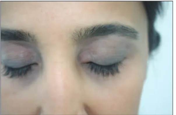
A 41-year-old woman presented with discrete painless localized swellings on her arms and calves that had appeared two weeks earlier and rapidly in-creased in number. Her medical history and sys-temic medication were unremarkable except for the chronic osteomyelitis that had occurred one year earlier. She received systemic antibiotics for approximately a year for the treatment-resistant os-teomyelitis on the fifth toe of her left foot. Then, she was treated with ozone therapy for ten sessions in an algology clinic, which resulted in an almost complete remission of the osteomyelitis. The der-matological examination revealed two regular con-toured, mobile, painless subcutaneous tumoral lesions of 2x2 cm in size on the extensor surface of her right and left arms (Figure 1a, b) and two subcutaneous nodules measuring 1x1 cm on the posterior surface of her right patella. An erythe-matosus, asymptomatic nodular lesion of 0,5 cm was also noted on the medial cantus of her right upper eyelid (Figure 2). She had no other symp-toms and was systemically well. The lesions ap-peared sequentially at an interval of 3-5 days with a marked increase in size in a few days. The pres-ence of systemic sarcoidosis with cutaneous and pulmonary involvement in her monozygotic twin was also noteworthy.
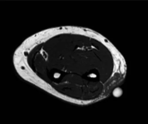
The axial and coronal T1W images of the fore-arm showed hypo-intense areas of subcutaneous fat tissue (Figure 3a, b). On palpation the lesions as appeared to be nodule-like masses, however the Magnetic resonance images showed ill-defined subcutaneous swellings. The axial and sagittal im-ages of the knee showed smaller but similar con-trast-enhanced ill-defined subcutaneous lesions as defined on the forearm. The laboratory work-up consisting of a hemogram and biochemistry were normal except for a mild chronic disease anemia. The tuberculin test was negative. Serum calcium, angiotensin-converting enzyme levels and urine
FIGURE 1a: Two regular contoured, mobile, painless subcutaneous tumoral
lesions of 2x2 cm’s size on the extensor surface of the patient’s right arm. (See color figure at http://www.turkiyeklinikleri.com/journal/dermatoloji-dergisi/1300-0330/)
FIGURE 1b: On the close examination of the subcutaneous tumoral lesion the
level difference is more evident.
(See color figure at http://www.turkiyeklinikleri.com/journal/dermatoloji-dergisi/1300-0330/)
FIGURE 2: An erythematosus asymptomatic nodular lesion of 0.5 cm on the
medial cantus of the patient’s right upper eyelid.
calcium were all elevated. Respiratory symptoms were not evident. The axial CT scan of the chest showed bilateral hilar lymph nodes. The lung parenchyma was normal. Informed consent was taken and finally, one of the nodular lesions on her arm was completely excised. The macroscopic ex-amination of the tumoral lesion showed tawny-col-ored soft tissue material with elastic viscosity, rich in lipids. Histological examination of the nodules on the left arm and right upper eyelid showed mul-tiple non-caseating granulomas accompanying with multinucleated giant cells that confirms the
diagnosis of granulomatous sarcoidosis (Figure 4). The Ziehl-Neelsen stain was negative for acid-fast bacilli. A bronchoscopic lung biopsy also confirmed the diagnosis of granulomatous sarcoidosis. The scintigraphic examination of the whole body showed no involvement of the vertebral spine or pelvis.The patient was referred to the ophthalmol-ogy department and the histopathological exami-nation of the incisional biopsy material confirmed the presence of granulomatous sarcoidosis. Uveitis was not detected.
The diagnosis of systemic sarcoidosis was made on the basis of the conventional criteria: a compat-ible clinical and radiological picture, a histological demonstration of non-caseating granulomas in-volving one or more tissues and exclusion of other granulomatous diseases. One month later, the pa-tient also presented with constitutional symptoms such as dyspnea and fatigue. She was treated with methylprednisolone (40 mg/day) and hydroxy-chloroquine (400 mg/day). A satisfactory response was achieved at three weeks of starting the treat-ment, when the systemic component of the disease disappeared. The subcutaneous nodules were grad-ually reduced in number and size. The patient had a complete remission after six months. No recur-rence was seen after the treatment at one year fol-low-up.
FIGURE 3a: The axial and coronal T1W images of the forearm showing the
hypo-intense areas of the subcutaneous fat tissue.
FIGURE 3b: The axial and coronal T1W images of the forearm showing the
hypo-intense areas of the subcutaneous fat tissue.
FIGURE 4: Non-caseating epithelioid granulomas in the dermis.
Micro-scopic examination revealed well-defined non-caseating granulomas including epitheloid histiocytes in the dermis with a considerable part of multinucleated giant cells. Lymphocytes and fibrosis were seen around the granulomas (HE, x200).
DISCUSSION
Sarcoidosis is a granulomatous inflammatory dis-ease that is induced by unknown antigen(s) in a ge-netically susceptible host.3 It is heterogeneous, showing geographic and racial variation in clinical presentation. The proposed mechanism is that a susceptible genotype is exposed to one or more po-tential antigens. Occupational and environmental factors, including microbial elements, may then affect the development of this disease. It was previously reported in the literature that a my-cobacterial infection triggered a sarcoid reaction in the infected subject and two other non-infected fam-ily members.3The possibility of the familial spread-ing of sarcoidosis may also point to Mycobacterium
tuberculosisas a potential etiological factor;
how-ever, it does not occur in all familial cases.
Subcutaneous sarcoidosis is the less frequent form of the specific cutaneous lesions of sarcoidosis with only few reports.1A strong association be-tween subcutaneous sarcoidosis and a systemic dis-ease has been acknowledged, mainly with bilateral hilar adenopathy. In our case, the subcutaneous nodules preceded the constitutional symptoms, with an interval of one month, similar to other re-cent reports suggesting that subcutaneous lesions usually appear at the beginning of the disease and lead to systemic involvement.
It is well-established that when ozone is in-fused into human blood, it produces reactive oxy-gen speciesor free radicals, the over-abundance of which is known to cause oxidative stressand cell damage, and is implicated in the progression of some degenerative diseases;such as osteoarthritis, osteoporosis and atherosclerosis.4Another topic of
discussion is that the development of the non-caseating granulomas in this case might also be as-sociated with the ozone therapy.
In this case, the rapid development of the lesions and the presence of the intractable osteomyelitis were remarkable. We believe that the underlying os-teomyelitis in this case might have also caused a predilection of the granulomas via producing a chronic inflammatory process. A sustained inflam-matory response followed, which ultimately resulted in the pathognomonic formation of granulomas. To our knowledge, this is so far the seventh case of sar-coidosis accompanied by a history of osteomyelitis.5,6 However, the association of cutaneous sarcoidosis with osteomyelitis is not clear since the location of the osteomyelitis was far from the sarcoidosis lesions. This case was also noteworthy due to the presence of the unusual involvement of the eyelid. To our knowledge, the eyelid involvement has not been re-ported except for a single case of scar sarcoidosis that developed as a result of an old scar from the previous removal of a basal cell carcinoma of the eyelid.7
The differential diagnosis of this case includes; subcutaneous granuloma annulare, panniculitis, tu-berculosis, atypical mycobacterial infections, deep fungal infections, multiple enchondromatosis, re-action to foreign bodies such as beryllium and zir-conium, rheumatoid nodules, lymphocytoma cutis, leiomyoma, lipomatosis and cutaneous metasta-sis.1,8,9This large variety of clinical conditions show that nodular lesions can easily be misdiagnosed as multiple lipomatosis, which is more frequently seen. Subcutaneous sarcoidosis should also be con-sidered in similar cases presenting with an unusual, asymptomatic isolated nodular lesion on the eye-lids.
1. Dalle Vedove C, Colato C, Girolomoni G. Sub-cutaneous sarcoidosis: report of two cases and review of the literature. Clin Rheumatol 2011;30(8):1123-8.
2. Mañá J, Marcoval J. Skin manifestations of sarcoidosis. Presse Med 2012;41(6 Pt 2):355-74.
3. Piotrowski WJ, Górski P, Duda-Szymańska J, Kwiatkowska S. Mycobacterium tuberculosis as a sarcoid factor? A case report of family sarcoidosis. Am J Case Rep 2014;16(15):216-20.
4. Bocci V, Valacchi G, Corradeschi F, Aldinucci C, Silvestri S, Paccagnini E, et al. Studies on
the biological effects of ozone: 7. Generation of reactive oxygen species (ROS) after expo-sure of human blood to ozone. J Biol Regul Homeost Agent 1998;12(3):67-75. 5. Jyonouchi H, Lien KW, Aguila H, Spinnato
GG, Sabharwal S, Pletcher BA. SAPHO os-teomyelitis and sarcoid dermatitis in a patient with DiGeorge syndrome. Eur J Pediatr 2006; 165(6):370-3.
6. Wildstein MS, Martin SM Jr, Glaser JA. Cryp-tococcal osteomyelitis in a 20-year-old male with sarcoidosis. Spine J 2005;5(4):467-70. 7. Tawfik HA, Assem M, Elkafrawy MH, Talib
NM. Scar sarcoidosis developing 16 years
after complete excision of an eyelid Basal car-cinoma. Orbit 2008;27(6):438-40.
8. Chokoeva AA, Tchernev G, Tana M, Tana C. Exclusion criteria for sarcoidosis: A novel ap-proach for an ancient disease? Eur J Intern Med 2014;25(10):e120.
9. Hunninghake GW, Costabel U, Ando M, Baughman R, Cordier JF, Du Bois R, et al: ATS/ERS/WASOG statement on sarcoidosis. American Thoracic Society/European Respi-ratory Society/World Association of Sarcoido-sis and other Granulomatous Disorders. Sarcoidosis Vasc Diffuse Lung Dis 1999;16: 149-73.