237 © 2017 Indian Journal of Radiology and Imaging | Published by Wolters Kluwer - Medknow
Differentiation of secondary
involvement of the breast by
lymphoreticular malignancy from
fibroadenoma using ultrasound
elastography: A report of two cases
Eren Abdulkadir, Ertan Gulhan, Sila Ulus1Department of Radiology, School of Medicine, Medipol University, 1Department of Radiology, School of Medicine, Acibadem University, Istanbul, Turkey
Correspondence: Dr. Sila Ulus, Kerem Aydinlar Kampusu, Kayisdagi Cad. No: 32, 34752 Atasehir, Istanbul, Turkey. E‑mail: [email protected]
Abstract
Extramedullary lymphoma infiltration of the breast by lymphoblastic lymphoma is very rare and most cases are of B‑cell lineage; T‑cell neoplasms represent less than 10% of all breast lymphomas. Here, we report one patient with lymphoblastic lymphoma and one patient with leukemia, who have similar lesions in breasts with different ultrasound elastography findings. Ultrasound‑guided tru‑cut biopsies were performed and the first lesion was confirmed as lymphoma infiltration and the second as fibroadenoma. In cases of breast mass presence in patients with a history of hematologic malignancies such as lymphoma or leukemia, breast infiltration should be kept in mind. Elastography findings can assist in the differentiation of these lesions and further investigations or biopsies can be avoided.
Key words: Breast; elastography; fibroadenoma; leukemia; lymphoma infiltration; tru‑cut biopsy
Introduction
Lymphoreticular malignancy infiltration of the breast is rare and may occur as an isolated tumor or as extramedullary manifestation in systemic disease.[1] Patients with lymphoblastic or leukemic infiltration of the breast usually present with a palpable lesion that is generally well circumscribed and rapidly enlarging. The lesion may be bilateral and/or multifocal simulating a fibroadenoma.[2] Despite the clinical and imaging similarities, the treatment
options differ. Thus, it is crucial to differentiate these lesions from other breast lesions accurately and pathology is the gold standard for this differentiation. Ultrasound (US) elastography findings can also assist in the differentiation of these lesions.It is an inexpensive, non‑invasive, and comfortable technique, which is used for the characterization of the breast masses and to differentiate benign vs. malignant lesions with reasonable confidence that have already been detected in B‑mode. We report the elastographic findings
Cite this article as: Abdulkadir E, Gulhan E, Ulus S. Differentiation of
secondary involvement of the breast by lymphoreticular malignancy from fibroadenoma using ultrasound elastography: A report of two cases. Indian J Radiol Imaging 2017;27:237‑40.
This is an open access article distributed under the terms of the Creative Commons Attribution‑NonCommercial‑ShareAlike 3.0 License, which allows others to remix, tweak, and build upon the work non‑commercially, as long as the author is credited and the new creations are licensed under the identical terms.
For reprints contact: [email protected]
Access this article online Quick Response Code:
Website:
www.ijri.org
DOI:
10.4103/ijri.IJRI_238_16
B
reast
Abdulkadir, et al.: Elastography in lymphoreticular breast malignancy
238 Indian Journal of Radiology and Imaging / Volume 27 / Issue 2 / April - June 2017
of breast involvement in a patient with lymphoblastic lymphoma and the elastographic findings of fibroadenoma in a patient with leukemia emphasizing the discrepancy.
Case History
Case 1
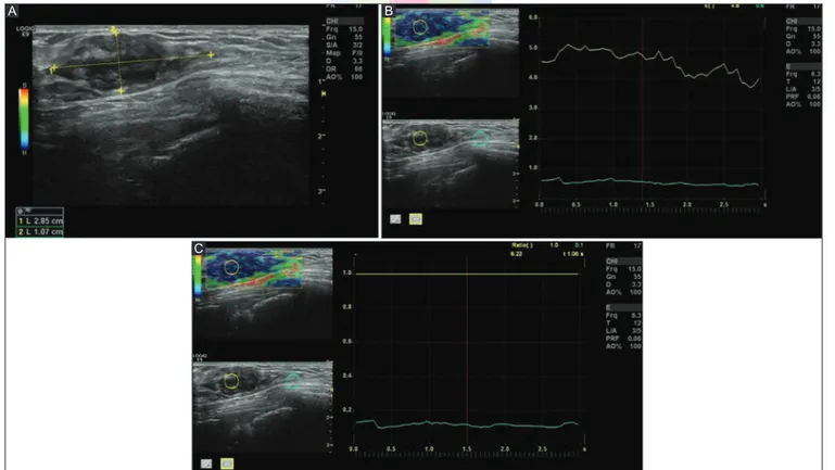
A 39‑year‑old female with a history of T‑ cell lymphoblastic lymphoma presented with multiple and bilateral painless breast lumps. B‑mode US (Logiq E9 GE Healthcare, Milwaukee, WI, USA) revealed multifocal ovoid shaped, hypoechoic solid masses with microlobulated borders in different sizes in both breasts [Figure 1A]. The biggest lesion was 29 × 11 mm in size. US elastography (Logiq E9 GE Healthcare, Milwaukee, WI, USA) was performed. On US elastography images, when we set the color‑coded scale ranging from red (softest components) to blue (hardest components), the biggest lesion was almost entirely blue with a green surrounding area. Elastographic strain index of the biggest lesion was 4.8 and elasticity ratio was 6.22 [Figures 1B and C]. According to the Tsukuba elasticity scoring system, she had point 4. The differential diagnosis included lymphoma infiltration, primary malignancies of the breast, and multiple fibroadenomatous lesions. Color‑coded elastography findings and high strain ratio (SR) values were in concordance with a malignant lesion rather than a benign lesion. Tru‑cut biopsy was performed for the biggest mass.
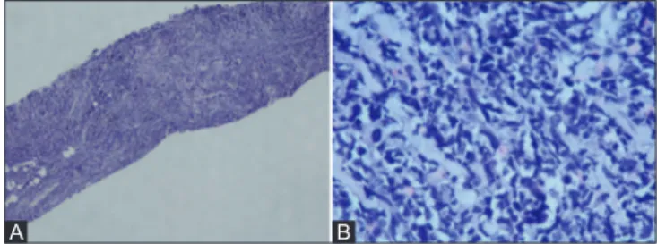
Histopathological assessment confirmed the diagnosis of T‑cell lymphoma infiltration [Figures 2A and B]. CD3, CD99, CD34, CD33, CD68, tdt, CD4, CD8, CD7, CD5, CD117, and CD68 were positive. Ki 67 index was % 80. PET‑CT showed no involvement in any other localization of the body. The patient went under chemotherapy. Currently, the patient is in remission and follow‑up.
Case 2
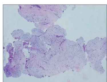
A 42‑year‑old female with a history of acute myeloblastic leukemia (AML) presented with multiple and bilateral painless breast lumps. B‑mode US examination was performed and multiple ovoid‑shaped hypoechoic solid masses with macrolobulated borders were seen [Figure 3A]. The biggest lesion was 15 × 7 mm in size. At US elastography examination, elastographic strain index and elasticity ratio were 2.3 and 0.71, respectively [Figures 3B and C]. Her Tsukuba elasticity score was 2. These values were concordant with benign lesions.[3] Tru‑cut biopsy of the biggest lesion confirmed the diagnosis of fibroadenoma [Figure 4]. She had no change in the size, appearance or the number of lesions in the follow‑up US examination performed 6 months later and is still in follow‑up.
Discussion
Involvement of breast by hematologic malignancies is a very rare condition.[4] In most described cases of AML involving
Figure 1 (A-C): B‑mode US (A) shows an ovoid shaped, hypoechoic solid mass with microlobulated borders. US elastography images (B and C) demonstrate an almost completely blue mass with green surrounding tissue. The elastographic strain index of the biggest lesion is 4.8 and elasticity ratio is 6.22
B
C A
Abdulkadir, et al.: Elastography in lymphoreticular breast malignancy
239 Indian Journal of Radiology and Imaging / Volume 27 / Issue 2 / April - June 2017
breasts, the disease appeared as bilateral multiple nodules.[5] Differential diagnosis of palpable breast lesions of a patient with hematologic malignancies includes involvement of the breast, multiple fibroadenomas, and primary breast carcinoma. Treatment of leukemic or lymphoblastic breast involvement is similar to other localizations of infiltration and chemotherapy and/or radiotherapy are the main treatment options.[6] On US, most masses are homogenously hypoechoic with microlobulated or indistinct margins and these are not specific.[7] Histopathologic assessment is the gold standard, but it is an invasive and expensive procedure. US elastography is an inexpensive, non‑invasive and comfortable technique. It is used for the characterization of the breast masses and to differentiate benign vs. malignant
lesions with reasonable confidence that have already been detected in B‑mode. In free‑hand elastography techniques, relative strain values can be obtained in comparison with surrounding tissues, with a color map or elasticity ratios via an ultrasound probe that calculates the strain produced by an externally applied force. Color maps according to the tissue stiffness are produced. Harder lesions appear blue, whereas softer lesions appear green on these maps. Malignant lesions are usually harder and blue, while benign lesions are usually softer and green. The data regarding tissue displacement prior and subsequent to compression is measured and combined with the B‑mode image.[8] The result of these two measurements is the so‑called “strain index” or “strain ratio” and it represents a semi‑quantitative assessment obtained by comparing the strain levels of normal areas of the breast with the strain level of the lesion on the elasticity maps. A higher strain index indicates less strain in the measured lesions. Many studies have shown that the use of elastography parameters in adjunct to ultrasound parameters can improve BI‑RADS score.[9] Also; Tsukuba elasticity scoring system has been established by Itoh et al. to categorize focal breast lesions in the elastography maps;[3] score 1, strain appears in the entire hypoechoic lesion; score 2, strain is not seen in part of the hypoechoic lesion; score 3, strain appears only in the peripheral areas of the hypoechoic lesion; score 4, no strain appears in the
Figure 3 (A-C): B‑mode USG (A) reveals an ovoid shaped, hypoechoic solid mass with macrolobulated regular borders. US elastography images (B and C) demonstrate a mostly green mass with green surrounding tissue and the elastographic strain index and elasticity ratio of the biggest lesion as 2.3 and 0.71, respectively
B
C A
Figure 2 (A and B): Histopathology slide revealing leukemic diffuse infiltration in breast tru‑cut biopsy (HE ×40) (A). Blasts have hyperchromatic nucleus with scant cytoplasm (HE ×400) (B)
B A
Abdulkadir, et al.: Elastography in lymphoreticular breast malignancy
240 Indian Journal of Radiology and Imaging / Volume 27 / Issue 2 / April - June 2017
entire hypoechoic lesion; score 5, no distortion appears either in the hypoechoic lesion or the surrounding areas. An elasticity score of 4 is usually an indicator of tumors that are circumscribed and homogeneously harder than the adjacent normal breast tissue. A score of 2 indicate lesions that are soft yet somewhat harder than normal breast tissue. This is often characteristic of lesions such as fibroadenoma. These are consistent with our findings. In our cases, the two patients had history of hematologic malignancies. B‑mode US and US elastography were performed for both patients, and while B‑mode US findings were quite similar, US elastography findings were disparate. US elastography findings of malign and benign breast lesions were defined before. A threshold SR value of 3.8 has been shown to differentiate benign breast lesions from malignant lesions with a sensitivity of 93.5% and specificity of 75.9%.[8] High SR was associated with malignancy.[10] In our cases, the lesion confirmed by biopsy as lymphoma involvement of the breast was stiffer than the other lesion, and Tsukuba scores of the first and second cases were 4 and 2, respectively. At an earlier study, elastographic findings of breast involvement in a patient with lymphoblastic leukemia were reported.[10] In that report, the lesion had a mean SR of ranging from 6.85 to 11.04 when compared with the normal appearing parenchyma, which is consistent with our findings.
In cases of breast mass presence in patients with a history of hematologic malignancies such as lymphoma or leukemia, breast infiltration should be kept in mind. Histopathologic evaluation is the gold standard in the differentiation of these lesions and we believe that US elastography findings can be used to assist in this differentiation.
Financial support and sponsorship
Nil.
Conflicts of interest
There are no conflicts of interest.
References
1. Karbasian‑Esfahani M, Wiernik PH, Yeddu M, Abebe L. Leukemic infiltration of the breast in acute lymphocytic leukemia (ALL). Hematology 2008;13:101‑6.
2. Mandal S, Jain S, Khurana N. Breast lump as an initial manifestation in acute lymphoblastic leukemia: An unusual presentation. A case report. Hematology 2007;12:45‑7.
3. Itoh A, Ueno E, Tohno E, Kamma H, Takahashi H, Shiina T,
et al. Breast disease: Clinical application of US elastography for
diagnosis. Radiology 2006;239:341‑50.
4. Memis A, Killi R, Orguc S, Ustun EE. Bilateral breast involvement in acute lymphoblastic leukemia: Color Doppler sonography findings. AJR Am J Roentgenol 1995;165:1011.
5. Asai S, Miyachi H, Kubota M, Koyanagi N, Ogawa Y, Hotta T, et al. In: Ueno E, Shiina T, Kubota M, Sawai K, editors. Ultrasonographic appearance and clinical implication of bilateral breast infiltration with leukemia cells. Research and development in breast ultrasound. Tokyo: Springer; 2005. p. 185‑9.
6. Cunningham I. A clinical review of breast involvement in acute leukemia. Leuk Lymphoma 2006;47:2517‑26.
7. Surov A, Wienke A, Abbas J. Breast leukemia: An update. Acta Radiol 2012;53:261‑6.
8. Ebner L, Bonel HM, Huber A, Ross S, Christe A. Diagnostic Performance and Additional Value of Elastosonography in Focal Breast Lesions: Statistical Correlation between Size‑Dependant Strain Index Measurements, Multimodality‑BI‑RADS Score, and Histopathology in a Clinical Routine Setting. ISRN Radiol 2014;2014:396368.
9. Sadigh G, Carlos RC, Neal CH, Dwamena BA. Accuracy of quantitative ultrasound elastography for differentiation of malignant and benign breast abnormalities: A meta‑analysis Breast Cancer Res Treat 2012;134:923‑31.
10. Pourbagher A. Breast Involvement in Acute Lymphoblastic Leukemia. Cukurova Med J 2015;40:64‑70.
Figure 4: Histopathology slide shows fibroadenoma with mild mixoid stroma (HE ×40)