ArchBronconeumol.2017;53(1):29
w w w . a r c h b r o n c o n e u m o l . o r g
Clinical
image
Pulmonary
Artery
Aneurysm
Ruptured
Into
Bronchus
in
a
Patient
With
Behc¸
et’s
Disease
夽
Ruptura
intrabronquial
de
un
aneurisma
de
arteria
pulmonar
en
un
paciente
con
enfermedad
de
behc¸
et
Ekrem
Cengiz
Seyhan,
a,∗Mehmet
Zeki
Gunluoglu,
bCengiz
Erol
caMedipolUniversity,MedicalFaculty,ChestDiseases,Estambul,Turkey bMedipolUniversity,MedicalFaculty,ThoracicSurgery,Estambul,Turkey cMedipolUniversity,MedicalFaculty,DepartmentofRadiology,Estambul,Turkey
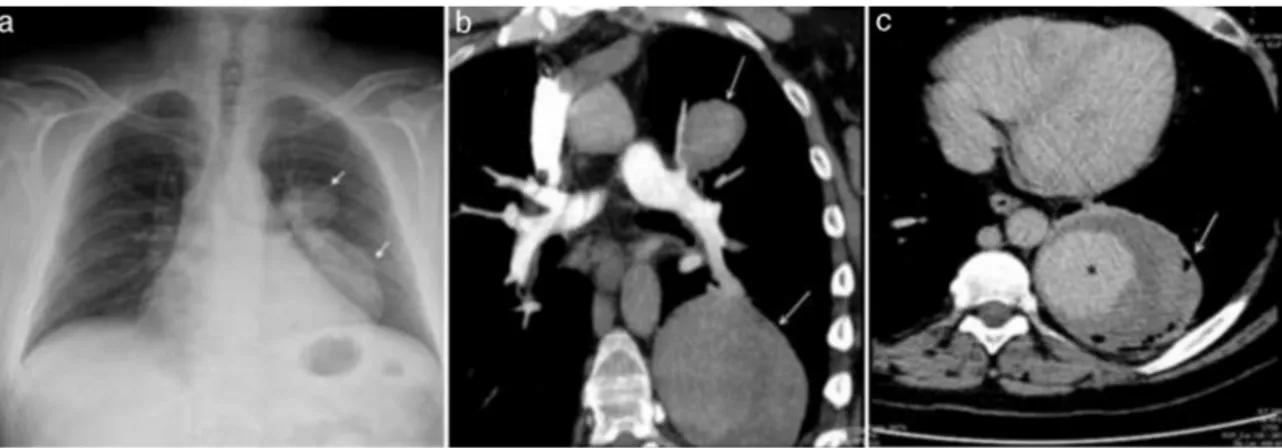
A54-year-oldmandiagnosedwithBehc¸et’sDisease(BD)5years previouslyatanotherhospitalwasadmitted toourcenter with massivehemoptysis. ThechestX-rayshowedbilobular,smooth edgedopacity localizedin theleft hilarand paracardiacregion that not conceal heart contours (Fig. 1a, arrow). Computerized tomographypulmonaryangiographyshowed2lesionsoriginating fromtheupperandlowerbranchesoftheleftpulmonaryartery, theircentralzones filled withcontrastmaterial, atearly phase ofimaging(Fig.1b,arrow).Contrastfillingwasenhancedatlate phaseoftheimaging(Fig.1c,asterisk).Theperipheryofthelower lesionwasless opaque, withsmallair bubbles(Fig.1c,arrow). Thesignssuggestedthrombosedpseudoaneurysmsrupturedinto thebronchus.Thepatientwasdiagnosedwitharuptureof pul-monaryarteryaneurysm(PAA)andhighdoseglucocorticoidand cyclophosphamidepulsetherapywasstarted.
Fig.1.ThechestX-rayshowedopacitylocalizedinthelefthilarandparacardiacregion(a,arrow).Computerizedtomographypulmonaryangiography(CTPA)showed2 lesions,originatingfromupperandlowerbranchesoftheleftpulmonaryartery(b,arrow).Contrastfillingwasenhancedatlatephaseofimaging(c,asterisk),withsmallair bubblesattheperipheryofthelowerlesion(c,arrow).
夽 Pleasecitethisarticleas:SeyhanEC,GunluogluMZ,ErolC.Ruptura intra-bronquialdeunaneurismadearteriapulmonarenunpacienteconenfermedad debehc¸et.ArchBronconeumol.2017;53:29.
∗ Correspondingauthor.
E-mailaddresses:drekremcs@gmail.com,drekremcs@yahoo.com(E.C.Seyhan).
BDisamulti-systeminflammatorydisorder,classifiedas vas-culitis.PAArepresentthemajorcomplicationofpulmonaryBDand hasapoorprognosis,beingassociatedwithmassivehemoptysis.1
Medical treatmentwithimmunosuppressive agentsis preferred oversurgerybecausearecurrentaneurysmorfistulaatthe ana-stomoticsiteisacommoncomplicationaftersurgicalresection.2
References
1.ErkanF,GulA,TasaliE.PulmonarymanifestationsinBehcet’sdisease.Thorax. 2001;56:572–8.
2.TrombatiN,SouabnyA,AichaneA,BahlaouiA,AfifH,BouayadZ.Pulmonary arte-rialaneurysmsrevealingBehcet’sdisease:fromdiagnosistotreatment.RevMed Interne.2002;23:334–41.
