ORIGINAL CONTRIBUTION
Vol. 27, No. 4, April 2015 199
C
ardiologists are responsible for about 40% of the en-tire cumulative radiation to the United States popula-tion from all medical sources excluding radiotherapy.1Radiation given during coronary angiography (CA) and per-cutaneous coronary intervention (PCI) may have some del-eterious effects. The dose area product (DAP), related to the effective dose, is a measure of stochastic risk and a potential quality indicator. Angiographic systems used for intervention-al procedures have a digitintervention-al acquisition or “cine” mode. A high radiation dose rate is used to obtain a series of high-resolu-tion images with reduced image noise. The radiahigh-resolu-tion dose per frame for digital acquisitions can be 15 times greater than for fluoroscopy. The number and length of digital acquisition or cine “runs” may be the greatest source of patient radiation dose in interventional cardiology procedures.
Last fluoroscopy hold (LFH) is a new advanced feature that dynamically stores only the last current sequence of fluoroscopy images for instant replay, editing, and storage in angiography systems without the need for operator pre-setting.2-9 LFH could reduce the fluoroscopy time to half
compared to when it is not used and enables the operator to examine the image as long as necessary without the use of radiation. There is no previous study about feasibility of LFH use in coronary angiography.
In our study, we compared cumulative DAP, cumulative air Kerma, fluoroscopy time, and interobserver variability in visual and quantitative coronary angiography (QCA) assess-ment of coronary stenoses during coronary angiography in LFH and cine angiography techniques.
Methods
In our prospective study, a total of 46 patients were en-rolled into the LFH group and 82 patients were enen-rolled into the cine angiography group according to operator de-cision during a 6-month period. All angiographic images were taken at similar angles by all operators. Frame rate for both cine and fluoroscopy was 15 frames/second and constant for all operators and all studies. The study was approved by the ethics committee of the institutional re-view board and informed consent was obtained from all patients. All investigators were experienced and the least experienced cardiologist had experience with >100 PCIs. Imaging data were digitized and stored in DICOM format. Each angiogram was reviewed independently by three in-terventional cardiologists using the Philips Inturis Suite Lite version 2.1.1 DICOM viewer on a high resolution 19˝ TFT flat screen and stenoses were visually assessed. The reviewers were blinded to clinical data and clinical outcome. Based on
Comparison of Fluoro and Cine Coronary Angiography:
Balancing Acceptable Outcomes With a Reduction in
Radiation Dose
Ayhan Olcay, MD1; Ekrem Guler, MD1; Ibrahim Oguz Karaca, MD1; Mehmet Onur Omaygenc, MD1;
Filiz Kizilirmak, MD1; Erkam Olgun, MD1; Esra Yenipinar, RN1; Huseyin Altug Cakmak, MD2; Dursun Duman, MD1
ABSTRACT: Use of last fluoro hold (LFH) mode in fluoroscopy, which enables the last live image to be saved and displayed, could reduce radiation during percutaneous coronary intervention when compared with cine mode. No previous study compared coronary angiography radiation doses and image quality between LFH and conventional cine mode techniques. Methods. We compared
cu-mulative dose-area product (DAP), cucu-mulative air kerma, fluoroscopy time, contrast use, interobserver variability of visual assessment between LFH angiography, and conventional cine angiography techniques. Forty-six patients were prospectively enrolled into the LFH group and 82 patients into the cine angiography group according to operator decision. Results. Mean cumulative DAP was higher
in the cine group vs the LFH group (50058.98 ± 53542.71 mGy•cm2 vs 11349.2 ± 8796.46 mGy•cm2; P<.001). Mean fluoroscopy times were higher in the cine group vs the LFH group (3.87 ± 5.08 minutes vs 1.66 ± 1.51 minutes; P<.01). Mean contrast use was higher in the cine group vs the LFH group (112.07 ± 43.79 cc vs 88.15 ± 23.84 cc; P<.001). Mean value of Crombach’s alpha was not statistically differ-ent between visual estimates of three operators between cine and LFH angiography groups (0.66680 ± 0.19309 vs 0.54193 ± 0.31046; P=.20). Conclusion. Radiation doses, contrast use, and fluoroscopy times are lower in fluoroscopic LFH angiography vs cine
angiog-raphy. Interclass variability of visual stenosis estimation between three operators was not different between cine and LFH groups. Fluoroscopic LFH images conventionally have inferior diagnostic quality when compared with cine coronary angiography, but with new angiographic systems with improved LFH image quality, these images may be adequate for diagnostic coronary angiography.
J INVASIVE CARDIOL 2015;27(4):199-202
KEY WORDS: coronary angiography, fluoroscopy time, radiation safety
For
Personal
Use.
Copyright
HMP
2015
Comparisonof Cineand fluoro angiography olCay, etal.
200 The JourNAlof iNVAsiVe CArdiology®
visual assessment alone, each investigator was asked to clas-sify the percent stenosis of each lesion. All procedures were undertaken on a Philips Integris Allura FD10 angiographic system. Prior to the study, quality control tests were car-ried out to assess the system performance and to calibrate the DAP meter installed on the machine. Cumulative DAP value, cumulative air kerma product, fluoroscopy time, total amount of contrast used, and operator’s name were collect-ed for each patient. Additional measurcollect-ed parameters were sex, age, weight, height, body mass index (BMI), presence of diabetes mellitus, creatinine level, history of coronary artery disease, history of PCI, history of coronary artery bypass grafting (CABG), in-hospital mortality and morbidity, and image quality as assessed by two cardiologists.
Definitions. Air kerma was defined as the energy
extract-ed from an x-ray beam per unit mass of air in a small irradiat-ed air volume. Air kerma is measurirradiat-ed in grays. For diagnostic radiographs, air kerma is the dose delivered to that volume of air. Fluoroscopy time (FT) was defined as the total time that flu-oroscopy was used during an imaging or interventional pro-cedure. Kerma-area product (PKA) was defined as the integral of air kerma across the entire x-ray beam emitted from the x-ray tube. PKA is a surrogate measurement for the entire amount of energy delivered to the patient by the beam, and is measured in Gy • cm2. Air kerma is measured at a specific
point 15 cm on the gantry side from iso-center. PKA is usually measured without scatter. This quantity was previously called dose-area product, and earlier publications used the abbreviations “KAP” and “DAP” for this quantity.
Statistical analysis. All statistical
analyses were done with the SPSS for Windows program, version 11.0 (SPSS, Inc). Continuous variables were com-pared by student t-test. Categorical vari-ables were compared by Chi-square test. Values are expressed as mean ± standard deviation and percentages. The agree-ment in lesion assessagree-ment between re-viewers was assessed by intraclass correla-tion coefficient and Cronbach’s alpha. Statistical measures were calculated at observer- and vessel-segment level. For every single observer, the diameter ste-nosis estimates were compared for all 13 vessel segments. All tests were two-tailed and differences were considered signifi-cant at P-value <.05.
Results
There was no difference between age, sex distribution, presence of diabetes mel-litus, creatinine level, acute coronary syn-drome presentation, or history of PCI or CABG between LFH and cine stenting groups. Patients with normal coronary arteries represented 34.8% of the LFH group and 23.17% of the cine group, and the difference was not statistically differ-ent (Table 1).
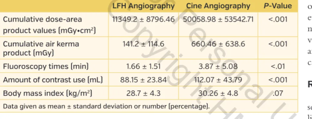
Mean cumulative air kerma was higher in the cine group vs the LFH group (660.46 ± 638.6 mGy vs 141.2 ± 114.6 mGy; P<.001). Mean cumulative DAP was higher in the cine group vs the LFH group (50058.98 ± 53542.71 mGy•cm2
vs 11349.2 ± 8796.46 mGy•cm2; P<.001). Mean
fluorosco-py times were higher in the cine group vs the LFH group (3.87 ± 5.08 minutes vs 1.66 ± 1.51 minutes; P<.01). Mean contrast use was higher in the cine group vs the LFH group (112.07 ± 43.79 cc vs 88.15 ± 23.84 cc; P<.001). Body mass indices were not different between cine and LFH groups (30.26 ± 4.8 kg/m2 vs 28.7 ± 4.3 kg/m2; P=.07) (Table 2).
There was no morbidity, mortality, or contrast-in-duced nephropathy in any of the patients. Cardiologists assessed LFH images sufficient for decision making and additional cine images were taken for better images in only 1 LFH case.
Mean value of intraclass correlation was not statistical-ly different between percent stenosis visual estimates of the three operators between cine and LFH angiography groups (0.69387 ± 0.18711 vs 0.62327 ± 0.30585; P=.45) (Table 3). Table 1. Patient demographic data.
LFH Angiography
(n = 46) Cine Angiography(n = 82) P-Value
Age (years) 58.74 ± 10.48 60.76 ± 10.98 .31
Male
Female 27 (58.69%) 19 (41.3%) 35 (42.68%)47 (57.32%) .56
Body mass index (kg/m2) 28.7 ± 4.3 30.26 ± 4.8 .07
Diabetes mellitus 14 (30.43%) 27 (32.9%) .84
Creatinine (mg/dL) 0.93 ± 0.7 0.84 ± 0.2 .32
History of PCI 6 (13%) 17 (20.7%) .34
History of CABG 1 (2.17%) 4 (4.88%) .65
Clinical presentation of ACS 5 (10.87%) 5 (6.1%) .31
Normal coronary arteries 16 (34.8%) 19 (23.17%) .21
Data given as mean ± standard deviation or number (percentage).
Table 2. Radiation doses and contrast use.
LFH Angiography Cine Angiography P-Value
Cumulative dose-area product values (mGy•cm2)
11349.2 ± 8796.46 50058.98 ± 53542.71 <.001 Cumulative air kerma
product (mGy) 141.2 ± 114.6 660.46 ± 638.6 <.001
Fluoroscopy times (min) 1.66 ± 1.51 3.87 ± 5.08 <.01 Amount of contrast use (mL) 88.15 ± 23.84 112.07 ± 43.79 <.001
Body mass index (kg/m2) 28.7 ± 4.3 30.26 ± 4.8 .07
Data given as mean ± standard deviation or number (percentage).
For
Personal
Use.
Copyright
HMP
2015
Vol. 27, No. 4, April 2015 201
Comparisonof Cineand fluoro angiography olCay, etal.
Average Cronbach’s alpha was 0.69 and 0.62, which is in the acceptable range for visual assessment:
Cronbach’s alpha Internal consistency
α ≥ 0.9 Excellent (high-stakes testing) 0.7 ≤ α < 0.9 Good (low-stakes testing) 0.6 ≤ α < 0.7 Acceptable
0.5 ≤ α < 0.6 Poor
α < 0.5 Unacceptable
Discussion
The increasing use and complexity of imaging and interven-tional techniques have not been matched by increasing awareness and knowledge by prescribers and practitioners. The majority of doctors — including cardiologists — grossly underestimate the radiation doses for most commonly requested tests.10,11
In-terventional cardiologists are competitive and perfectionist-type people, but a recent publication about brain and neck tumors in interventional cardiologists should warn and encourage them to reduce radiation doses and perfection during procedures.12
The significant increase in the cumulative exposure of patients and population to ionizing radiation, which is an important and potentially avoidable public health threat, is likely to increase the incidence of cancer in the future.13
Quantitative coronary angiography improved our ability to more accurately estimate the percent stenosis of a lesion and its length. Although this technique is a well-validat-ed tool for accurately and reproducibly defining coronary
lesion severity, these validations were done mostly in the cine film era using high-dose radiography and high-speed filming rates (60 frames/second). Its use, as originally validated, remains mostly in specialized research using experimental models and in clinical trials. With the transition to so-called lossless compression digital angiography, the use of lower-dose ra-diography, and the lower cine capture rates (15 frames/second), the information captured has been compromised.14,15
In our study, radiation doses were about 4 times higher in the cine group vs the LFH group. Fluoroscopy times were 2 times higher in the cine group vs the LFH group. Contrast use was prominently higher in the cine group vs the LFH group. Operators independent-ly assessed LFH images adequate for decision making. With recent improvements in imag-ing equipment and software, fluoroscopic LFH image quality has greatly improved. Even per-fect angiographic images on their own do not provide enough functional information with-out intravascular ultrasound, fractional flow re-serve, or thallium scintigraphy; therefore, the need for a “perfect” angiographic image for clinical decision-making is disputable.
Visual stenosis estimates of three operators were tested statistically for intraclass variability for different vessels in the cine and LFH groups. Mean intraclass variability was not different between groups in the analysis, which supports our hypothesis that the LFH angiography technique can be used as reliably as the cine angiography technique. Extra radiation doses did not produce a difference in variability.
Consistent collimation, adequately low-level acquisition modes, fewer irradiating angulations, reduced magnification and full inspiration during radiography whenever possible, long source-to-skin, and short patient-to-detector distanc-es are other important and effective techniqudistanc-es.16 We did
not use these effective techniques during this study, which would reduce radiation doses much more in both the LFH and cine groups.
LFH technique for coronary angiography should be test-ed in a larger population with different angiographic de-vice brands despite interventional cardiologists’ reluctance to select an examination protocol that intentionally sacri-fices image quality. Management decisions given by cine and LFH angiographic images should be tested by fractional flow reserve, intravascular ultrasound, or thallium scintigra-phy; if concordant results are obtained, the low-dose LFH technique for coronary angiography should universally be accepted by the clinicians in interventional cardiology and new industry standards in imaging should be established by device manufacturers.
Table 3. Cine and last fluoroscopy hold intraclass correlation between visual assessment of percent stenosis.
Cine Intraclass
Correlation P-Value LFH Intraclass Correlation P-Value
LMCA 1.000 <.001 1.000 <.001 Proximal LAD 0.804 <.001 0.749 <.001 Mid LAD 0.879 <.001 0.918 <.001 Distal LAD 0.755 <.001 0.849 <.001 D1 0.570 <.001 0.579 <.001 D2 0.642 <.001 0.593 <.001 Proximal CX 0.551 <.001 0.808 <.001 Distal CX 0.557 <.001 0.611 <.001 OM1 0.748 <.001 0.789 <.001 OM2 0.689 <.001 0.582 <.001 Proximal RCA 0.885 <.001 0.864 <.001 Mid RCA 0.854 <.001 0.590 <.001 Distal RCA 0.678 <.001 0.477 <.01 RPD 0.554 <.001 0.053 .41 RPL 0.242 .69 -0.113 .64
LMCA = left main coronary artery; LAD = left anterior descending coronary artery; D = diagonal artery; CX = circumflex artery; RCA = right coronary artery; RPD = right posterior descending artery; RPL = right posterolateral artery.
For
Personal
Use.
Copyright
HMP
2015
Comparisonof Cineand fluoro angiography olCay, etal.
202 The JourNAlof iNVAsiVe CArdiology®
In clinical practice, stenosis severity is typically determined during or shortly after the procedure and most commonly re-lies on visual estimation by physicians. This approach, howev-er, has well-known limitations.17,18 Older studies, conducted a
decade or more ago, described interobserver and intraobserver variation in visual estimations of stenosis severity and inac-curacies compared with computer-assisted techniques, expert panel review, autopsy results, or simulations.19-28
Study limitations. Some operators switched to cine
mode when performing radial angiography because cathe-ter engagement was weak and visualization was suboptimal. Some operators were dissatisfied with image quality and re-sisted using LFH angiography. One cardiac surgeon was not satisfied with images and requested cine images in his patient, but three other cardiac surgeons found images acceptable. In one case, additional cine images were obtained when there was a doubtful image. We propose that these techniques be used especially by experienced operators, and when in doubt additional cine images should be taken. There are also po-tential legal issues, wherein LFH images may cause problems in lawsuits. LFH, quantitative coronary angiography, fractional flow reserve, and thallium correlations should be performed to determine the safety of the LFH technique for diagnostic coronary angiography.
Conclusion
Radiation doses, contrast use, and fluoroscopy times are lower in fluoroscopic LFH angiography than cine angiography. Intraclass variability of visual stenosis estimation between three operators was not different between cine and LFH groups. Four times higher radiation dose does not give any extra accuracy in the interpretation of coronary stenoses. Fluoroscopic LFH images conventionally have inferior diagnostic quality when compared with cine coronary angiography, but with new an-giographic systems with improved LFH image quality, these images may be adequate for diagnostic coronary angiography.
References
1. Picano E, Vano E. The radiation issue in cardiology: the time for action is now. Cardiovasc Ultrasound. 2011;9:35.
2. Scanavacca M, d’Avila A, Velarde JL, Reolao JB, Sosa E. Reduction of radi-ation exposure time during catheter ablradi-ation with the use of pulsed fluo-roscopy. Int J Cardiol. 1998;63(1):71-74.
3. Nicholson R, Tuffee F, Uthappa MC. Skin sparing in interventional radiolo-gy: the effect of copper filtration. Br J Radiol. 2000;73(865):36-42. 4. Steffenino G, Rossetti V, Ribichini F, et al. Short communication: staff dose
reduction during coronary angiography using low framing speed. Br J
Ra-diol. 1996;69(825):860-864.
5. Geijer H, Beckman KW, Andersson T, Persliden J. Image quality vs radiation dose for a flat-panel amorphous silicon detector: a phantom study. Eur
Radiol. 2001;11(9):1704-1709.
6. Tsapaki V, Kottou S, Kollaros N, et al. Comparison of a conventional and a flat-panel digital system in interventional cardiology procedures. Br J
Ra-diol. 2004;77(919):562-567.
7. Tsapaki V, Kottou S, Kollaros N, Dafnomili P, Kyriakidis Z, Neofotistou V. Comparison of a CCD and a flat panel digital system in an Interventional cardiology laboratory. Radiat Prot Dosim. 2005;117(1-3):93-96.
8. Vano E, Geiger B, Schreiner A, Back C, Beissel J. Dynamic flat panel detec-tor versus image intensifier in cardiac imaging: dose and image quality.
Phys Med Biol. 2005;50(23):5731-5734.
9. Soudack M, Hirschenbein A, Sosna J, Dafne E, Engel A. Last fluoroscopy hold in paediatric fluoroscopy: dynamic capture of physiological events
and a potential for radiation exposure time reduction. J Med Imaging
Radi-at Oncol. 2012;56(1):93-95.
10. Correia MJ, Hellies A, Andreassi MG, Ghelarducci B, Picano E. Lack of radio-logical awareness among physicians working in a tertiary care cardiologi-cal centre. Int J Cardiol. 2005;103(3):307-311.
11. Einstein AJ, Tilkemeier P, Fazel R, Rakotoarivelo H, Shaw LJ; American Soci-ety of Nuclear Cardiology. Radiation safSoci-ety in nuclear cardiology — current knowledge and practice: results from the 2011 American Society of Nucle-ar CNucle-ardiology member survey. JAMA Intern Med 2013;173(11):1021-1023. 12. Roguin A, Goldstein J, Bar O, Goldstein JA. Brain and neck tumors
among physicians performing interventional procedures. Am J Cardiol. 2013;111(9):1368-1372. Epub 2013 Feb 16.
13. Berrington de González A, Darby S. Risk of cancer from diagnos-tic X-rays: estimates for the UK and 14 other countries. Lancet. 2004;363(9406):345-351.
14. Goldberg RK, Kleiman NS, Minor ST, Abukhalil J, Raizner AE. Comparison of quantitative coronary angiography to visual estimates of lesion severity pre and post PTCA. Am Heart J. 1990;119(1):178-184.
15. Kerensky RA, Cusma JT, Kubilis P, et al. American College of Cardiology/ European Society of Cardiology international study of angiographic data compression phase I: the effect of lossy data compression on recognition of diagnostic features in digital coronary angiography. J Am Coll Cardiol. 2000;35(5):1370-1379.
16. Tuinenburg JC, Koning G, Hekking E, et al. American College of Cardiology/ European Society of Cardiology international study of angiographic data compression phase II: the effects of varying JPEG data compression levels on the quantitative assessment of the degree of stenosis in digital coro-nary angiography. J Am Coll Cardiol. 2000;35(5):1380-1387.
17. Kuon E, Empen K, Weitmann K, et al. Long-term efficacy of a mini-course in radiation-reducing techniques in invasive cardiology. Rofo. 2013;185(8):720-725. Epub 2013 May 21.
18. Marcus ML, Skorton DJ, Johnson MR, Collins SM, Harrison DG, Kerber RE. Visual estimates of percent diameter coronary stenosis: “a battered gold standard.” J Am Coll Cardiol. 1988;11(4):882-885.
19. Raphael MJ, Donaldson RM. A “significant” stenosis: thirty years on.
Lan-cet. 1989;1(8631):207-209.
20. Detre KM, Wright E, Murphy ML, Takaro T. Observer agreement in evaluat-ing coronary angiograms. Circulation. 1975;52(6):979-986.
21. DeRouen TA, Murray JA, Owen W. Variability in the analysis of coronary arteriograms. Circulation. 1977;55(2):324-328.
22. Galbraith JE, Murphy ML, de Soyza N. Coronary angiogram interpretation. Interobserver variability. JAMA. 1978;240(19):2053-2056.
23. Fisher LD, Judkins MP, Lesperance J, et al. Reproducibility of coronary ar-teriographic reading in the Coronary Artery Surgery Study (CASS). Cathet
Cardiovasc Diagn. 1982;8(6):565-575.
24. Goldberg RK, Kleiman NS, Minor ST, Abukhalil J, Raizner AE. Comparison of quantitative coronary angiography to visual estimates of lesion severity pre and post PTCA. Am Heart J. 1990;119(1):178-184.
25. Fleming RM, Kirkeeide RL, Smalling RW, Gould KL. Patterns in visual inter-pretation of coronary arteriograms as detected by quantitative coronary arteriography. J Am Coll Cardiol. 1991;18(4):945-951.
26. Desmet W, Willems J, Van Lierde J, Piessens J. Discrepancy between vi-sual estimation and computer-assisted measurement of lesion sever-ity before and after coronary angioplasty. Cathet Cardiovasc Diagn. 1994;31(3):192-198.
27. Folland ED, Vogel RA, Hartigan P, et al. Relation between coronary artery stenosis assessed by visual, caliper, and computer methods and exercise capacity in patients with single-vessel coronary artery disease: the Veter-ans Affairs ACME Investigators. Circulation. 1994;89(5):2005-2014. 28. Leape LL, Park RE, Bashore TM, Harrison JK, Davidson CJ, Brook RH. Effect
of variability in the interpretation of coronary angiograms on the appro-priateness of use of coronary revascularization procedures. Am Heart J. 2000;139(1 Pt 1):106-113.
From the 1Istanbul Medipol University, Department of Cardiology, Istanbul, Turkey; and 2Mehmet Akif Ersoy Thoracic and Cardiovascular Surgery Training and Re-search Hospital, Department of Cardiology, Istanbul, Turkey.
Disclosure: The authors have completed and returned the ICMJE Form for Disclo-sure of Potential Conflicts of Interest. The authors report no conflicts of interest regarding the content herein.
Manuscript submitted April 16, 2014 and accepted June 15, 2014.
Address for correspondence: Ayhan Olcay, MD, Istanbul Medipol University, De-partment of Cardiology, Istanbul, Turkey. Email:[email protected]
For
Personal
Use.
Copyright
HMP
2015
View publication stats View publication stats