Turk J Phys Med Rehab 2016;62(3):286-287 DOI: 10.5606/tftrd.2016.24483
©Copyright 2016 by Turkish Society of Physical Medicine and Rehabilitation - Available online at www.ftrdergisi.com Scientific Letter / Bilimsel Mektup
Resorption of extruded intervertebral disc herniation:
Effect of physical therapy modalities
Mehmet Ağırman, Oğuz Durmuş, İlknur Saral, Engin Çakar
Department of Physical Medicine and Rehabilitation, İstanbul Medipol University, İstanbul, Turkey Received / Geliş tarihi: January 2016 Accepted / Kabul tarihi: February 2016
Corresponding author / İletişim adresi: Mehmet Ağırman, MD. İstanbul Medipol Üniversitesi, Fiziksel Tıp ve Rehabilitasyon Anabilim Dalı, 34214 Bağcılar, İstanbul,
Turkey. e-mail / e-posta: [email protected] Cite this article as:
Ağırman M, Durmuş O, Saral İ, Çakar E. Resorption of extruded intervertebral disc herniation: Effect of physical therapy modalities. Turk J Phys Med Rehab 2016;62(3):294-5.
We would like to share our experience with extruded lumbar disc herniation which was treated with non-surgical spinal decompression and physical therapy.
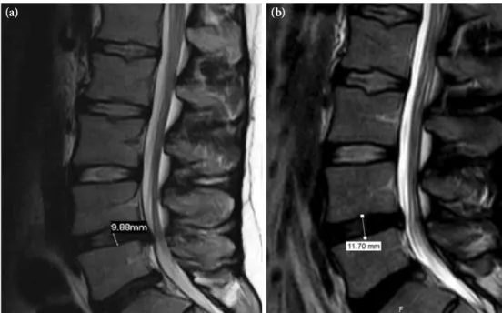
A 29-year-old man presented with a six-month history of lower back and right leg pain. He could not recall experiencing any trauma or initiating factor before the pain began, and the pain had increased over the last month. His pain increased when he was moving and he experienced numbness in his right foot. Medication had not been beneficial, and as a result surgery for a herniated disc was proposed. Upon examination, he had right L4 and L5 dermatomal hypoesthesia. The strength of the L5 muscle was 4/5 on the right side, and the right Achilles tendon reflex was hypoactive. Magnetic resonance imaging (MRI) performed six months prior to this exam showed L4-L5 extruded disc herniation and right L5 root compression (Figure 1).
The patient refused to have surgery, so a physical therapy program consisting of non-surgical lumbar spinal decompression (28 min) (with a Integrity Spinal Care System® machine), cold packs (20 min), and transcutaneous electrical nerve stimulation (TENS; 20 min) was initiated. After a total of 20 sessions which took place over a four-week period, the patient’s lower back and leg pain had gradually decreased. The visual analog scale score was eight initially and two after the last physiotherapy session. Neurological deficits persisted but did not progress. An MRI, which was taken at the end of the treatment, showed almost
complete resolution of the extruded fragment and no evidence of compression of the nerve root. In addition, the maximum L5-S1 disc height increased from 9.88 to 11.70 mm (Figure 1).
Regression of herniated discs in patients who have
been treated conservatively has been reported[1] and
can be demonstrated by MRI. The three main theories explaining spontaneous regression of a herniated nucleus pulposus (HNP) are dehydration, retraction and inflammation-related resorption. In instances of extruded fragments, the main mechanism is
resorption of the HNP.[2] Spontaneous reduction of
HNP begins in the first year and may continue for up
to seven years.[3] With neurological loss of function
due to root compression, surgical treatment is usually recommended. However, conservative approaches (exercise, medication, injections, or active physical therapy) are as effective as surgery over the
long-term (e.g., two-year period).[4] In a retrospective
cohort study, non-surgical spinal decompression were found to be effective in pain reduction and increased disc height in patients with lumbar disc
herniation.[5]
In this report, the increased disc space might have resulted in improvements in the clinical findings and resorption of HNP. It is difficult to say whether the resorption was spontaneous or due to the physiotherapy which contained non-surgical spinal decompression, cold pack and TENS; nevertheless, clinical improvements were observed after the physiotherapy. In this article,
287 Extruded lumbar disc herniation treatment
we aimed to highlight that well-planned physical therapy modalities with spinal decompression can be the preferred for patients with major lumbar disc herniation.
Declaration of conflicting interests
The authors declared no conflicts of interest with respect to the authorship and/or publication of this article.
Funding
The authors received no financial support for the research and/or authorship of this article.
REFERENCES
1. Kim ES, Oladunjoye AO, Li JA, Kim KD. Spontaneous regression of herniated lumbar discs. J Clin Neurosci 2014;21:909-13.
2. Liu JT, Li XF, Yu PF, Li XC, Qian Q, Liu GH, et al. Spontaneous resorption of a large lumbar disc herniation within 4 months. Pain Physician 2014;17:803-6.
3. Masui T, Yukawa Y, Nakamura S, Kajino G, Matsubara Y, Kato F, et al. Natural history of patients with lumbar disc herniation observed by magnetic resonance imaging for minimum 7 years. J Spinal Disord Tech 2005;18:121-6.
4. Weinstein JN, Tosteson TD, Lurie JD, Tosteson AN, Hanscom B, Skinner JS, et al. Surgical vs nonoperative treatment for lumbar disk herniation: the Spine Patient Outcomes Research Trial (SPORT): a randomized trial. JAMA 2006;296:2441-50.
5. Apfel CC, Cakmakkaya OS, Martin W, Richmond C, Macario A, George E, et al. Restoration of disk height through non-surgical spinal decompression is associated with decreased discogenic low back pain: a retrospective cohort study. BMC Musculoskelet Disord 2010;11:155.
Figure 1. Sagittal T2-weighted magnetic resonance imaging of the lumbar spine shows L4-L5 disc herniation with caudal migration (a) and (b) resorption in the second image. In addition, the maximum L5-S1 disc height increased from 9.88 to 11.70 mm.