SLEEP BREATHING PHYSIOLOGY AND DISORDERS• ORIGINAL ARTICLE
The role of antioxidant vitamins and selenium in patients
with obstructive sleep apnea
Ercan Saruhan1 &Erdim Sertoglu2&Yasemin Unal3&Semai Bek3&Gulnihal Kutlu3
Received: 15 May 2020 / Revised: 23 August 2020 / Accepted: 26 September 2020 # Springer Nature Switzerland AG 2020
Abstract
Purpose Obstructive sleep apnea (OSA) is a disorder characterized by recurrent episodes of obstruction of the upper respiratory tract during sleep often accompanied by oxygen desaturations. Antioxidant defense mechanisms are important to prevent OSA-associated diseases and decrease mortality. We aimed to determine the levels of selenium and vitamins A, C, and E in patients with OSA but without any comorbidities and compare the results with a control group, theorizing that the findings may be helpful to understand the antioxidant mechanisms in the pathogenesis of OSA and associated diseases.
Methods We designed a case-control study with 146 subjects. Subjects were categorized into four groups by apnea-hypopnea index (AHI) scores: control (n = 32; AHI < 5), mild OSA (n = 32; 5≤ AHI < 15), moderate OSA (n = 34; 15 ≤ AHI < 30), and severe OSA (n = 48; AHI≥ 30) groups. Serum levels of selenium were measured by atomic absorption spectrometer. Vitamin A, C, and E levels were measured by high-performance liquid chromatography and ultraviolet (HPLC-UV) detector. Results After adjusting for age, BMI, and gender, serum selenium and vitamin A levels were found to be higher in patients with OSA compared with controls (ANCOVA, p < 0.008, and p = 0.014 respectively), and levels of these markers increased with the severity of the disease. AHI was positively correlated with selenium (r = 0.289; p < 0.001), and vitamin A levels (r = 0.276; p < 0.001).
Conclusion These results demonstrated that antioxidant response with increased vitamin A, and selenium concentrations, may be important defense mechanisms in patients with OSA patients who do not have other comorbidities. Antioxidant nutrients or supplements may be implemented as a complementary treatment of OSA to support antioxidant defense.
Keywords Obstructive sleep apnea . Selenium . Vitamin . Antioxidant
Introduction
Obstructive sleep apnea (OSA) is a disorder characterized by recurrent episodes of obstruction of the upper respiratory tract during sleep and commonly associated with oxygen desaturations [1]. OSA is a common sleep disorder with a 20 to 50% prevalence in older adults. OSA patients had a greater risk of developing life-threatening complications
such as hypertension, coronary artery disease, and stroke [2]. OSA-associated diseases develop slowly and increase mortality that makes OSA an important public health con-cern. The antioxidant defense mechanism is important to prevent OSA-associated diseases and decrease mortality [3].
Vitamins A, C, and E are the main antioxidant vitamins that protect cells against lipid peroxidation and decrease the risk of oxidative stress-related diseases including cardiovascular dis-eases and diabetes [4,5]. Selenium is also known for its bio-logical functions as an enzymatic cofactor of selenoproteins that participate in antioxidant defense and immune responses [6]. Previous studies demonstrated that vitamins and selenium are associated with OSA pathogenesis [3,7–9]. Vitamin A and vitamin E levels were lower compared with healthy con-trols [3,9]. In addition, serum selenium levels were also lower in patients with OSA [7].
* Ercan Saruhan [email protected]
1
Department of Biochemistry, Faculty of Medicine, Mugla Sitki Kocman University, Mugla, Turkey
2
Department of Biochemistry, Gulhane Faculty of Medicine, University of Health Sciences, Ankara, Turkey
3 Department of Neurology, Faculty of Medicine, Mugla Sitki
Kocman University, Mugla, Turkey
These data suggest that diminished antioxidant defense mechanisms such as vitamins and selenium may contribute to pathogenesis of OSA. We therefore aimed to determine the levels of selenium and vitamins A, C, and E in patients with OSA who did not have comorbidities and to compare the results with a control group. We theorized that the results may be helpful to understand the antioxidant mechanisms in OSA pathogenesis and associated diseases. We hypothesized that patients with OSA without any comorbidities may have higher antioxidant response compared with healthy controls.
Material and methods
Study design
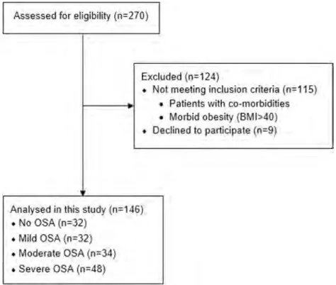
We enrolled 270 participants who were referred to Mugla Sitki Kocman University Training and Research Hospital Sleep Laboratory between July 2018 and April 2019 for the first polysomnography (PSG) for suspicion of OSA. Exclusion criteria were age under 18 and morbid obesity (BMI > 40), vitamin supplement usage in 3 months, previous-ly diagnosed OSA and using a continuous positive airway pressure (CPAP) device, history or diagnosis of narcolepsy, parasomnia, coronary artery disease, diabetes mellitus, hyper-tension, and chronic obstructive pulmonary disease. After ex-clusions, the remaining 114 newly diagnosed, without any comorbidities, untreated OSA patients and 32 subjects with-out OSA were included in our study (Fig.1).
Polysomnography
P o l y s o m n o g r a ph y w a s p e r f o r m ed i n t h e Cl i n i c a l Neurophysiology Laboratory of Mugla Sitki Kocman University Research and Training Hospital for patients with OSA symptoms by using an Embla N7000 (Natus, Kanata, Canada) PSG system. PSG was performed during the patient’s spontaneous sleep by a technician. Electroencephalogram (EEG), electrooculogram (EOG), electromyogram (EMG), nasal airflow, body position, and blood oxygen saturation (pulse oximetry) were recorded all night. This data was scored manually by the certified neurologist according to the American Academy of Sleep Medicine (AASM) manual [10]. The apnea-hypopnea index (AHI) is the total number of apneas and hypopneas per hour of sleep. We categorized all participants by using AHI scores as No OSA (AHI < 5), mild OSA (AHI between 5 and 14.9), moderate OSA (AHI between 15 and 29.9), and severe OSA (AHI≥ 30).
Biochemical analysis
After overnight fasting, venous blood samples were collected into gel-separated blood tubes by venipuncture before patients were discharged from the hospital. The blood tubes were left at room temperature for 10–20 min for separation of serum followed by centrifugation at 2000×g for 10 min. The follow-ing biochemical parameters were analyzed in the same day: glucose, C-reactive protein (CRP), urea, creatinine, alanine transaminase (ALT), aspartate aminotransferase (AST), cholesterol, and triglycerides, and high-density lipoprotein
cholesterol (HDL-C) concentrations were determined by enzymatic methods on a COBAS 8000 (c702) biochemical a n a l y z e r ( R o c h e D i a g n o s t i c s G m b H ; M a n n h e i m , Germany). Low-density lipoprotein cholesterol (LDL-C) levels were calculated using the Friedewald equation [11]. Remaining serums were aliquoted and stored at– 80 °C until selenium and vitamin A, C, and E analyses.
Serum vitamin C (ascorbic acid) levels were measured by using high-performance liquid chromatography and ultravio-let (HPLC-UV) detector [12].L-Ascorbic acid was purchased from Shinyo Pure Chemical Co. (Osaka, Japan).D-Isoascorbic acid, metaphosphoric acid, and all other reagents were of an-alytical grade and were purchased from Sigma-Aldrich (St. Louis, MO, USA). Ascorbic acid analysis was performed at 245-nm wavelengths. Chromatographic separations were per-formed using a Zorbax Eclipse XDB-C18 (4.6μm × 150 mm, particle size 3.5 μm). The mobile phase consisted of 1.8 mmol/L sulfuric acid in diH2O. The flow rate was kept constant at 0.8 mL/min.
Serum vitamin E (α-tocopherol) and vitamin A (β-carotene) levels were measured by the HPLC-UV method. Serum samples were deproteinized with ethanol/chloroform (4:3), and chromatographic separation of analytes was per-formed by Agilent 1260 Infinity HPLC system, using Nucleosil® C18, 10μm, 125 mm × 4 mm, and isocratic elu-tion of methanol:isopropanol:diH2O (90:14:6). The detecelu-tion wavelength was set at vitamin A: 325 nm and vitamin E: 300 nm, with a flow rate of 1 mL/min.
Serum selenium levels were determined using a graphite furnace atomic absorption spectrometer (Thermo Scientific, iCE 3300 AAS, Waltham, MA USA).
Outcome questionnaires
Demographic information like age, sex, body mass index (BMI), medications, and comorbidities were obtained from the patient’s medical record. Excessive daytime sleepiness (EDS) was evaluated using the Epworth Sleepiness Scale (ESS) [13], which is an eight-item questionnaire. The total possible score ranges from 0 to 24. ESS scores greater than 16 indicate severe sleepiness during daily activities.
Statistical analysis
Statistical analysis was done using SPSS software (IBM SPSS Statistics, version 22.0. Armonk, NY: IBM Corp). We hy-pothesized (H0) that there are no differences in the levels of selenium and vitamins between controls and OSA patients. The sample size and power calculations were carried out using the G*Power software version 3.1 (Düsseldorf University, Germany). The effect size was determined as 0.85 and sample size as 140 by using data of a previous study [3].
The Shapiro-Wilk test was used to evaluate the distribution of variances. Variables with normal distribution were presented as mean ± SD and non-normally distributed variables were presented as median and quartiles (25–75th percentiles). Differences among the groups for each parameter were ana-lyzed by analysis of covariance (ANCOVA) using Tukey cor-rection for normally distributed parameters and Kruskal-Wallis test for non-normally distributed parameters. The correlation between variables was explored by using the Spearman’s cor-relation analysis. All p values less than 0.05 were considered statistically significant.
Results
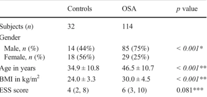
A total of 146 subjects (47 female, 99 male) who underwent PSG were included in this study. Post hoc power calculations showed that our sample size gave 0.952 power and 1.093 effect size for selenium at the level ofα error probability = 0.05. One hundred fourteen subjects were diagnosed as OSA (AHI≥ 5), while 32 subjects were negative (AHI < 5). The average sleep efficiency percentage of the controls was 87.47% ± 9.71, and the average percentage of REM to total sleep time was 16.15% ± 5.39. They had no complaints of excessive daytime sleepi-ness, and their ESS scores were lower than 10. The mean age of OSA patients was 46.5 ± 10.7 (range 18–69 years), while the mean age for controls was 34.9 ± 10.8 (range 18–64 years). The mean BMI of the OSA patients was significantly higher than the control group (30 ± 4.5 kg/m2vs 24 ± 3.3 kg/m2, p < 0.001). OSA patients were prevalently male (85 males, 75%; 29 fe-males, 25%) and older than the control group (47 vs 35 years, p < 0.001). There was no statistically significant difference be-tween the groups in terms of ESS scores. The descriptive sta-tistics of OSA patients and controls are shown in Table1.
After adjustment for age, BMI, and gender, serum selenium levels were found lower in the control group compared with the severe (ANCOVA, Tukey adjusted p = 0.008), the moder-ate (p = 0.041), and the mild OSA groups (p = 0.027). Although there was no significant difference in selenium levels between OSA subgroups, severe OSA group had the highest levels. Regarding vitamin concentrations, serum vita-min A levels were found higher in OSA patients compared with controls (ANCOVA, p = 0.014). By contrast, there was no significant difference in vitamin E and C levels between OSA patients and controls (ANCOVA, p = 0.075, p = 0.209, respectively) (Table2, Fig.2).
Other routine biochemical parameters were also compared to evaluate the possible confounding effect of these parameters in OSA patients. After adjustment for age, BMI, and gender, it was observed that serum levels of glucose, urea, creatinine, cholesterol, LDL-C, HDL-C, triglycerides, CRP, and ESR did not show any significant difference between groups (ANCOVA, p > 0.05) (Table3).
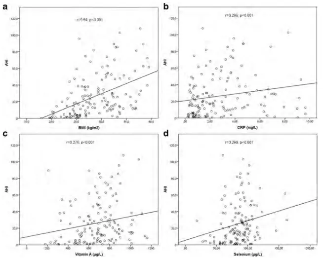
AHI was positively correlated with BMI (r = 0.54; p < 0.001), cholesterol (r = 0.349; p < 0.001), CRP (r = 0.286; p < 0.001), vitamin A (r = 0.276; p < 0.001), and sele-nium levels (r = 0.289; p < 0.001). Vitamin E levels were not significantly correlated with AHI (Fig.3). There was no cor-relation between the antioxidant (vitamins A, C, and E and selenium) and routine biochemical parameters (glucose, urea, creatinine, cholesterol, LDL-C, HDL-C, triglycerides, CRP, and ESR).
An association between obesity (BMI > 30) and OSA was observed, as usual,χ2(1) = 10.87, p < 0.001. We also ob-served an association between male sex and OSA,χ2(3) = 18.43, p < 0.001.
Discussion
In this study, we evaluated the antioxidant mechanisms by measuring serum levels of vitamins A, E, and C, and selenium
in patients with OSA. The main finding of our study was the higher levels of selenium, and vitamin A in OSA patients compared with the control group, and levels of these antioxi-dants increase with the severity of the disease. The literature is very limited regarding the studies investigating the role of selenium and antioxidant vitamins in OSA pathogenesis.
Selenium deficiency decreases antioxidant activity and im-pairs the scavenging of free radicals especially in systemic inflammatory diseases [14]. Chen et al. found lower concen-trations of selenium that reflect oxidative damage in patients with newly diagnosed mild-to-moderate OSA patients [7]. Our results do not support this mechanism. The significance of increased selenium in our patients is not known. A reason-able explanation for conflicting results reported in the litera-ture might be due to the adaptation mechanism of selenoproteins against chronic hypoxia and oxidative stress. Besides, our patients had no medications, and comorbidities, so selenium levels were not affected by other diseases. The number of patients with severe OSA in our study was much higher than this study which may cause contradictory results. Previous studies showed a relationship between OSA and antioxidant response, but their results have been challenged. Some authors provided findings suggesting that OSA patients had lower antioxidant levels [15–18]. Singh et al. found that antioxidant glutathione levels decreased in OSA patients which got restored after CPAP therapy followed by oral anti-oxidant supplementation (vitamin E 400 IU and vitamin C 100 IU for 45 days) [19]. On the other hand, some studies showed no significant differences [20, 21]. Christou et al. found that antioxidant capacity was not different between OSA patients and the healthy group [22]. In this respect, the relationship between OSA and antioxidant response remains unclear.
Regarding vitamin levels in OSA patients, Barcelo et al. found significantly decreased levels of vitamin A and vitamin E in patients with OSA compared with controls [3]. Sales et al. also found lower levels of vitamin E in OSA patients [9]. By contrast with these studies, Day et al. reported higher levels of vitamin E and lower levels of vitamin A in OSA patients [8]. In our study, we found significantly higher levels of vitamin A
Table 2 Comparison of selenium and vitamin levels between OSA subgroups and controls
Controls Mild OSA Moderate OSA Severe OSA p value*
Subjects (n) 32 32 34 48
Selenium (μg/L) 69.6 ± 20.1 87.2 ± 18.3 87.9 ± 21.0 92.7 ± 22.1 0.008 Vitamin C (mg/L) 7.69 ± 0.95 7.88 ± 0.95 8.10 ± 0.92 7.77 ± 1.25 0.209 Vitamin A (μg/L) 534 ± 196 737 ± 202 735 ± 169 708 ± 197 0.014 Vitamin E (mg/L) 8.89 ± 2.25 9.67 ± 1.93 10.51 ± 1.75 9.91 ± 2.29 0.075 Data are presented as mean ± SD for normally distributed variables and as median and quartiles (25th–75th percentiles) for non-normally distributed variables
*ANCOVA test. Italicized p values indicate statistical differences between groups Table 1 Descriptive statistics of OSA patients and controls
Controls OSA p value Subjects (n) 32 114 Gender Male, n (%) 14 (44%) 85 (75%) < 0.001* Female, n (%) 18 (56%) 29 (25%) Age in years 34.9 ± 10.8 46.5 ± 10.7 < 0.001** BMI in kg/m2 24.0 ± 3.3 30.0 ± 4.5 < 0.001** ESS score 4 (2, 8) 6 (3, 10) 0.081*** Data are presented as mean ± SD for normally distributed variables and as median and quartiles (25th–75th percentiles) for non-normally distributed variables
BMI, body mass index; ESS, Epworth Sleepiness Scale; SD, standard deviation
*Chi-square test. Italicized p values indicate statistical differences be-tween groups
**Student’s t test ***Mann-Whitney U test
in severe OSA patients, and a positive correlation between AHI and vitamin A. Vitamin E levels were also found higher, but the change was not significant. The sample sizes of these studies were very restricted which may cause irrelevant
results. Our findings are more reliable and suggest that OSA patients presented sufficiency to increase their vitamin A, and E, but not vitamin C levels by different mechanisms. Fat-soluble vitamins can be stored in tissues such as liver and
Fig. 2 Comparison of antioxidant levels between groups a selenium, b vitamin A, c vitamin E, and d vitamin C concentrations. Outliers between 1.5 and 3 box lengths are depicted by white circle
Table 3 Comparison of biochemical parameters between OSA subgroups and controls
Controls Mild OSA Moderate OSA Severe OSA p value
Subjects (n) 32 32 34 48 Glucose (mg/dL) 89.3 ± 10.2 93.2 ± 11.6 90.8 ± 9.3 95.7 ± 13.4 0.221 Urea (mg/dL) 25.3 ± 8.9 29.3 ± 8.3 31.1 ± 6.6 30.1 ± 6.8 0.209 Creatinine (mg/dL) 0.77 ± 0.18 0.79 ± 0.18 0.82 ± 0.16 0.83 ± 0.12 0.347 Cholesterol (mg/dL) 168.7 ± 35.8 196.9 ± 36.4 204.5 ± 31.7 207.2 ± 41.8 0.096 LDL-C (mg/dL) 96.6 ± 34.2 115.7 ± 30.6 125.1 ± 27.3 126.5 ± 37.7 0.420 HDL-C (mg/dL) 50.4 ± 12.0 47.5 ± 17.1 47.3 ± 13.9 40.6 ± 10.4 0.308 Triglycerides (mg/dL) 94 (69, 130) 140.5 (92.5, 238.5) 137.5 (99, 181) 182.5 (135, 256) 0.183 CRP (mg/L) 1.1 (0.7, 2.0) 2.2 (0.8, 3.9) 1.9 (1.0, 4.2) 3.1 (1.5, 4.3) 0.966 ESR (mm/h) 9 (6, 13) 15 (9, 29) 12 (9, 18) 14 (9, 22) 0.236 Data are presented as mean ± SD for normally distributed variables and as median and quartiles (25th–75th percentiles) for non-normally distributed variables
LDL-C, low-density lipoprotein cholesterol; HDL-C, high-density lipoprotein cholesterol; CRP, C-reactive pro-tein; ESR, erythrocyte sedimentation rate
adipose tissue. Increased oxidative stress and inflammation in OSA could contribute to increased intestinal absorption or release of vitamins from adipose tissue or liver. This mecha-nism can explain the higher levels of fat-soluble vitamins in response to OSA-related oxidative stress. This adaptive mech-anism for vitamin E is maintained byα-tocopherol transfer protein (αTTP) that facilitates the incorporation of the vitamin into lipoproteins in the liver for transport to other tissues. Oxidative stress stimulates the expression ofαTTP mRNA and protein in cells and increases the levels of vitamin E in blood [23]. These results are probably the consequence of oxidative stress, not the cause, and underline the importance of antioxidant activity in OSA.
We found no dyslipidemia in patients with OSA in contrast to the literature. Gündüz et al. found an association between OSA severity and lipid parameters [24]. In our study, serum concentrations of triglycerides, cholesterol, LDL-C, and HDL-C remained unchanged between groups, after adjust-ment for age, BMI, and gender.
There was no significant difference in CRP levels of OSA patients. Our results were not in line with the findings of previous studies, indicating the role of inflammation in OSA
[25,26]. Although the etiology remains unclear, chronic in-flammation was shown as a pathogenetic factor in previous OSA studies.
The risk factors of OSA are age, male gender, and high BMI. Our results were consistent with other prevalence stud-ies [27,28]. It should be noted that, although we enrolled OSA patients without any comorbidities, these patients presented a trend towards increased cholesterol, triglycerides, and BMI compared with controls. We used ANCOVA statistical anal-ysis to exclude the effects of these factors in our study.
The main strength of our study is the well-designed, large samples of controls, and newly diagnosed OSA subgroups without any treatment and comorbidities. However, our study has some limitations to be acknowledged. First, the dietary habits of patients could not be controlled in this study. The effect of diet on antioxidant response was not assessed, and this should be taken into account while interpreting our re-sults. The second factor that could have affected our results is age and obesity. In our study, age and BMI of OSA patients were higher than the control group. Thus, we analyzed our results with covariance test to exclude the effects of these factors. Lipid parameters should also be evaluated while
interpreting vitamin results. Despite limitations, our study is of value by determining vitamin and selenium differences be-tween OSA subgroups and controls.
Conclusions
Although the antioxidants have been the subject of many clin-ical trials, researches on antioxidant vitamins in OSA is in earlier stages. The researches in sleep medicine should focus on nutrient and supplement intake to determine whether higher antioxidant status contributes to better OSA outcomes, and antioxidant supplements can decrease OSA-associated complications, such as cardiovascular events.
The present study demonstrated that antioxidant response with increased vitamin A, and selenium concentrations, is an important defense mechanism in OSA patients without any comorbidities. OSA is mostly treated with mechanical (CPAP) or surgical interventions. Our study suggests that an-tioxidant nutrients or supplements can be implemented as a complementary treatment of OSA to support antioxidant de-fense and used to reduce cardiovascular events in patients with OSA. Further prospective studies with vitamins and selenium supplemented OSA patients are needed to fully elucidate the relationship and mechanisms of antioxidant vitamins in OSA and any need for supplementation. Future studies should also include examining the effects of CPAP therapy on antioxidant changes observed in our study.
Authors’ contributions Ercan Saruhan: conceptualization, methodology, funding acquisition, formal analysis, investigation, writing—original draft, project administration. Erdim Sertoglu: investigation, resources, writing—review and editing. Yasemin Unal: writing—review and editing, data curation. Semai Bek: writing—review and editing, data curation. Gulnihal Kutlu: supervision, editing, writing—review and editing
Funding Muğla Sitki Kocman University Scientific Research Projects Office supported this work under grant number 19/087/06/3/4.
Compliance with ethical standards
Conflict of interest The authors declare that they have no conflict of interest.
Ethics approval The Clinical Research Ethical Board of Muğla Sitki Kocman University reviewed and approved this study (28/06/2018-10/ IV). This study was performed in accordance with the ethical standards laid down in the 1964 Declaration of Helsinki and its later amendments. Informed consent was obtained from all individual participants prior to their inclusion in the study.
Consent to participate Informed consent was obtained from all individ-ual participants included in the study.
Consent for publication Patients signed informed consent regarding publishing their clinical and laboratory data.
References
1. Jelic S, Le Jemtel TH (2008) Inflammation, oxidative stress, and the vascular endothelium in obstructive sleep apnea. Trends Cardiovasc Med 18(7):253–260. https://doi.org/10.1016/j.tcm. 2008.11.008
2. Young T, Peppard PE, Gottlieb DJ (2002) Epidemiology of ob-structive sleep apnea. Am J Respir Crit Care Med 165(9):1217– 1239.https://doi.org/10.1164/rccm.2109080
3. Barcelo A, Barbe F, de la Pena M, Vila M, Perez G, Pierola J, Duran J, Agusti AG (2006) Antioxidant status in patients with sleep apnoea and impact of continuous positive airway pressure treat-ment. Eur Respir J 27(4):756–760. https://doi.org/10.1183/ 09031936.06.00067605
4. Saremi A, Arora R (2010) Vitamin E and cardiovascular disease. Am J Ther 17(3):e56–e65. https://doi.org/10.1097/MJT. 0b013e31819cdc9a
5. Shahidi F, de Camargo AC (2016) Tocopherols and tocotrienols in common and emerging dietary sources: occurrence, applications, and health benefits. Int J Mol Sci 17(10).https://doi.org/10.3390/ ijms17101745
6. Lu J, Holmgren A (2009) Selenoproteins. J Biol Chem 284(2):723– 727.https://doi.org/10.1074/jbc.R800045200
7. Chen PC, Guo CH, Tseng CJ, Wang KC, Liu PJ (2013) Blood trace minerals concentrations and oxidative stress in patients with ob-structive sleep apnea. J Nutr Health Aging 17(8):639–644.https:// doi.org/10.1007/s12603-013-0023-x
8. Day RM, Matus IA, Suzuki YJ, Yeum KJ, Qin J, Park AM, Jain V, Kuru T, Tang G (2009) Plasma levels of retinoids, carotenoids and tocopherols in patients with mild obstructive sleep apnoea. Respirology 14(8):1134–1142. https://doi.org/10.1111/j.1440-1843.2009.01623.x
9. Sales LV, Bruin VM, D’Almeida V, Pompeia S, Bueno OF, Tufik S, Bittencourt L (2013) Cognition and biomarkers of oxidative stress in obstructive sleep apnea. Clinics (Sao Paulo) 68(4):449– 455.https://doi.org/10.6061/clinics/2013(04)03
10. (1992) EEG arousals: Scoring rules and examples: a preliminary report from the sleep disorders atlas task force of the American Sleep Disorders Association. Sleep 15(2):173–184
11. Friedewald WT, Levy RI, Fredrickson DS (1972) Estimation of the concentration of low-density lipoprotein cholesterol in plasma, without use of the preparative ultracentrifuge. Clin Chem 18(6): 499–502
12. Kim Y, Kim M-G (2016) HPLC-UV method for the simultaneous determinations of ascorbic acid and dehydroascorbic acid in human plasma. Transl Clin Pharmacol 24(1):37–42
13. Johns MW (1991) A new method for measuring daytime sleepi-ness: the Epworth sleepiness scale. Sleep 14(6):540–545.https:// doi.org/10.1093/sleep/14.6.540
14. Steinbrenner H, Sies H (2009) Protection against reactive oxygen species by selenoproteins. Biochim Biophys Acta 1790(11):1478– 1485.https://doi.org/10.1016/j.bbagen.2009.02.014
15. Chung S, Yoon IY, Lee CH, Kim JW (2011) The effects of nasal continuous positive airway pressure on vascular functions and se-rum cardiovascular risk factors in obstructive sleep apnea syn-drome. Sleep Breath 15(1):71–76. https://doi.org/10.1007/s11325-009-0323-x
16. Oktay B, Akbal E, Firat H, Ardic S, Akdemir R, Kizilgun M (2008) Evaluation of the relationship between heart type fatty acid binding protein levels and the risk of cardiac damage in patients with ob-structive sleep apnea syndrome. Sleep and Breathing 12(3):223– 228.https://doi.org/10.1007/s11325-007-0167-1
17. Chung S, Yoon IY, Shin YK, Lee CH, Kim JW, Ahn HJ (2009) Endothelial dysfunction and inflammatory reactions of elderly and
middle-aged men with obstructive sleep apnea syndrome. Sleep Breath 13(1):11–17.https://doi.org/10.1007/s11325-008-0210-x
18. Christou K, Markoulis N, Moulas AN, Pastaka C, Gourgoulianis KI (2003) Reactive oxygen metabolites (ROMs) as an index of oxida-tive stress in obstrucoxida-tive sleep apnea patients. Sleep Breath 7(3): 105–110.https://doi.org/10.1007/s11325-003-0105-9
19. Singh TD, Patial K, Vijayan VK, Ravi K (2009) Oxidative stress and obstructive sleep apnoea syndrome. Indian J Chest Dis Allied Sci 51(4):217–224
20. Wali SO, Bahammam AS, Massaeli H, Pierce GN, Iliskovic N, Singal PK, Kryger MH (1998) Susceptibility of LDL to oxidative stress in obstructive sleep apnea. Sleep 21(3):290–296.https://doi. org/10.1093/sleep/21.3.290
21. Grabska-Kobylecka I, Kobylecki A, Bialasiewicz P, Krol M, Ehteshamirad G, Kasielski M, Nowak D (2008) No evidence of enhanced oxidant production in blood obtained from patients with obstructive sleep apnea. J Negat Results Biomed 7:10.https://doi. org/10.1186/1477-5751-7-10
22. Christou K, Moulas AN, Pastaka C, Gourgoulianis KI (2003) Antioxidant capacity in obstructive sleep apnea patients. Sleep Med 4(3):225–228. https://doi.org/10.1016/s1389-9457(02) 00253-8
23. Ulatowski L, Dreussi C, Noy N, Barnholtz-Sloan J, Klein E, Manor D (2012) Expression of the alpha-tocopherol transfer protein gene is regulated by oxidative stress and common single-nucleotide poly-morphisms. Free Radic Biol Med 53(12):2318–2326.https://doi. org/10.1016/j.freeradbiomed.2012.10.528
24. Gunduz C, Basoglu OK, Hedner J, Zou D, Bonsignore MR, Hein H, Staats R, Pataka A, Barbe F, Sliwinski P, Kent BD, Pepin JL, Grote L (2018) Obstructive sleep apnoea independently predicts lipid levels: data from the European Sleep Apnea Database. Respirology 23(12):1180–1189. https://doi.org/10.1111/resp. 13372
25. Kim J, Yoon DW, Lee SK, Lee S, Choi KM, Robert TJ, Shin C (2017) Concurrent presence of inflammation and obstructive sleep apnea exacerbates the risk of metabolic syndrome: a KoGES 6-year follow-up study. Medicine (Baltimore) 96(7):e4488.https://doi.org/ 10.1097/md.0000000000004488
26. Kim J, Lee SJ, Choi KM, Lee SK, Yoon DW, Lee SG, Shin C (2016) Obstructive sleep apnea is associated with elevated high sensitivity C-reactive protein levels independent of obesity: Korean Genome and Epidemiology Study. PLoS One 11(9): e0163017.https://doi.org/10.1371/journal.pone.0163017
27. Peppard PE, Young T, Barnet JH, Palta M, Hagen EW, Hla KM (2013) Increased prevalence of sleep-disordered breathing in adults. Am J Epidemiol 177(9):1006–1014.https://doi.org/10.1093/aje/ kws342
28. Young T, Peppard PE, Gottlieb DJ (2002) Epidemiology of ob-structive sleep apnea: a population health perspective. Am J Respir Crit Care Med 165(9):1217–1239.https://doi.org/10.1164/ rccm.2109080
Publisher’s note Springer Nature remains neutral with regard to jurisdic-tional claims in published maps and institujurisdic-tional affiliations.