T H E P R O T E C T I V E E F F E C T S O F N - A C E T Y L C Y S T E I N E I N PARAQUAT-I N D U C E D T O X PARAQUAT-I C PARAQUAT-I T Y
P A R A K U A T T O K S İ S İ T E S İ N D E N-ASETİLSİSTEİNİN K O R U Y U C U ETKİSİ
Şebnem Ş. ÇEÇEN Gülhan CENGİZ Tülin S Ö Y L E M E Z O Ğ L U
Ankara Üniversitesi, Adli Tıp Enstitüsü, 06260, Dikimevi, ANKARA-TÜRKİYE
ABSTRACT
İn this study, ameliorating effects of N-Acetylcysteine, a potent free radical scavenger, on paraquat-induced oxidative stress were investigated. N-Acetylcysteine (NAC, 50 mg/kg, i.p.) was administered one hour prior to the administration of paraquat (PQ, 40 mg/kg, i.p.). Malondialdehyde (MDA) and gluthathione (GSH) levels were determined as markers of oxidative stress. After PQ treatment, significant changes were observed in both MDA and GSH levels (p<0.001). Pretreatment with NAC, prevented PQ-induced increases in plasma (p<0.01); liver, kidney, lung (p<0.001) and brain (p<0.05) MDA levels. NAC when administered prior to PQ injection, caused significant amelioration in liver, lung, brain (p<0.01) and kidney (p<0.05) GSH levels.
Key Words: Paraquat, N-Acetylcysteine, Lipid peroxidation, Malondialdehyde, Gluthathione.
Ö Z E T
Bu çalışmada, güçlü bir serbest radikal tutucusu olan N-Asetilsistein'in (NAC) parakuatın (PQ) indüklediği oksidatif strese karşı iyileştirici etkileri araştırıldı. NAC, (50 mg/kg, i.p.) PQ enjeksiyonundan (40 mg/kg, i.p.) bir saat önce uygulandı. Oksidatif stresin göstergesi olarak malondialdehit (MDA) ve glutatyon (GSH) düzeyleri ölçüldü. PQ uygulamasından sonra, MDA ve GSH düzeylerinde önemli değişiklikler (p<0.001) saptandı. Plazmada (p<0.01); karaciğerde, akciğerde, böbreklerde (p<0.001) ve beyinde (p<0.05), PQ'in neden olduğu MDA düzeyindeki artışın NAC ön-uygulamasıyla düştüğü gözlendi. PQ+NAC uygulanan grupta karaciğer, akciğer, beyin (p<0.01) ve böbrek (p<0.05) GSH düzeylerinde önemli derecede iyileşmeler görüldü.
260 Şebnem Ş. ÇEÇEN, Gülhan CENGİZ, Tülin SÖYLEMEZOĞLU
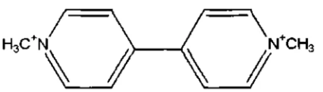
Figure 1. Chemical structure of the herbicide, paraquat (5).
If compared with other xenobiotics, PQ is only moderately toxic, but the occurence of cases of poisoning in human makes it of great toxicological interest. Paraquat is believed to show its toxic effect by inhibiting the conversion of NADP to NADPH and by interfering with the electron transport system reducing itself to PQ radical (6,7). The reduced PQ radical immediately reacts with molecular oxygen to form superoxide (02 - 0), hydroperoxy (HO2) radicals and hydrogen peroxide (H202). Reactive oxygen species formed, are catabolized; and lipid hydroperoxydes are reduced to less toxic lipid alcohols by the cellular enzymes gluthathione reductase (GSH-Rx) and superoxide dismutase (SOD) (8,9,10). Cell death is a consequence of the loss of NADPH and resulting oxidative stress or lipid peroxidation initiating from the formation of reactive oxygen radicals, or both of these processes taking place together (6). Lipid peroxidation (LPO) caused by the free radicals formed during the redox cycling of PQ, is simply considered as the deterioration of membrane phospholipids.
Gluthathione (GSH), a tripeptide consisting of glutamic acid, cysteine, and glycine being one of the most important intracellular defence systems (11) maintains a reducing environment through the thiol group of cysteine (12) and therefore protects the intracellular constituents from oxidative stress (10,13). A dramatic fall of hepatic GSH overwhelms the cellular defences against oxiadative stress; resulting in the development of LPO, which in turn would produce loss of protein thiols; leading into disturbed homeostasis of cellular calcium and cell death.
INTRODUCTION
Paraquat (PQ, metil viologen; l-1'-dimethyl-4-4'-bipyridilium) is a very effective and widely used nonselective, contact and toxic herbicide that can easily enter the human body through the skin, with the inspired air or by food contamination (1) (Figure 1). Paraquat causes tissue damage, mainly in the lungs and on most other organs such as heart, gastrointestinal tract, liver, kidneys and brain (2,3,4).
Recognition of the protective role played by GSH, NAC is the first choice as an antidote because of its ability to counteract oxidative stress and replenish GSH through the sulphydryl groups in its structure (14).
In this study, amelorating effects of NAC on paraquat-induced oxidative stress were evaluated.
MATERIAL AND METHOD Animals
Local breed albino male mice were purchased from Refik Saydam Hıfzısıhha Institute, Animal Care Unit, Ankara. The animals weighing 20-25 g were housed in group cages under normal laboratory conditions (temperature 20-22°C, natural day-night cycle), and free access to commercial chow and water but the food was withdrawn 12-16 h before the experiment. Procedures involving animals and their care were conducted in conformity with international laws and policies and the studies on animals accepted by Ethic Council of Ankara University (11.07.2001, No: 2001/35).
Treatment
Animals were divided into three groups consisting six mice each and exposed as follows: (1) control group received corn oil; (2) received 40 mg/kg PQ; (3) received 50 mg/kg NAC+40 mg/kg PQ. All administrations were done intraperitonally. NAC was administered 1 hour prior to the administration of PQ. Mice were sacrified by an overdose of diethyl ether 6 hours after the last injections. Blood was withdrawn by intracardiac punction and then lungs, brains, livers and kidneys were removed.
LPO in plasma
MDA, a decomposition product of LPO, was determined by measuring the thiobarbituric acid reactive substances (TBARs) in plasma and tissues. MDA levels in plasma were measured according to the method of Buege and Aust (15). One ml of plasma was combined with 2.0 ml of trichloroacetic acid (TCA; 15% w/v )-thiobarbituric acid (TBA; 0.375 % w/v )-0.25 N HC1 and centnfuged at 10 000 x g for 5 min. The supernatant was mixed with 20 ml butylated hydroxytoluene (BHT; 0.02% in 95% ethanol w/v) to prevent further oxidation and heated for 15 min in a boiling water bath. The precipitate was removed by centrifugation at 10 000 x g for 5 min after cooling under tap water. The absorbance of the plasma sample was read at 532 nm
262 Şebnem Ş. ÇEÇEN, Gülhan CENGİZ, Tülin SOYLEMEZOĞLU
against blank. The plasma TBA reactive products were expressed as nmoles of MDA/ml. 1.1,3,3-tetraethoxypropan was used as standard for calibration of the curve.
Homogenization
Tissues of animals were immediately excised and chilled in ice-cold 0.9 % NaCl. After washing with 0.9% NaCl, 0.5 g of wet tissue was weighed exactly and homogenized in 4.5 ml of 0.25 M sucrose to obtain a %10 w/v suspension in order to measure LPO in tissues. However 0.2 g tissue is homogenized with 8 ml 0.02 M Na2EDTA to measure GSH level.
LPO in tissues
The method of Jamall and Smith (16) was used to determine lipid peroxidation in tissue samples. The cytosolic fraction was obtained by a two step-centrifugation first at 1000 x g for 10 min and then at 2000 x g for 30 min at 4 °C. 0.20 ml of supernatant was transferred to a vial and was mixed with 0.20 ml sodium dodecyl sulfate solution (SDS; 8.1%), 1.50 ml of acetic acid solution (CH3COOH; 20% v/v, adjusted to pH 3.5 with NaOH), 1.50 ml of 0.8%(w/v) solution of TBA and the final volume was adjusted 4.0 ml with distilled water. Each vial was tightly capped and heated in a boiling water bath for 60 min. The vials were then cooled under running water. Equal volumes of tissue blank or test sample and 10% trichloroacetic acid (TCA) were centrifuged at 1000 x g for 10 min.The absorbance of the supernatant was measured at 532 nm against tissue blank. Tissue blank was processed using the same experimental procedure except the TBA solution was replaced with distilled water. 1,1,3,3-tetraethoxypropan was used as standard for the calibration curve. The tissue TBA reactive products were expressed as nmoles of MDA/g wet weight.
GSH in tissues
GSH was measured by Sedlak and Lindsay's method (17). Aliquots of 5.0 ml of the homogenates were mixed in 15.0 ml test tubes with 4.0 ml distilled H20 and 1.0 ml of 50% TCA. The tubes were centrifuged for 15 min at approximately 3000 x g. Two ml of supernatant was mixed with 4.0 ml of 0.4 M Tris buffer (pH: 8.9), 0.1 ml Ellman's reagent, (5,5'-dithiobis-2-nitro-benzoic acid) - DTNB added, and the sample was shaken. The absorbance was read within 5 minutes after the addition of DTNB at 412 nm against a reagent blank with no homogenate. The tissue GSH levels were expressed as mol/g wet tissue.
Statistical Analysis
The data obtained were analyzed by one-way analysis of variance (ANOVA) and Student's t-test for the possible significant interrelation between the various groups with the help of Instat computer software.
RESULTS and DISCUSSION
Lipid peroxidation is known to be effective in the toxicity mechanism of many xenobiotics (18,19). Oxygen or free radicals and xenobiotics like PQ, that is able to produce free radicals, attack polyunsaturated fatty acids (PUFA) and destroy membrane structure. Lipid peroxidation in membranes results in cell death (20,21). The exact mechanism of PQ is still a subject of much debate. However, the toxicity appears to be associated with one electron reduction of PQ to PQ monocation radical (1).
Kornbrust and Mavis (7) reported that mouse lung microsomes contain three-to five fold lower concentrations of vitamin E than rat, rabbit or human lung microsomes. This explains the reason why PQ stimulates peroxidation more in mouse microsomes compared with rat, rabbit or human microsomes. Therefore in order to observe LPO, mice is considered to be the best experimental animal for this study.
Paraquat accumalates selectively in lungs, however it causes the formation of MDA, one of the end products of LPO, in brain, lung, liver and kidneys (1,22,23). Keeping in mind the multitude toxic effects of PQ on many organs, liver, brain and kidney is also taken under evaluation in this study.
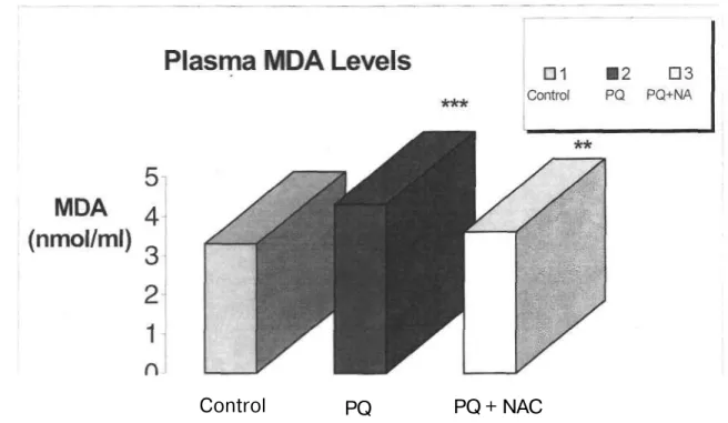
Fukushima et al. (24) demonstrated that increase in LPO may be observed by only high dose PQ exposure (40 mg/kg body weight) in rat lung but not by low dose exposure (10 mg/kg body weight). In another study performed Suntres and Shek (25) proved that blood, lung and liver peroxides increase after 6 hours of PQ exposure, decrease after 12 and 24 hours and increase again after 48 hours. Taking these studies into account, PQ is administered to mice at a single dose (40 mg/kg, i.p.). No mortality is observed in PQ intoxicated mice after 6 hours of exposure. In this study, a significant increase in MDA levels in plasma and liver of PQ-intoxicated mice is observed comparedc with the control group (p<0.001) (Figure 2, 3).
264 Şebnem Ş. ÇEÇEN, Gülhan CENGİZ, Tülin SÖYLEMEZOĞLU
Control
PQ
PQ + NAC
Figure 2. Effects of PQ, NAC and PQ+NAC on plasma MDA levels.
Results are obtained as the mean (±SEM) of nine mice per group. PQ: Paraquat; NAC: N-Acetylcysteine. MDA levels in plasma is expressed as nmol/ml.
***p<0.001, **p<0.01.
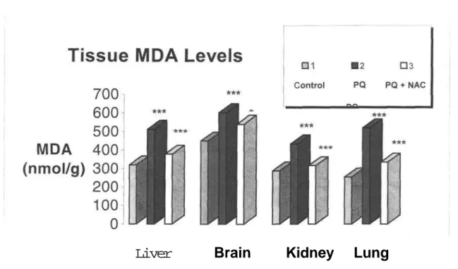
The lung is the target organ for toxicity due to its accumulation through a process of active transport in the Clara cells and type I and type II alveolar epithelial cells (7,26). The high concentration of PQ in lung is reasoned as the mistakenly accumulation of PQ because of its structural similarity to endogenous polyamines such as putrescine and spermidine (27). Shu et al. (26) indicated that high dose exposure of PQ decreases lipid soluble antioxidant levels in lungs which results in LPO. Our results showing correlation with the above results, indicates a very significant increase in lung MDA levels (p<0.001) (Figure 3).
Liver
Brain Kidney Lung
Figure 3. Effects of PQ, NAC and PQ+NAC on MDA levels in mice tissue.
Results are obtained as the mean ( SEM) of nine mice per group. PQ: Paraquat; NAC: N-Acetylcysteine. Lipid peroxidation (LPO) levels are expressed as nmol MDA/g wet weight.
***p<0.001,*p<0.05.
Paraquat also damages brain. The reason for the remarkable increase observed in brain MDA levels in the group administered PQ only (p<0.001) may be as follows:
(a) Brain is a highly oxygenated organ taking most of its energy from oxidative metabolism of the mitochondrial respiratory chain.
(b) It may not be probable to scavenge all oxygen radicals formed because of its poor antioxidant enzyme activities such as SOD, catalase and gluthathione peroxidase. (c) Brain membrane lipids are very rich in polyunsaturated fatty acids (28).
Paraquat also may induce Parkinsonism in workers exposed to it (4,29).
A big portion of PQ elimination is done by kidneys. Recent studies have shown that PQ induces renal tubuler necrosis (30,31). The same significancy in the increases in MDA levels determined in brain and lung is also observed (p<0.001).
These results suggest that, PQ is not organospecific and is effective on all the organs taken into consideration in this study.
266 Şebnem Ş. ÇEÇEN, Gülhan CENGİZ, Tülin SÖYLEMEZOĞLU
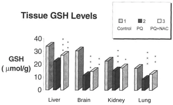
Glutathione is the most important and widely occuring nonprotein thiol in living systems (11). Mice dosed with PQ showed a significant decrease in GSH levels in all tissues studied (p<0.001).
There may be several reasons which induce and/or cause depletion in GSH levels in tissues:
(a) Enchanced oxidation of redox cycling xenobiotics such as PQ. (b) Either decreased synthesis or increased degradation of cellular GSH.
(c) Excessive usage of GSH in detoxification of foreign compounds and in removal of peroxides (12,13).
Sandy et al. (32) reported that, the rate of redox cycling of PQ and other bipyridilium derived herbicides is directly related to the depletion of GSH and the cytotoxicity caused by these compounds.
In normal physiological conditions, antioxidant defence systems found in the organisms are efficient to prevent the damage in cell membranes. However, several xenobiotics such as PQ, induces the formation of free radicals via decreasing the capacity of cell to protect itself. Melchiorri et al. (33) indicated that PQ decreases the amount of reduced GSH while increasing the oxidized one. These results correlate well with our current findings.
N-Acetylcysteine, a known antioxidant, a precursor of GSH and a free radical scavenger seems a perfect antidote for PQ intoxications. It is readily used as an antidote in various intoxications such as acetaminophen toxicity by inducing the increase of GSH levels in plasma and tissues (2,34,35). Wegener et al. (35) reported that the effectiveness of NAC was markedly diminished when it was given after the administration PQ while, pretreatment with it had a significant protection. It is conceivable that by NAC pretreatment the oxidant chain reaction is blocked and time is gained for the clearence of PQ from the blood by kidneys.
In the current study, NAC pretreatment induced a remarkable decrease in lung MDA levels (p<0.001) and a significant increase in GSH levels (p<0.01) which was increased by PQ administration. While the exact protective mechanism of NAC pretreatment in PQ toxicity is uncertain, there are some hypothesis such as:
(a) directly scavenges toxic oxygen free radicals (3).
(c) enchances the resistance of cells against oxidative damage especially type II cells by replenishing intracellular GSH stores by increasing intracellular substrates for the GSH reductase pathways (34).
(d) act as a precursor for cysteine which is used in GSH biosynthesis (2,8).
(e) delays lung inflammation by supressing PQ-induced release of chemoattractants (3). (f) interferes PQ uptake system and prevent mistakenly accumulation of it instead of
endogenous polyamines (34).
Intraperitoneal administration of PQ+NAC significantly decreased MDA levels in plasma (p<0.01), liver and kidney (p<0.001). There was a marked decrease in MDA levels (p<0.05) and a significant amelioration in GSH levels in brain (p<0.01) compared to PQ-treated group. However, these results are not as significant as the results obtained in liver. In the same group a significant amelioration is observed in liver, brain (p<0.01) and kidney (p<0.05) GSH levels (Figure 4). As a result, the strong antioxidant activity in liver is obvious which is confirmed by many researchers whereas the neuroprotective effect of this antidote still needs further
Liver
Brain
Kidney Lung
Figure 4. Effects of PQ, NAC and PQ+NAC on GSH levels in mice tissue.
Results are obtained as the mean (±SEM) of nine mice per group. PQ: Paraquat; NAC: N-Acetylcystcir Glutathione levels are expressed mol/g wet weight.
268 Şebnem Ş. ÇEÇEN, Gülhan CENGİZ, Tülin SÖYLEMEZOĞLU
Present results suggest that NAC pretreatment caused benefical changes in oxidative stress markers in PQ intoxication. However, the details of its mechanism of action and the neuroprotective effect of this antidote still needs further investigations. In addition, considering other enzymatic levels in further investigations may lead us to improve an effective protective way in PQ intoxications.
REFERENCES
1. Hara, S., Endo, T., Kuriwa, F. and Kano, S. "Different effects of paraquat on microsomal lipid peroxidation in mouse, brain, lung and liver", Pharmacol. Toxicol., 68,260-265(1991).
2. Dawson, J. R., Norbeck, K., Amundi, I. and Moldeus, P. "The effectiveness of N-acetylcysteine in isolated hepatocytes, against the toxicity of paracetamol, acrolein, and paraquat", Arch. Toxicol., 55, 11-15 (1984).
3. Eisenmann, A., Armali, Z., Eisenkraft, B., Bentur, L., Bentur, Y., Guralnik, L. and Enat, R. "Nitric oxide inhalation for paraquat-induced lung injury" Clin. Toxicol.. 36(6), 575-584 (1998).
4. Yang, W. and Sun, A. Y. "Paraquat-induced free radical reaction in mouse brain microsomes", Neurochem. Res., 23(1), 47-53 (1998).
5. Goldfrank, L. R., Flomenbaum, N. E., Lewin, N. A., Weisman, R. S., Howland, M. A. and Hoffman, R. S. Herbicides:Paraquat and Diquat. Goldfrank's Toxicologic Emergencies. 6 th ed. Appleton & Lange. U.S.A., p.: 1472-1484. (1998).
6. Smith, L. L. "Mechanism of paraquat toxicity in lung and its relevance to treatment", Hum. Toxicol., 6,31-36(1987).
7. Kornbrust, D. J. and Mavis, R. D. "The effect of paraquat on microsomal lipid peroxidation in vitro and in vivo", Toxicol. Appl. Pharmacol., 53, 323-332 (1980). 8. Drost, E., Lannan, S, Bridgeman, M., M., E., Brown, D., Selby, C, Donaldson, K.
and MacNee, W. "Lack of effect of N-Acetylcysteine on the release of oxygen radicals from neutrophils and alveolar macrophages", Eur. Respir. J. 4, 723-729 (1991).
9. Sies, H. and De Groot H. "Role of reactive oxygen species in cell toxicity", Toxicol Lett., 64/65,547-551 (1992).
10. Ward, P. A. "Host defence mechanisms responsible for lung injury", J. Allergy. Clin.
Immunol, 78(3), 373-378 (1986).
11. Reed, J. D. "Glutathione: Toxicological implications", Annu. Rev. Pharmacol. Toxicol.,
30,603-631 (1990).
12. De Vries, N. and De Flora, S. "N-acetylcysteine" J. Cell. Biochem., suppl 17F:
270-277(1993).
13. Hazelton, G.A. and Lang, C. A. "Glutathione contents in the aging mouse" Biochem.
Pharmacol, 188,25-30(1980).
14. Benrahmoune, M., Therand, P. and Abedinzadeh, Z. "The reaction of superoxide
radical with N-acetylcysteine", Free Rad. Biol. Med., 29(8), 775-782 (2000).
15. Buege, J. A. and Aust, S. D. "Microsomal lipid peroxidation", Meth. Enzymol., 52,
303-310(1978).
16. Jamall, I. S.and Smith, J. C. "Effects of cadmium on glutathione peoxidase,
superoxide dismutase, and lipid peroxidation in the rat heart: A possible mechanism of cadmium cardiotoxicity", Toxicol. Appl. Pharmacol, 80, 33-42 (1985).
17. Sedlak, J. and Lindsay, R. H. "Estimation of total protein-bound and nonprotein
sulfudryl groups in tissue with Ellman's reagent", Anal. Biochem., 25, 192-205 (1968).
18. Halliwell, B. "Reactive oxygen species in living systems: Source, biochemistry, and
role in human disease", Am. J. Med., 91, 14-22 (1991).
19. Halliwell, B. "Oxidants and human disease some new concepts", Faseb J., 1, 358-364,
(1987).
20. Basaga, H. S. "Biochemical aspects of free radicals", Biochem. Cell. Biol, 68, 989-998 (1989).
21. Kapus, H. and Sies, H. "Toxic drug effects associated with oxygen metabolism: Redox cycling and lipid peroxidation" Experentia., 37(12), 1233-1241 (1981).
22. Bagchi, D., Bagchi, M., Hassoun, E., Moser, J. and Stohs, S. J. "Effects of carbon
tetrachloride, menadione, and paraquat on the urinary excretion of MDA, formaldehyde, acetaldehyde, and acetone in rats", J. Biochem. Toxicol, 8(2), 101-106 (1993).
270 Şebnem Ş. ÇEÇEN, Gülhan CENGİZ, Tülin SÖYLEMEZOĞLU
23. Wolfgang, G.H.I., Jolly, R. A., Donarski, W. J. and Petry, T. W. "Inhibition of diquat-induced lipid peroxidation and toxicity in precision-cut rat liver slices by novel antioxidants", Toxicol. Appl. Pharmacol, 108, 321-329. (1991).
24. Fukushima, T., Yamada, K., Hojo, N., Isobe, A., Shikawu, K. and Yamane, Y. "Mechanisms of cytotoxicity of paraquat.III. The effects of acute paraquat exposure on the electron transport system in rat mitochondria", Exp. Toxicol. Pathol., 46(6), 437-441 (1994).
25.~Suntress, Z. E. and Shek, P. N. "Liposomal alpha -tocopherol alleviates the progression of paraquat-induced lung damage", J. Drug. Target., 2(6), 493-500. (1995). 26. Shu, H., Talcot, R. E., Rice, S. A. and Wei, E. T. "Lipid peroxidation and paraquat
toxicity", Biochem. Pharmacol., 28, 327-331 (1979).
27. Bayoumi, A. E., Perez-pertejo, Y., Ordonez, C, Reguera, R. M., Cubria, J. C. Balana-fouce, R. and Ordonez, D. "Alterations on polyamine content and gluthathione metabolism induced by different concentrations of paraquat in CHO-K1 cells", Toxicol. In vitro., 14,211-217(2000).
28. Pellegreni-Giampietro, D. E. "Free radicals and the pathogenesis of neuronal death cooperative role of excitatory amino acids. Free radicals in diagnostic medicine" Plenum Press, New york, p:59-71 (1994).
29. Ellenhorn, J. M., Barceloux, D. G. "Chemical Products. Medical Toxicology: Diagnosis and Treatment of human poisoning" Elsevier Science Publishing Comp. New York., p.: 1088-1092(1988).
30. Chan, B. S. H., Seale, J. P. and Duggin, G. G. "The mechanism of excretion of paraquat in rats", Toxicol. Lett., 90, 1-9. (1997).
31. Murray, R.E. and Gibson, J. E. "Paraquat disposition in rats, guinea pigs and monkeys", Toxicol. Appl. Pharmacol., 21, 283-291 (1974).
32. Sandy, M. S., Moldeus, P., Ross, D. and Smith, M. T. "Role of redox cycling and lipid peroxidation in bipyridil herbicide cytotoxicity", Biochem. Pharmacol, 35(18), 3095-3101 (1986).
33. Melchiorri, D., Retter, R, J., Sewerynek, E., Hara, M., Cheen, L. and Nistico, G. "Paraquat toxicity and oxidative damage. Reduction by melatonin", Biochem. Pharmacol., 51(8), 1096-1099 (1996).
34. Hoffer, E., Baum, Y., Tabak, A. and Taitelman, U.. "N-Acetylcystein increases the glutathione content and protects rat alveolar type II cells against paraquat-induced cytotoxicity", Toxicol. Lett., 84, 7-12 (1996)
35. Wegener, T., Sandhagen, B., Chan, K. W. and Saldeen, T. "N-Acetylcystein in paraquat toxicity: Toxicological and histological evaluation in rats", Upsala. J. Med. Sci, 93,81-89(1988).
Başvuru Tarihi: 12.09.2002 Kabul Tarihi: 07.10.2002