Relationship between Thyroid Volume and
Baseline Vitamin D Levels in New-Onset Graves
Disease
Gülşah Yenidünya Yalın
1, Hüseyin Çelik
2, Fuat Cenik
3, Serkan Feyyaz Yalın
4 1Division of Endocrinology and Metabolism Disorders, Başkent University, School of Medicine,İstanbul, Turkey
2Clinic of Endocrinology and Metabolism Disorders, Kanuni Sultan Süleyman Training and Research
Hospital, İstanbul, Turkey
3Clinic of Internal Medicine, Kanuni Sultan Süleyman Training and Research Hospital, İstanbul, Turkey 4Clinic of Nephrology, Kartal Dr. Lütfi Kırdar Training and Research Hospital, İstanbul, Turkey
ABSTRACT
Objective: Serum vitamin D is shown to be decreased and associated with higher thyroid volumes in Graves disease (GD). We aimed to
investigate the relationship between thyroid volume and baseline serum vitamin D status in newly diagnosed GD patients.
Methods: This was a single-center cross-sectional study with a total of 61 new-onset GD patients (n=61, F: 40, M: 21) who were
divided into two groups, according to baseline serum vitamin D levels, as Group-1 (vitamin D <20; n: 42) and Group-2 (vitamin D ≥20; n=19). Thyroid volume (mL) and isthmus measurements (mm) were compared between the two groups.
Results: There was an inverse correlation between the baseline serum vitamin D levels and thyroid volume, thyroid receptor
au-toantibodies (TRAb), free triiodothyronine (fT3), and parathyroid hormone (PTH) levels (p=0.02, r=-0.31; p=0.005, r=-0.36; p=0.04, r=-0.26; p=0.02, r=-0.32, respectively). Thyroid volume was also correlated with serum free thyroxine (fT4), fT3, TRAb, and thyroid peroxidase autoantibodies (TPOAb; p=0.001, r=0.426; p=0.001, r=0.50; p=0.04, r=0.26; p=0.001, r=0.42, respectively). Low vitamin D and high thyroglobulin antibody (TgAb) levels were significantly associated with thyroid volume based on a regression analysis (p=0.03, odds ratio [OR]:18.7, 95% confidence interval [CI]: 1.34-260.91 and p=0.04, OR: 16.6, 95% CI: 1.07-255.64, respectively).
Conclusion: Baseline serum vitamin D levels are inversely related with thyroid volumes, fT3, and TRAb levels in new-onset GD.
In addition to several advantages, optimization of vitamin D levels would also be beneficial in the surveillance of these patients. However, larger scale studies are required to make further suggestions.
Keywords: Autoimmune disorders, 25 (OH) vitamin D, TRAb, Graves disease, thyroid volume
ORCID IDs of the authors: G.Y.Y. 0002-9013-5237; H.Ç. 0003-0775-5978 ; F.C. 0003-2063-541X; S.F.Y.
0000-0002-8146-6966.
Corresponding Author: Gülşah Yenidünya Yalın E-mail: [email protected] Received: 20.03.2018 • Accepted: 02.04.2018
Original Research
173
INTRODUCTION
Vitamin D is as a pro-hormone in the regulation of calcium and phosphate levels, and therefore it is particularly essential for bone and mineral metabolism. However, in recent years, many studies have been demonstrating an effect of vitamin D deficien-cy in several diseases, such as cancer, hypertension, cardiovascu-lar diseases, and diabetes mellitus as well as autoimmune thy-roid disorders (1). Although vitamin D levels of over 20 ng/mL are considered sufficient for its skeletal effects, values over 30 ng/mL are required to avoid manifestation of vitamin D deficiency in the organs outside the skeletal system (2).
Vitamin D mediates its effects on autoimmune disorders by triggering immune responses through its receptors on macro-phages, dendritic cells, and T and B lymphocytes. Thus, the re-lationship between vitamin D deficiency and autoimmune thy-roid disorders may also be related with vitamin D receptor (VDR)
gene polymorphisms as well as environmental factors. Recently, Graves disease (GD) has been associated with vitamin D deficien-cy (3-6). Serum 25(OH)D levels were shown to be significantly lower and inversely correlated with thyroid volume in patients with new-onset GD compared to control subjects (7). Vitamin D deficiency was also reported to be associated with lower remis-sion rates in GD (8, 9). In this study, we aimed to investigate the impact of baseline vitamin D levels on thyroid volume in patients with new-onset GD.
METHODS
Study Group
The patients who were admitted to the endocrinology and me-tabolism outpatient clinic with a diagnosis of new-onset GD were included in the study (n=61, F: 40, M: 21). The serum vitamin D levels were measured using the competitive protein- binding
assay. The vitamin D status was defined as deficient, inadequate, and sufficient for vitamin D levels of <20 ng/mL, 20-30 ng/mL, and >30 ng/mL, respectively (1). According to the baseline se-rum vitamin D status at the time of diagnosis, the patients were divided into three groups: vitamin D deficient group (Group-1; n: 42, F/M: 31/11), vitamin D inadequate group (Group-2; n=10, F/M: 4/6), and vitamin D sufficient group (Group-3; n: 9, F/M: 5/4). The diagnosis of GD was based on the standard clinical criteria, thyroid function tests with autoantibody levels, and thyroid scin-tigraphy imaging. The serum free triiodothyronine (fT3) and free thyroxine (fT4) levels were measured using a competitive en-zyme immunoassay. Serum thyroid stimulating hormone (TSH), thyroid receptor autoantibodies (TRAb), thyroid peroxidase au-toantibodies (TPOAb), and thyroglobulin antibody (TgAb) levels were measured using a two-site immunoenzymetric assay. Thy-roid volume (mL) and isthmus measurements (mm) were com-pared between each group. Thyroid volume was calculated with using the following standardizsed formula (10).
Thyroid Volume (mL)=0.479×[Right lobe depth × width × length(cm)]+0.479 × [Left lobe depth × width × length(cm)] Thyroid ultrasonography imaging for the calculation of thyroid volumes (mL) and isthmus measurements (mm) were performed by the same physician using Logic 7, General Electric, Milwau-kee, Wiskonsin. Patients with a history of thyroidectomy, renal disease, hepatic disease, or malignancy or those under medica-tions that affect vitamin D status and having prior replacement of vitamin D in the last 6 months were excluded from the study. The patients younger than 18 years and older than 75 years were also not included in the study groups.
This study was approved by the ethics committee of Kanuni Sultan Süleyman Training and Research Hospital on 17.06.2016 (2016/16) and written informed consent was obtained from all patients. All procedures were performed in accordance with the Declaration of Helsinki.
Statistical Analysis
The distribution of variables was evaluated using the Kolmogor-ov-Smirnov test and a histogram analysis. The mean±standard er-ror of the mean (SEM) and frequency values are reported for each data. The three groups were compared using ANOVA, Kruskal-Wal-lis, and Mann-Whitney U tests, with the post-hoc Bonferroni adjust-ment as appropriate. for statistically significant results. Categorical variables were processed using the chi-square test. The Pearson and Spearman correlation analysis was performed for normally and non-normally distributed data, respectively. A statistical signif-icance was accepted at a p value of <0.05. Statistical calculations were performed using the Statistical Package for Social Sciences 22.0 software (SPSS IBM Corp.; Armonk, New York, USA).
RESULTS
A total of 61 patients were included in the study (F: 40, M: 21). The serum TSH levels were below 0.005 µIU/mL, and the fT4 and fT3 levels were above upper limit of normal (1.71 ng/dL, 4.4 pg/mL) in all the patients. The mean age and vitamin D levels of the patients were 34.8±1.2 years (18-61) and 13.8±1.3 (2.5-39), respectively,
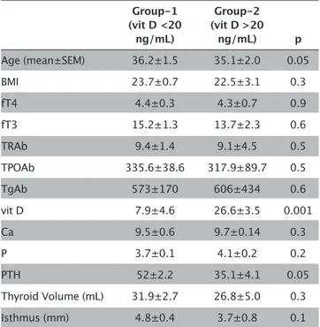
(mean±SEM [min-max]). The distribution of the clinical features according to vitamin D levels are shown in Table 1. Thyroid vol-umes and isthmus measurements did not show significant differ-ence according to the baseline serum vitamin D levels at the time of diagnosis (p=0.8 and p=0.1, respectively; Table 1).
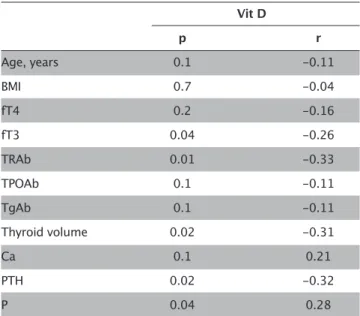
The presence of Graves ophthalmopathy was similar between the three groups (p=0.7). Thyroid volume was higher in males and in patients with body mass index (BMI) >25 (p=0.001 and 0.04, re-spectively). The thyroid volume did not show significant differ-ence according to presdiffer-ence of Graves ophthalmopathy (Table 2). There was a positive correlation between thyroid volume and serum fT4, fT3, TRAb, TPOAb, and TgAb levels (p=0.001, r=0.426; p=0.001, r=0.50; p=0.04, r=0.26; p=0.001, r=0.42; p=0.001 r=0.42; respectively; Table 3). There was an inverse correlation between serum vitamin D levels and thyroid volume, TRAb, fT3, and para-thyroid hormone (PTH) levels (p=0.02, r=-0.31; p=0.005, r=-0.36; p=0.04, r=-0.26; p=0.02, r=-0.32; respectively; Table 4).
Low baseline vitamin D and high TgAb levels were significantly as-sociated with higher thyroid volume in a logistic regression analy-sis (p=0.03, odds ratio [OR]: 18.7, 95% confidence interval [CI]:1.34-260.91; p=0.04, OR: 16.6, 95% CI: 1.07-255.64, respectively; Table 5).
DISCUSSION
The major effect of vitamin D is on the regulation of bone and mineral homeostasis; however, it has been recently shown that hypovitaminosis D is also associated with extraskeletal disorders,
Table 1. Distribution of clinical features according to the
vitamin D status Group-1 Group-2 (vit D <20 (vit D >20 ng/mL) ng/mL) p Age (mean±SEM) 36.2±1.5 35.1±2.0 0.05 BMI 23.7±0.7 22.5±3.1 0.3 fT4 4.4±0.3 4.3±0.7 0.9 fT3 15.2±1.3 13.7±2.3 0.6 TRAb 9.4±1.4 9.1±4.5 0.5 TPOAb 335.6±38.6 317.9±89.7 0.5 TgAb 573±170 606±434 0.6 vit D 7.9±4.6 26.6±3.5 0.001 Ca 9.5±0.6 9.7±0.14 0.3 P 3.7±0.1 4.1±0.2 0.2 PTH 52±2.2 35.1±4.1 0.05 Thyroid Volume (mL) 31.9±2.7 26.8±5.0 0.3 Isthmus (mm) 4.8±0.4 3.7±0.8 0.1
BMI: body mass index, fT4: free thyroxine; fT3: free triiodothyronine; TRAb: thyroid receptor antibody; TPOAb: thyroid-peroxidase antibody; TgAb: thyroglobulin antibody; vit D: vitamin D; Ca: calcium, P: phosphorus; PTH: parathyroid hormone; SEM: standard error mean
such as hypertension, diabetes mellitus, malignancy, cardiovas-cular and autoimmune thyroid diseases (1). Vitamin D regulates inflammatory cytokine production and inhibits the proliferation of proinflammatory cells through its receptors on lymphocytes and macrophages (11). The effects of vitamin D on monocytes and macrophages is in favor of activating the innate immune sys-tem;, however, there is an inhibitory effect on the acquired im-mune response (12). These effects represent the
immunomod-ulatory action, which results in an association between vitamin D deficiency and autoimmune disorders. Vitamin D inhibits the production of Th1 cells by suppressing the function of interleu-kin (IL)-2, IL-12, and interferon (IFN)-γ and stimulating IL-4, there-by shifting the polarization of T cells towards the Th2 phenotype (13). The decrease in this inhibitory effect on Th1 production is related to an increase in the autoimmune thyroid disorders with vitamin D deficiency (1).
The relationship between low vitamin D levels and GD has been previously reported (14, 15). GD is characterized by a loss of
im-Table 2. Relationship of thyroid volume with categorical
variables Thyroid Volume (mean±SEM) p Gender Female 25±2.8 0.001 Male 40±2.7 Ophthalmopathy Absent 28.7±2.5 0.1 Present 35.8±4.3 Age, years <40 31.1±2.8 0.8 >40 30.2±3.4 BMI <25 28.3±2.5 0.04 >25 39.4±5.2
Chi-square; SEM: standard error mean; BMI: body mass index
Table 3. Correlation of thyroid volume and isthmus thickness
with clinical features
Thyroid Volume Isthmus Thickness
p r p r Age, years 0.7 0.04 0.6 0.07 BMI 0.2 0.15 0.04 0.27 fT4 0.001 0.45 0.001 0.52 fT3 0.001 0.50 0.001 0.55 TRAb 0.04 0.26 0.001 0.40 TPOAb 0.001 0.42 0.1 0.21 TgAb 0.001 0.42 0.8 -0.02 Vit D 0.02 -0.31 0.3 -0.12 Ca 0.1 0.21 0.6 0.06 PTH 0.3 0.13 0.9 -0.004 P 0.7 0.03 0.1 -0.21
Pearson, Spearman; BMI: body mass index; fT4: free thyroxine; fT3: free triiodothyronine; TRAb: thyroid receptor antibody; TPOAb: thyroid-peroxi-dase antibody; TgAb: thyroglobulin antibody; vit D: vitamin D; Ca: calcium; P: phosphorus; PTH: parathyroid hormone
Table 4. Correlation of vit D levels with clinical features Vit D p r Age, years 0.1 -0.11 BMI 0.7 -0.04 fT4 0.2 -0.16 fT3 0.04 -0.26 TRAb 0.01 -0.33 TPOAb 0.1 -0.11 TgAb 0.1 -0.11 Thyroid volume 0.02 -0.31 Ca 0.1 0.21 PTH 0.02 -0.32 P 0.04 0.28
BMI: body mass index; fT4: free thyroxine; fT3: free triiodothyronine; TRAb: thyroid receptor antibody; TPOAb: thyroid-peroxidase antibody; TgAb: thyroglobulin antibody; vit D: vitamin D; Ca: calcium, P: phosphorus; PTH: parathyroid hormone
Table 5. Logistic regression analysis of risk factors
asociatedassociated with thyroid volume
Thyroid volume p OR CI 95% Vitamin D 0.03 18.7 1.34-260.91 Age, years 0.8 1.19 0.19-7.2 BMI 0.1 0.14 0.18-1.11 Gender 0.002 142.85 0.0-0.15 fT4 0.5 0.38 0.01-9.33 fT3 0.1 13.9 0.68-285,67 TRAb 0.4 0.30 0.02-4.25 TPOAb 0.4 0.47 0.07-3.04 TgAb 0.04 16.6 1.07-255.64
BMI: body mass index; fT4: free thyroxine; fT3: free triiodothyronine; TRAb: thyroid receptor antibody; TPOAb: thyroid-peroxidase antibody; TgAb: thyroglobulin antibody; vit D: vitamin D; PTH: parathyroid hormone; OD: odds ratio; CI: confidence interval
mune tolerance to thyroid antigens leading to the inflammation of thyroid gland (3). Thus, the decreased inhibitory effect on the immune system caused by hypovitaminosis D is thought to act as a further reinforcement in the development of GD. Vitamin D levels were shown to be decreased and related with thyroid vol-ume in patients with new-onset GD (7). Furthermore, lower levels of vitamin D were reported to be associated with lower rates of remission in patients with GD (8). To the best of our knowledge, this is the first study investigating the change in thyroid volumes according to the baseline serum vitamin D levels among patients with new-onset GD. In our study, the mean thyroid volume and isthmus measurements did not show significant difference be-tween the three groups according to the vitamin D status. How-ever, thyroid volumes had an inclination to be higher in patients with lower vitamin D levels revealing a negative correlation be-tween the two parameters. In contrast, thyroid volumes showed positive correlation with thyroid hormones and autoantibody levels. Higher autoantibody levels causing enhanced stimulation of thyroxine synthesis, accompanied with a further increase in the volume of thyroid gland, is the possible explanation of this relation.
In an animal in vivo autoimmune thyroiditis model, vitamin D treatment is shown to reduce the severity of inflammatory le-sions in the thyroid gland (16). It is also reported that lower vi-tamin D levels are associated with higher thyroid autoantibody levels (17, 18). In our study, there was an inverse correlation between baseline vitamin D levels and serum TRAb titers. This may be associated with the lack of an inhibitory action on the immune system due to decreased vitamin D levels. In our study, there was a tendency to higher fT3 levels in patients with low-er levels of vitamin D, which may be related with the increased TRAb titers and further stimulation of the inflammatory changes in the thyroid gland.
In addition to vitamin D deficiency due to environmental factors, vitamin D receptor (VDR) polymorphism is also reported to be an important cofactor in the development of GD (19). In a recent meta-analysis, significant difference in the association of vitamin D levels and GD has been reported in African and Asian patients, while no significant difference was found in the European popu-lation (20). These differences might be related with the VDR poly-morphisms among different ethnic populations. The increased prevalence of autoimmune thyroid disorders according to VDR polymorphisms has been reported in several studies (19, 21). The most common polymorphisms are reported as Apal and Fokl polymorphisms in Hashimoto disease and Taql polymorphism in GD (22). In a study performed in the Turkish population, Taql and Fokl genotypes were shown to be associated with higher prevalence of Hashimoto thyroiditis (23). Despite the negative correlation of vitamin D and thyroid volumes, the lack of differ-ence in the mean thyroid volumes between the three groups in our study may be due to the presence of these polymorphisms; however, further studies are needed to demonstrate the VDR genotypes related with GD in the Turkish population.
In our study, thyroid volume was higher in male patients accom-panied with higher BMI values, which was also related with an
increased gland size. There was no significant difference in the rates of ophthalmopathy development according to the vita-min D levels, and the thyroid volumes were similar in patients with and without Graves ophthalmopathy. Previous studies have shown that there was no difference in the severity of GD accord-ing to the vitamin D status (7, 24). However, higher fT3 and TRAb levels were observed with lower vitamin D levels in our study. Nevertheless, the ophthalmopathy presence was similar be-tween the three groups. The higher fT3 and TRAb levels may also be the reason for lower remission rates of GD in vitamin D defi-ciency, which has been previously reported (8).
The limitation to our study is that it was a single-center cross-sec-tional study with a limited number of patients. Prospective larg-er scale studies with vitamin D treatment and follow- up for the onset of remission are needed to make definitive suggestions. Studies of vitamin D receptor polymorphisms associated with GD among different population groups would also be beneficial for further conclusions.
CONCLUSION
Vitamin D deficiency is an important risk factor in the develop-ment of autoimmune thyroid disorders. Baseline serum vitamin D levels are inversely related to fT3, TRAb levels, and thyroid vol-umes, which are related to the adverse outcomes in GD. There-fore, in addition to several advantages, optimization of vitamin D levels would also be beneficial on the surveillance of these patients. However, larger scale studies on different ethnic popu-lations are required to make further suggestions.
Ethics Committee Approval: Ethics committee approval was received for
this study from the ethics committee of Kanuni Sultan Süleyman Training and Research Hospital.
Informed Consent: Written informed consent was obtained from all
pa-tients who participated in this study.
Peer-review: Externally peer-reviewed.
Author Contributions: Concept - G.Y.Y.; Design - H.Ç.; Supervision - G.Y.Y.;
Materials - F.C.; Data Collection and/or Processing - S.F.Y.; Analysis and/or Interpretation - G.Y.Y.; Literature Search - H.Ç.; Writing Manuscript - G.Y.Y.; Critical Review - G.Y.Y.
Conflict of Interest: The authors have no conflicts of interest to declare. Financial Disclosure: The authors declared that this study has received
no financial support.
REFERENCES
1. Bizzaro G, Shoenfeld Y. Vitamin D and autoimmune thyroid diseases: facts and unresolved questions. Immunol Res 2015; 61: 46-52. [CrossRef]
2. Vondra K, Starka L, Hampl R. Vitamin D and Thyroid Diseases. Physiol Res 2015; 64: 95-100.
3. Kivity S, Agmon-Levin N, Zisappl M, Shapira Y, Nagy EV, Danko K et al. Vitamin D and autoimmune thyroid diseases. Cell Mol Immunol 2011; 8: 243-7. [CrossRef]
4. Kurylowicz A, Ramos-Lopez E, Bednarczuk T, Badenhoop K. Vitamin D-binding protein (DBP) gene polymorphism is associated with Graves’ disease and the vitamin D status in a Polish population study. Exp Clin Endocrinol 2006; 114: 329-35. [CrossRef]
5. Abd el Gawad SS, Samee Er Abdul, Metwali AA, Abd el Gawad MS. Vita-min D receptor gene polymorphism and its association with 1,25-dihy-droxyvitamin D in patients with Graves disease in an Egyptian popula-tion: a pilot study. Endocr Pract 2012; 18: 132-9. [CrossRef]
6. Ramos-Lopez E, Kurylowicz A, Bednarczuk T, Paunkovic J, Seidl C, Badenhoop K. Vitamin D receptor polymorphisms are associated with Graves’ disease in German and Polish but not in Serbian pa-tients. Thyroid 2015; 15: 1125-30. [CrossRef]
7. Yasuda T, Okamoto Y, Hamada N, Miyashita K, Takahara M, Saka-moto F. Serum vitamin D levels are decreased and associated with thyroid volume in female patients with newly onset Graves’ disease. Endocrine 2012; 42: 739-41. [CrossRef]
8. Yasuda T, Okamoto Y, Hamada N, Miyashita K, Takahara M, Sakamo-to F, et al. Serum vitamin D levels are decreased in patients without remission of Graves’ disease. Endocrine 2013; 43: 230-2. [CrossRef]
9. Hollick MF. Vitamin D deficiency. N Engl J Med 2007; 357: 266-81.
[CrossRef]
10. Shabana W, Peeters E, De Maeseneer M. Measuring thyroid gland volume: should we change the correction factor? AJR Am J Roent-genol 2006; 186: 234-6. [CrossRef]
11. Yin K, Agrawal DK. Vitamin D and inflammatory diseases. J Inflamm Res 2014; 7: 69-87.
12. Bouillon R. Vitamin D: from photosynthesis, metabolism and action to clinical applications. In: Endocrinology, Jameson JL, De Groot LJ, Saunders Elsevier, Philadelphia 2010.
13. Baeke F, Takiishi T, Korf H, Gysemans C, Mathieu C. Vitamin D: modu-lator of the immune system. Curr Opin Pharmacol 2010; 10: 482-96.
[CrossRef]
14. Li YB, Xue XH, Liu SW, Xi GX, Zhao LX, Zhang XL. Clinical research of se-rum vitamin D in early Graves disease. Chin Rem Clin 2014; 14: 242-3. 15. Miao W, Ma J, Guo R, Wang YJ, Wang G, Guan HX. Research about
the correlation between Serum 25(OH)D with Graves’ disease. Chin. J Pract Internal Med 2013; 33: 394-95.
16. Fournier C, Gepner P, Sadouk M. In vivo beneficial effects of cyclo-sporin A and 1,25-dihydroxyvitamin D3 on the induction of experi-mental autoimmune thyroiditis. Clin Immunol Immunopathol 1990; 54: 53-63. [CrossRef]
17. Choi YM, Kim WG, Kim TY. Low levels of serum vitamin D3 are associ-ated with autoimmune thyroid disease in pre-menopausal women. Thyroid 2014; 24: 655-61. [CrossRef]
18. Zhang H, Liang L, Xie Z. Low vitamin D status is associated with in-creased titers of thyroid stimulating hormone receptor antibodies in Graves disease. Endocr Pract 2015; 21: 258-63. [CrossRef]
19. Feng M, Li H, Chen SF. Polymorphisms in the vitamin D receptor gene and risk of autoimmune thyroid diseases: a meta-analysis. En-docrine 2013; 43: 318-26. [CrossRef]
20. Xu MY, Cao B, Yin J, Wang DF, Chen KL, Lu QB. Vitamin D and Graves’ Disease: A Meta-Analysis Update.Nutrients 2015; 7: 3813-27. [CrossRef]
21. Effraimidis G, Wiersinga WM. Mechanisms in endocrinology: au-toimmune thyroid disease: old and new players. Eur J Endocrinol 2014; 170: 241-52. [CrossRef]
22. Inoue N, Watanabe M, Ishido N. The functional polymorphisms of VDR, GC and CYP2R1 are involved in the pathogenesis of autoimmune thy-roid diseases. Clin Exp Immunol 2014; 178: 262-9. [CrossRef]
23. Yazici D, Yavuz D, Tarcin O. Vitamin D receptor gene ApaI, TaqI, FokI and BsmI polymorphisms in a group of Turkish patients with Hashimoto’s thyroiditis. Minerva Endocrinol 2013; 38: 195-201. 24. Pantazi H, Papapetrou PD. Changes in parameters of bone and
min-eral metabolism during therapy for hyperthyroidism. J Clin Endocri-nol Metab 2000; 83: 1099-106. [CrossRef]
How to cite:
Yenidünya Yalın G, Çelik H, Cenik F, Yalın SF. Relationship between Thyroid Volume and Baseline Vitamin D Levels in New-Onset Graves Disease. Eur J Ther 2018; 24(3): 173-7.