Corresponding author: Atilla KIRCELLI E-mail: [email protected]
Original Investigation
Received: 02.11.2015 / Accepted: 28.12.2015 Published Online: 12.08.2016
Halil CAN
1, Atilla KIRCELLI
2, Gulis KAVADAR
3, Erdinc CIVELEK
1, Tufan CANSEVER
2, Aydın AYDOSELI
4,
Mehmet Bulent ONAL
5, Cem YILMAZ
21Private Medicine Hospital, Neurosurgery Clinic, Istanbul, Turkey 2Baskent University, Department of Neurosurgery, Ankara, Turkey
3Private Medicine Hospital, Physical Medicine and Rehabilitation Clinic, Istanbul, Turkey 4Istanbul University, Istanbul School of Medicine, Department of Neurosurgery, Istanbul, Turkey 5Private Dunya Hospital, Neurosurgery Clinic, Batman, Turkey
Lumbosacral Conjoined Root Anomaly: Anatomical
Considerations of Exiting Angles and Root Thickness
ABSTRACT
by showing different courses or making anastomoses between the nerve roots.
The first classification of CNR was suggested in 1962 by Cannon et al. (7) who distinguished 3 groups according to their courses: conjoined roots, transverse course of the root and anastomoses between roots. Even though this classification was further developed by Postacchini (21), Kadish and Simmons (13), Kikuchi (16), the most commonly used is the classification by Neidre and MacNab (17).
█
INTRODUCTION
C
onjoined nerve roots (CNRs) are congenital anomalies of the spine. The incidence has been reported to be 14% in postmortem studies and ranges between 0.3% and 30% in different series. An anatomical description of CNR was first provided by Zagnoni (24) in 1949, and subsequently, similar cases were reported by Ethelberg and Riished, Fineschi, Deyerle and May (9-11). The term CNR refers to the roots that emerge from the dura in a common root sleeve that bifurcates after leaving the dura and exits through the foramenAIm: Conjoined nerve roots (CNR) can be damaged during lumbar disc surgery and lead to neuropathic pain due to excessive retraction. The purpose of this study was to investigate the factors that facilitate the identification of CNRs that can lead to the loss of fragments in the secondary axilla, nerve root injury or unpleasant surprises during lumbar disc surgery. Accordingly, we aimed to measure the thicknesses and exit angles of the nerve roots close to the pedicle to obtain scientific data regarding rare double roots. mATERIAl and mEThODS: The data of 612 patients who were operated for lumbar disc disease in our hospital between 2012 and 2014 were reviewed retrospectively. Twenty one cases of CNR were detected in this series.
RESUlTS: The mean nerve root thickness was 1.92 ± 0.45 mm for medially located roots and 3.33 ± 0.95 mm for laterally located roots. The comparison of medially located roots versus laterally located roots revealed a significant difference in mean values (p<0.0001). The mean exit angle was 12.290± 4.890 for medially located roots, and 22.110 ± 5.420 for laterally located roots (p<0.0001).
In addition, the exit angles of the medially and laterally located roots increased as going down to caudal levels, (p=0.005, p=0.042). CONClUSION: CNRs are congenital anomalies that are usually diagnosed during the surgical procedure and affect the success of discectomy. The presence of a more medially located or thinner root during surgical exploration and the absence of the fragment in the axilla in extruded or sequestered discs usually indicate a conjoined nerve root closer to the pedicle.
According to the classification proposed by Neidre and MacNab, the nerve arises from a single dural sheath emerges from the lower foramen in Type 1 anomalies. Type 2 in which the main root bifurcates and exits through one foramen is the most common type. In this case, the adjacent foramen may be unoccupied. In Type 3, adjacent nerve roots are connected in the form of a vertical or transverse anastomosis. In Type 4, a thinner nerve root origin at the caudal section of a nerve root of normal thickness and then these two nerve roots connect at the distal and arise from the foramen as a single nerve root. This condition, described by Burke et al. (6), was first proposed by Keon-Cohen in 1968 (15), but was not included in the Neidre and Macnab classification (Figure 1).
Double roots can be damaged during lumbar disc surgery, which is a common procedure in neurosurgical practice, or can lead to neuropathic pain due to excessive retraction. The purpose of this study was to investigate the factors that facilitate the identification of CNRs that can lead to the loss of fragments in the secondary axilla, nerve root injury or unpleasant surprises during lumbar disc surgery. Accordingly, we aimed to measure the thicknesses and exit angles of the nerve roots close to the pedicle, as commonly mentioned in the literature, and to obtain scientific data regarding rare double roots.
█
mATERIAl and mEThODS
Study Design and Settings
A total of 612 lumbar disc cases operated on in our hospital between 2012 and 2014 were reviewed retrospectively and cases of conjoined nerve root anomalies in this series were presented in this series. This study was conducted as a single-center trial in our hospital. Prior to the study, an informed patient consent form was obtained from each patient.
Participants
Out of all patients who presented to our clinic with complaints
of at least a one-month history of low-back pain and unilateral-bilateral leg pain, where magnetic resonance imaging (MRI) confirmed lumbar disc hernia and who underwent microsurgical discectomy, patients with documented CNRs were included in this study. The patients having an instability problem in addition to lumbar disc herniation were not included in the study. For this purpose, medical records of the patients were reviewed retrospectively and 612 cases of lumbar disc herniation were investigated, leaving a total of 21 patients with CNRs.
Neurologic examination was performed in all cases of lumbar disc herniation detected by radiological imaging and those patients meeting the inclusion criteria were operated on.
Inclusion Criteria
All patients with extruded or sequestered lumbar disc hernia indicating lumbar radiculopathy radiologically and who had signs and symptoms of radiculopathy were included in the study.
Patients who met the inclusion criteria were operated on. Out of these patients, 21 patients with documented CNRs were included in this study. The procedure was performed under the surgical microscope in all patients and the surgical procedure was recorded. Patients undergoing surgery for recurrent lumbar disc herniation and those with spinal tumors were excluded from the study. Even though it is difficult to differentiate the double roots due to epidural fibrosis in recurrent cases, a neurological examination in the postoperative period can be misleading.
Interventions
Lumbar disc hernia was documented via the microscope by performing hemipartial laminectomy, flavectomy and forami-notomy respectively, and was followed by microdiscectomy surgery. During surgery, high-resolution images were ob-tained with the surgical microscope and a tape measure of known length was used at the surgery site in each procedure.
The patients were mobilized the next day and discharged. No patients had procedure-related complications or neurological deficits.
Data Source & measurements
Images were obtained from video recordings during surgery in patients with documented CNRs and disc herniations were evaluated according to the exit levels of the nerve roots from the dural sheath, their sites and courses, based on the MacNab classification. However, images of the dura and root were obtained from surgical recordings in the TIFF and JPEG formats and were transferred to the ImageJ program. Using the ImageJ image processing program (Rasband, WS. ImageJ, U.S. National Institutes of Health, Bethesda, Maryland, USA, http://imagej.nih.gov/ij/, 1997–2014) the measurement was performed by using the reference tape measure on the surgery site and the pixel size of an image was converted to millimeter (Figure 2). The exit sites of dual roots from the dura were verified using the program, and regardless of the exit site, the root close to the dura was called the medially located root, whereas, the root that arises laterally at the exterior of this root was called the laterally located root. In this study, the thicknesses and the angles of these roots with each other were measured using this program.
Objectives
CNR is a very rare anomaly. As mentioned in previous literature, the laterally located root that is closer to the pedicle is longer than the medially located root. In this study, a possible correlation between the thicknesses of the medially and laterally located roots and a possible relationship between root exit angles were investigated.
Outcome measurements
The results of preoperative and postoperative 1 day and 6 month examinations, the visual numeric scale (VNS-leg) and the Douleur Neuropathique 4 questionnaire (dn4) assessing neuropathic pain were evaluated and presented in this study (4,5,19). VNS is a measure of pain rated on a numerical scale from 0 (no pain) to 10 (severest pain). After patients were given necessary information about the scale, they were asked to rate their current leg and radicular pain. The DN4 survey consists of 4 questions including 10 items related to the characteristics of pain and scored as 0 or 1. A score of 4 out of 10 indicates the presence of neuropathic pain with a sensitivity of 83% and a specificity of 90%. These results were presented as means in the cohort.
Statistical Analysis
Statistical analysis of data was performed using SPSS v.21 for Windows (IBM corp., Armonk, NY, USA). The normally distributed continuous variables were reported as means± standard deviations (p>0.05 in the Kolmogorov-Smirnov test or Shapiro-Wilk test (n<30)), whereas non-normally distributed variables were described as medians. The paired T test was used for the comparison of normally distributed data among groups, and the Kruskal-Wallis test was used for non-normally distributed data. Pearson’s correlation coefficient was used to investigate a relationship between the factors. A p value of <0.05 was considered statistically significant.
█
RESUlTS
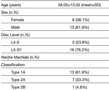
Out of a total of 612 cases, 21 cases of CNR were included in this study. Of these patients, 211 were operated for lumbar disc herniation at the level of L5-S1, 194 at the level of L4-5, 133 at the level of L3-4 and 91 at the level of L2-3. In this study, cases of CNR accounted for 3.4% of the whole series and lumbar disc herniation was most common at the level of L4-5 and L5-S1. The mean age of the patients with CNRs was 38.05±13.05 years, with 8 (38.1%) females and 13 (61.9%) males. Though most common at the L5-S1, the exit level of CNR was L4-5 in 5 (23.8%) patients and L5-S1 in 16 (76.2%). According to the MacNab classification, 13 (61.9%) patients had type 1A, 7 patients (33.3%) had type 2A and 1 patient (4.8%) had type 2B conjoined nerve roots whereas the most common was type 1A. These data as well as demographic data are presented in Table I.
Nerve root thickness was measured using the ImageJ program. Accordingly, the mean nerve root thickness was 1.92 mm± 0.45 mm for medially located roots and 3.33 mm±0.95 mm
Figure 2: Roots angle and thickness measurements with ImageJ program from the operation records.
Table I: Patients with Conjoined Root Anomaly and Demographic Factors
Age (years) 38.05±13.05 (mean±SD)
Sex (n,%) Female 8 (38.1%) Male 13 (61.9%) Disc Level (n,%) L4-5 5 (23.8%) L5-S1 16 (76.2%) Neidre MacNab (n,%) Classification Type 1A 13 (61.9%) Type 2A 7 (33.3%) Type 2B 1 (4.8%)
█
DISCUSSION
In this study, a statistically significant difference was found between the thicknesses and exit angles of the medially and laterally located roots in 21 patients with CNRs. However, it was also noted that exit angles of the medially and laterally located roots at the L5-S1 level were significantly higher than those at the L4-5 level.
The incidence of lumbosacral nerve root anomalies has been reported to range between 0.3% and 30% in different series (13-16). The incidence of CNR was reported to be 1.3% in a surgical study by White et al. (23), and CNR anomaly was observed in 2% of 8000 patients undergoing lumbosacral CT by Peyster et al. (20), and in 0.25% of 1200 patients undergoing MR and CT by Artico et al. (2). On the contrary, the incidence of CNR was reported to be 30% in a study of 60 fresh cadavers by Chotigavanich and Swanganatra (8). In our series of 612 cases of CNR, CNR anomaly was noted in 3.4%, which is consistent with other studies.
The diagnosis of lumbar disc herniation is usually established based on clinical findings and the results of MRI in micro discectomy, which is a common procedure in neurosurgical practice. Agnoli (1) emphasized that these anomalies should not be considered as the cause of low back pain and radicular pain. The findings of the physical examination are gathered in at least one or several dermatomal areas, which constitutes the main problem in patients with ruptured discs. In this case, the presence of anatomical variation should be a clue for the surgeon. On the other hand, a negative Laseque’s sign was present in 40% of patients with herniated disc. Similarly, 60% of our patients had negative Laseque’s sign. The presence of root anomalies may affect the success of discectomy. CNR can sometimes be mistaken for an extruded disc or can lead to severe radicular pain in mildly herniated discs or protrusions (18). Lumbar root anomalies are commonly observed at the L4-5 and L5-S1 levels. The L5-S1 is the most commonly involved spinal level, which is also consistent with our study in which the L5-S1 level was more common. It can also be suggested that at the level of L5-S1, which is anatomically flat with a wide interpeduncular distance, root exit angles are greater than other distances.
The findings from the study by White were contrary to a case study of 12 patients by Bouchard et al. (3) in 1978. The Laseque’s sign was positive in most patients and was suggested by Bouchard that the asymmetry of the root sleeves and the close course of the roots emerging from the dural sheath to each other by metrizamide lumbar myelogram might indicate the presence of CNR. Similar to the findings from the study by White, 40% of the patients had a positive SLR in our series, which indicates that extruded or sequestered herniated discs on radiological imaging can be a CNR anomaly, or in other words, CNR can worsen the appearance of disc herniation radiologically.
CNRs are usually diagnosed intraoperatively. Sequestered disc material may be hidden in the secondary axilla, thus complicating the identification of the fragment. In addition, for laterally located roots. The comparison of medially located
roots versus laterally located roots revealed a significant difference in mean values (p<0.0001). In addition, there was a significant correlation between medially and laterally located roots (Pearson’s correlation coefficient 0.907, p<0.0001). The exit angles of the roots were also measured using the same program. The mean exit angle was 12.290±4.890 for medially
located roots, and 22.110±5.420 for laterally located roots
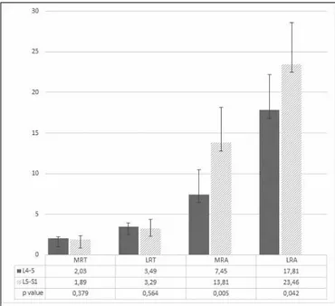
(p<0.0001). There was a high correlation between the exit angles of medially located roots and laterally located roots (Pearson’s correlation coefficient 0.801 and p<0.0001). In addition, the exit angles of the medially and laterally located roots increased as going down to caudal levels, (p=0.005, p=0.042). Figure 3 presents exit angles and thicknesses for the medially and laterally located roots with respect to disc levels.
The results of preoperative and postoperative VNS scale and DN4, a neuropathic pain scale, in patients with CNRs were recorded. The mean preoperative pain score on the VNS was 9.09 ± 0.77 and the mean pain score on the VNS at postoperative 6 month decreased to 1.86± 0.73. However, the mean DN4 score increased from 2.90±1.22 to 5.05±2.59, whereas patients with a DN4 score of 4, indicating the presence of neuropathic pain, at postoperative 6 month examination accounted for 52.4% of the whole cohort (23.8% of all patients had neuropathic pain preoperatively).
There was an increase in neuropathic pain scores of the CNR cohort at postoperative 6 months. A statistically significant relationship was noted between the thicknesses (p<0.0001 and p=0.30) and exit angles (p<0.0001 and p<0.0001) of medially located roots and lateral located roots in terms of neuropathic pain.
Figure 3: The measurement results of medial root thickness (mm)(MRT), lateral root thickness (mm) (LRT), medial roots angle (degree) (MRA) and lateral root angle (degree) (LRA) comparisons between L4-5 and L5-S1 disc levels.
small group of patients with CNRs in this study, the number of patients was larger than those in other rare CNR series. Finally, in this study, 82% of all patients with CNRs are from the eastern regions of our country, and thus, a possible genetic disposition to this anomaly should be investigated. We are planning to conduct a genetic study in this context.
█
CONClUSION
Conjoined nerve roots are congenital anomalies that are usually diagnosed during surgical procedure and affect the success of discectomy. The presence of a more medially located or thinner root during surgical exploration and the absence of the fragment in the axilla in extruded or sequestered discs usually indicate a conjoined nerve root closer to the pedicle.
█
REFERENCES
1. Agnoli AL: Anomalies of the pattern of lumbosacral nerve roots and its clinical significance (author’s transl). J Neurol 211:217-228, 1976
2. Artico M, Carloia S, Piacentini M, Ferretti G, Dazzi M, Franchitto S, Bronzetti E: Conjoined lumbosacral nerve roots: Observations on three cases and review of the literature. Neurocirugia (Astur) 17:54-59, 2006
3. Bouchard JM, Copty M, Langelier R: Preoperative diagnosis of conjoined roots anomaly with herniated lumbar disks. Surg Neurol 10:229-231, 1978
4. Bouhassira D, Attal N, Alchaar H, Boureau F, Brochet B, Bruxelle J, Cunin G, Fermanian J, Ginies P, Grun-Overdyking A, Jafari-Schluep H, Lanteri-Minet M, Laurent B, Mick G, Serrie A, Valade D, Vicaut E: Comparison of pain syndromes associated with nervous or somatic lesions and development of a new neuropathic pain diagnostic questionnaire (DN4). Pain 114:29-36, 2005
5. Bouhassira D, Attal N, Fermanian J, Alchaar H, Gautron M, Masquelier E, Rostaing S, Lanteri-Minet M, Collin E, Grisart J, Boureau F: Development and validation of the Neuropathic Pain Symptom Inventory. Pain 108:248-257, 2004
6. Burke SM, Safain MG, Kryzanski J, Riesenburger RI: Nerve root anomalies: Implications for transforaminal lumbar interbody fusion surgery and a review of the Neidre and Macnab classification system. Neurosurg Focus 35:E9, 2013 7. Cannon BW, Hunter SE, Picaza JA: Nerve-root anomalies in
lumbar-disc surgery. J Neurosurg 19:208-214, 1962
8. Chotigavanich C, Sawangnatra S: Anomalies of the lumbosacral nerve roots. An anatomic investigation. Clin Orthop Relat Res (278):46-50, 1992
9. Deyerle WM, May VR Jr: Sciatica; etiology and treatment. Clin Orthop 4:166-179, 1954
10. Ethelberg S, Riishede J: Malformation of lumbar spinal roots and sheaths in the causation of low backache and sciatica. J Bone Joint Surg Br 34-b:442-446, 1952
11. Fineschi G: Anomalie anatomiche delle radici lombari. Arch Putti Chir Org Mov 2:222-236, 1952 (In Italian)
12. Gomez JG, Dickey JW, Bachow TB: Conjoined lumbosacral nerve roots. Acta Neurochir (Wien) 120:155-158, 1993 CNRs are less mobile than normal roots. This makes them
more difficult to retract. In this circumstance, neuropathic symptoms can develop as a result of nerve injury due to excessive retraction. In a study by White et al. in 1982, a total of 4726 patients with herniated discs were operated on and CNRs were detected in 63 patients (1.3%). The authors suggested that neuropathy was more common in the secondary root, which is easily damaged because of being too close to the vertebral pedicle, and standard laminectomy should be performed in combination with pediculectomy in these patients. In this study, the nerve root located close to the pedicle was thicker than the medially located root (23). In the light of these data, we demonstrated that an increase in the thickness and exit angles of the roots was associated with increased neuropathic pain. The greater root thickness and root exit angle make the roots much more difficult to be retracted medially, thus leading to later onset neuropathy and root injury.
The embryology of root anomalies remains to be obscure. One possible mechanism for the emergence at a more caudal level is an abnormal migration of the roots during embryonic development. Bilateral anomalies of one or multiple roots as well as emergence of the roots at a more caudal level are likely to be caused by an abnormal emergence of roots from the spinal cord. Abnormal anastomosis between roots is likely to be caused by a link due to a band of nerve fibers or to be associated with a complete distal union in a common sheath. Magnetic Resonance (MR) is the gold standard for the differentiation of CNRs. A study by Gomez et al. (12) reported that the X-ray myelogram and the coronal T2 weighted MR image had equal sensitivity. Coronal MR Images provide definite data about the course of each nerve root. In addition, T1- and T2- weighted images enable differentiating between conjoined nerve roots and other space-occupying processes and the use of gadolinium contrast media in MRI enables better visualization of scar tissues in operated lumbar discs or tumors in doubtful cases. Several pathognomonic signs associated with CNRs have been described. In a study by Kang et al., the sagittal shoulder sign, a vertical structure connecting two consecutive nerve roots, was identified with a mean frequency of 90.9% on both T1 and T2 weighted sagittal MR images in herniated lumbar discs (14). In addition, Song et al. described 3 radiological signs in patients with CNRs. These signs should be detected at the level of the disc on standard axial MRI. The “corner sign” refers to an asymmetric structure of the anterolateral corner of the dural sac on T1-weighted axial MR images. The “Fat crescent sign” is the extradural fat between the asymmetric dural sac and the CNR. The “Parallel sign” is described as a nerve root running parallel to the disc plane at the disc level (22). A retrospective review of the results of MRI revealed coronary sign in 3 patients and parallel sign in 2 patients in this cohort.
There are several limitations to our study. Because of the retrospective design of the study, we were unable to carry out a comparison with the control group as in prospective, randomized or case control studies. Even though there was a
19. Perez C, Galvez R, Huelbes S, Insausti J, Bouhassira D, Diaz S, Rejas J: Validity and reliability of the Spanish version of the DN4 (Douleur Neuropathique 4 questions) questionnaire for differential diagnosis of pain syndromes associated to a neuropathic or somatic component. Health Qual Life Outcomes 5:66, 2007
20. Peyster RG, Teplick JG, Haskin ME: Computed tomography of lumbosacral conjoined nerve root anomalies. Potential cause of false-positive reading for herniated nucleus pulposus. Spine (Phila Pa 1976) 10:331-337, 1985
21. Postacchini F, Urso S, Ferro L: Lumbosacral nerve-root anomalies. J Bone Joint Surg Am 64:721-729, 1982
22. Song SJ, Lee JW, Choi JY, Hong SH, Kim NR, Kim KJ, Chung SK, Kim HJ, Kang HS: Imaging features suggestive of a conjoined nerve root on routine axial MRI. Skeletal Radiol 37:133-138, 2008
23. White JG 3rd, Strait TA, Binkley JR, Hunter SE: Surgical treatment of 63 cases of conjoined nerve roots. J Neurosurg 56:114-117, 1982
24. Zagnoni C: Reperto di un tipo non conosciuto di anastomosi nervosa delle radici spinali. Padova. Atti Soc Med-chir 27: 48-52, 1949 (In Italian)
13. Kadish LJ, Simmons EH: Anomalies of the lumbosacral nerve roots. An anatomical investigation and myelographic study. J Bone Joint Surg Br 66:411-416, 1984
14. Kang CH, Shin MJ, Kim SM, Lee SH, Kim HK, Ryu JA, Lee CS, Kim SS: Conjoined lumbosacral nerve roots compromised by disk herniation: Sagittal shoulder sign for the preoperative diagnosis. Skeletal Radiol 37:225-231, 2008
15. Keon-Cohen B: Abnormal arrangement of the lower lumbar and first sacral nerves within the spinal canal. J Bone Joint Surg Br 50:261-265, 1968
16. Kikuchi S, Hasue M, Nishiyama K, Ito T: Anatomic and clinical studies of radicular symptoms. Spine (Phila Pa 1976) 9:23-30, 1984
17. Neidre A, MacNab I: Anomalies of the lumbosacral nerve roots. Review of 16 cases and classification. Spine (Phila Pa 1976) 8:294-299, 1983
18. Pamir MN, Ozek MM, Ozer AF, Keles GE, Erzen C: Surgical considerations in patients with lumbar spinal root anomalies. Paraplegia 30:370-375, 1992