Address for Correspondence: Attila Dağdeviren, Professor Doctor, Baskent University Faculty of Medicine, Head of Histology and Embryology Department, Ankara, Turkey E-mail: [email protected]
©Telif Hakkı 2019 Gazi Üniversitesi Tıp Fakültesi - Makale metnine http://medicaljournal.gazi.edu.tr/ web adresinden ulaşılabilir. ©Copyright 2019 by Gazi University Medical Faculty - Available on-line at web site http://medicaljournal.gazi.edu.tr/
doi:http://dx.doi.org/10.12996/gmj.2019.63
Intussusceptive Growth of Vascular Bed in Human Placenta
İnsan Plasenta Damar Yatağının İntussusseptif Olarak Büyümesi
Pınar Ayran Fidan, Fatma Helvacioğlu, Attila Dağdeviren
Department of Histology and Embryology, Faculty of Medicine, Baskent University, Ankara, Turkey
ABSTRACT
Objective: Normal embryonic and fetal development is strictly bound to maternal health and functioning placenta. Besides the invasion and differentiation of trophoblastic cell lineage; development of effective vasculature is crucial for the function of placenta. Placental vessels first arise by vasculogenesis in early development of villi and then succeeded by angiogenesis during fetal life. In the recent decades a new form of angiogenesis, “intussusceptive angiogenesis”, besides classical sprouting angiogenesis is well documented. The presence of intussusception was shown at multiple organs but in placenta, in recent literature. We aimed to determine whether intussusceptive angiogenesis is present in human placenta to obtain further evidence on the development of vascular bed.
Methods: The term placenta samples were obtained from 10 healthy pregnancies following caesarean sections. Tissues were processed using routine plastic embedding technique; thin sections were contrasted with uranyl acetate & lead citrate; observed and photographed by transmission electron microscope.
Results: Our examinations revealed that both sprouting and intussusceptive angiogenesis is present in floating villi of term placenta. Phases of intussusception were documented in various samples.
Conclusion: The presence of intussusceptive angiogenesis will help our understanding of microvascular bed remodeling during pregnancy. We believe that this new finding will help us to determine the relation of microvascular bed development in normal and abnormal placentas.
Key Words: placenta, intussusceptive angiogenesis, sprouting angiogenesis, transmission electron microscopy
Received: 12.05.2017 Accepted: 03.23.2019
ÖZET
Amaç: Embriyo ve fetus gelişiminin normal olması anne sağlığı ve plasentanın işlevi ile doğrudan ilişkilidir. Trofoblast hücre dizisinin farklanması ve invazyonunun yanı sıra, etkin damar yatağının gelişmiş olması plasentanın fonksiyonu için kritik öneme sahiptir. Fetal hayat boyunca villusların erken gelişim döneminde plasenta damarları ilk olarak vaskülogenezle oluşur ve daha sonra anjiogenezle desteklenir. Son on yılda klasik anjiogenezin yanı sıra “intussusseptif anjiogenez” olarak tanımlanan yeni bir anjiogenez modeli bildirilmiştir. Literatürde birçok organda intussusseptif anjiogenez varlığı gösterilmiştir ancak plasentada böyle bir çalışma bulunmamaktadır. Çalışmamızda insan plasentasında intussusseptif anjiogenezin var olup olmadığını belirleyerek damar yatağının gelişimine katkısı ile ilgili kanıt elde etmeyi hedefledik.
Hastalar ve Yöntem: On adet sağlıklı, term gebeden sezeryan doğumu takiben plasenta örnekleri toplandı. Doku örneklerine rutin plastiğe gömme yöntemi uygulandı; ince kesitler alınarak uranil asetat & kurşun sitratla kontrastlandı; geçirimli elektron mikroskopta incelendi ve fotoğraflandı.
Bulgular: İncelemelerimiz sırasında term plasenta yüzen villuslarında anjiogenezin hem tomurcuklanma ile hem de intussusseptif olarak gerçekleştiği gözlendi. Birçok örnekte intussusseptif anjiogenezin çeşitli evreleri görüntülendi.
Sonuç: İntussusseptif anjiogenezin varlığı gebelik boyunca mikrovasküler yatağın yenilenme mekanizmasını anlamamıza yardımcıdır. İnanıyoruz ki bu yeni gözlem normal ve anormal plasentalarda mikrovasküler yatağın gelişim ilişkisini daha iyi anlamamıza yardımcı olacaktır.
Anahtar Sözcükler: Plasenta, intussusseptif anjiogenez, tomurcuklanma anjiogenez, geçirimli elektron mikroskop
Geliş Tarihi: 05.12.2017 Kabul Tarihi: 23.03.2019
Original Investigation
/
Özgün Araştırma
INTRODUCTION
Placenta represents one of the highly vascularized organs to establish a critical efficient feto-maternal interface for the exchange of nutrients and waste products for normal development.
Formation of new blood vessels (neovascularization) is essential to achieve a functional microvascular bed thus many investigators focused on the issue. Classically it is described that first blood vessels together with primitive erythrocytes arise within extraembryonic mesoderm de novo from mesenchymal cells during early embryonic life starting from 2nd week of development and this process is termed as vasculogenesis. Such blood vessels in developing placenta are first detected in tertiary villi during the 3rd week (1). Once primitive capillary network is formed and their lumen become prominent, another mechanism for the enlargement of capillary bed is on charge termed angiogenesis (formation of new blood vessels from existing ones) (2-6). During early stages of vessel development, proliferated endothelial cells establish intercellular junctions (desmosomes, tight junctions) sealing a narrow cleft-like lumen both in vasculogenesis and angiogenesis (7). By the time lumina of young vessels gradually enlarge and become filled with blood. Several investigators introduced a number of terms trying to describe the details of the formation of new vessels like branching angiogenesis, branching angiogenesis, sprouting angiogenesis, non-sprouting angiogenesis and finally intussusceptive angiogenesis (5-7). Branching angiogenesis term is used to describe formation of new short capillary loops from existing capillaries to enlarge vascular bed during 5th to 25th weeks of development. It is distinguished by the presence of numerous branching hemangioblastic cords induced by vascular endothelial growth factor (VEGF) and placenta growth factor (PlGF). By 25th week an abrupt decrease on VEGF level and diminished PlGF level results in a switch from branching angiogenesis to non-branching angiogenesis. This latter term is used to describe not the formation but also the elongation of capillary loops during mid to late pregnancy interval. Sprouting angiogenesis describes the migration of endothelial cells to form side branches along the lateral walls of parent capillaries and non-sprouting angiogenesis is used just for the elongation of migrated endothelial cells in side branches without proliferation or migration (8-15).
In addition to all angiogenesis types described above an additional mechanism for the development of new capillaries is also described as “intussusceptive angiogenesis”. In this process of angiogenetic expansion of vascular bed, partition then elongation of existing capillaries takes place as reviewed by Djonov et al in detail (16). First findings on intussusception were reported by Short et al in 1950, by Ogawa et al in 1977 and Appell et al in 1980 in rabbit lung and skeletal muscle respectively (17-19). In the following years Caduff et al studied expansion of vascular bed in rat lung postnatally using scanning electron microscope and they described this phenomenon as intussusceptive microvascular growth (20). Burri and Tarek also studied this mechanism in rat lung and described four phases during this process as: phase I, creation of a zone of contact between opposite capillary walls (formation of an interendothelial bridge); phase II, reorganization of the intercellular junctions of the endothelium, with central perforation of the capillary layer; phase III, formation of an interstitial post core, with successive invasion by cytoplasmic extensions of myofibroblasts, pericytes and finally interstitial fibers; and phase IV, growth of the slender pillar to a normal full size capillary mesh (21).
Several investigators described intussusceptive angiogenesis using different techniques and approach in a variety of tissue samples like myocardium of developing heart (22), chicken chorio-allantoic membrane (23-28), human colon adeno cancer (29), in the peritoneal cavity and mesenteric tissue induced by tumor ascites fluid (30), ventricular muscle wall in developing heart in mouse embryos (31), in retina (27, 32), in mouse model of tissue repair (33), in tumor angiogenesis (34, 35), in human endometrium (36), kidney (37) and several other organs (38, 39).
However, this process is not studied in placenta representing a major organ where intensive neovascularization takes place. Besides its biological significance and therapeutic potential our understanding on this process is still limited as it cannot be easily observed by light microscopy (40). Thus we planned to study human term placentas at electron microscopic level to determine whether signs of intussusceptive angiogenesis and other angiogenetic mechanisms are present or lacking to improve our understanding on expansion and remodeling of microvascular bed in this special organ.
METHODS
In this study 10 placenta samples that were obtained from healthy term pregnants (age 20-40) who were followed in Department of Gynecology and Obstetrics, Baskent University Faculty of Medicine by cesarean section (C/S) with their informed concent approved by the Review Board and Ethical Committee of Baskent University (No: 14/56; dated 04/09/2014). The placental tissue samples were taken immediately after delivery and placed in 2% glutaraldehyde solution as first step. Samples were cut in to small pieces (1 mm3) and kept in 2% glutaraldehyde solution for 24 hours. Following
primary fixation samples were washed with Sorenson phosphate buffer, and post-fixed in 1% osmium tetroxide for about an hour. Tissue samples were then dehydrated using graded ethanol series, substituted by propylene oxide and embedded in epoxy resin. Ultrathin sections (70-90 nm) were taken on copper grids using an ultramicrotome (Leica) and precipitated with uranyl acetate and lead citrate. The specimens were observed and photographed by LEO 906E (Zeiss, Germany) electron microscope.
RESULTS
We focused on the capillaries and related structures/cells in the floating villi of term placentas. All villi were rich in capillaries most of which exhibiting the fine structural features of mature capillaries with slender endothelial cells connected to each other by intercellular junctions, continuous basal lamina and associated pericytes.
Sprouting angiogenesis
In some capillaries sprouting angiogenetic endothelial cell clusters were present with a cleft-like narrow lumen. These cuboidal endothelial cells significantly rich in cytoplasmic filaments were connected to each other by tight junctions at their basal compartments (Figure 1). In most cases we observed cross sections of these sprouts with similar structural features though it was not always possible to outline their original communication with parent capillaries due to section plane. These were frequently observed along the trophoblastic basal lamina at the peripheral compartments of floating villi. Their basal lamina was laminated in some cases, and some were associated with pericytes (Figure 2). More mature forms of these newly developing capillaries with a broader lumen were also observed. The thickness of endothelial cells in this vasculature gradually diminished surrounding a moderately developed lumen. However, these maturing endothelial cells were still relatively rich in cytoplasmic microfilaments. Lamination of the basal lamina surrounding these vessels and external lamina of pericytes were less prominent and lumina of some of them were occupied by red blood cells (Figure 3).
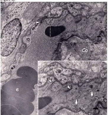
Figure 1: Electron micrograph of the peripheral compartment of a floating villus in term placenta. Longitudinal section of a capillary with several erythrocytes (e) in the lumen is seen. At upper end of this capillary an endothelial sprout extending towards right is distinguished. Villous stroma consists of mesenchymal cells and collagen fibers (Co). Inset: Higher magnification of the area where the angiogenetic sprout marked by dashed line. Interendothelial junctions (arrow head) are more abundant in this region. A cleft like narrow lumen (arrows) is present with in the sprout. (Uranyl acetate and lead citrate; original magnification X 3597 - 6000)
Figure 2: a) Peripheral compartment of another floating villus lined by syncytiotrophoblast (Syn), with a cytotrophoblast (Cyt) neighboring the trophoblast basal lamina (*) is seen. A newly developing capillary section close to the vasculosyncytial membrane is marked with dashed line. b) inset: Higher magnification of developing capillary. Cleft like lumen (arrows) is extremely narrow like the previous sample. Interendothelial junctions (arrow head) are abundant also in this developing capillary. A pericyte (p) is associated with this vessel (Uranyl acetate and lead citrate; original magnification a x3597 – b x6000).
Figure 3: Section through a maturing capillary which has a gradually enlarged lumen (arrow) occupied almost entirely by an erythrocyte. Endothelial cells rich in intracytoplasmic filaments (+) are attached to each other by intercellular junctions (arrowheads). These cells did not yet gain the typical structure of the endothelial cells of mature patent capillaries. Processes of pericytes are also distinguished (p). Basal and external lamina of the endothelial lining and pericyte process are clearly determined respectively. Basal infoldings of syncytiotrophoblast along the trophoblast basal lamina (*) are present. (Uranyl acetate and lead citrate; original magnification X 7750).
Intussusceptive angiogenesis
Besides sprouting angiogenetic figures described above we also observed structural features of intussusceptive angiogenesis in many examples representing phase I to IV of this remodeling process. In Phase-I samples, endothelial cells were observed to be extending towards the opposite wall some of which are already attached to each other by intercellular junctions forming pillars. In such newly established contact areas, endothelial cells were richer in cytoplasmic filaments like the endothelial cells of angiogenetic sprouts (Figure 4).
In other samples, processes of pericytes were projected between the attached endothelial cells crossing the original lumen of the parent capillaries representing a further stage of intussusception (Phase II-III). Endothelial cells crossing the lumen were similarly rich in cytoplasmic filaments reflecting a sign in their change in shape (Figure 5). We also determined migrating pericytes in intussusception areas surrounded by tiny intercellular matrix components resulting in relocation of intercellular junctions to allow this enlargement of the pillar area (Figure 6).
Vascular bed growth
Ayran Fidan et al.
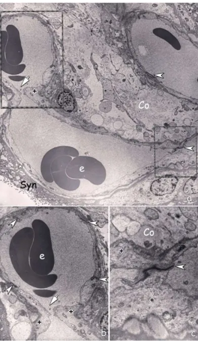
Figure 4: a) Electron micrograph of capillary within the villous stroma consisting of collagen fibers (Co) and cell processes. Upper right part of the capillary reflects the usual fine structural features mature patent capillary. However, the left side of the capillary has endothelial bridges (arrow) in the areas marked by dashed lines (Stage 1-2 of intussusceptive angiogenesis). Trophoblast basal lamina (*), interendothelial junctions (arrowhead), erythrocytes (e), Syncytiotrophoblast (Syn). b) Higher magnification of stage 1. Interendothelial bridge (arrow). The cytoplasm of the endothelial cells at this special location is rich in cytoplasmic filaments (+). Interendothelial cellular junctions (arrow heads), erythrocytes (e). c) Higher magnification of the marked are on the right (Stage II). Endothelial cells forming the bridge are rich in cytoplasmic filaments (+) similarly. Intercellular junction (arrow head) is also clearly distinguished. Collagen fibers (Co). (Uranyl acetate and lead citrate; original magnification a x2156, b x4646, c x10000)
Figure 5: a) Another capillary adjacent to vasculosyncytial membrane. Intussusception area marked by dashed line.
b) Higher magnification intercellular junction marked by arrow head and processes of pericytes (arrows) extend through the pillar. Syncytiotrophoblast (Syn), Cytotrophoblast (Cyt). Endothelial cells in the region are distinctly rich in intracytoplasmic filaments (+). (Uranyl acetate and lead citrate; original magnification a x2784, b x7750)
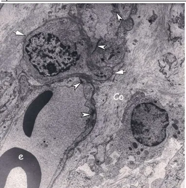
Figure 6: Section through capillary intussusception area. The stromal core of the pillar is occupied by interstitial tissue through which a pericyte (arrow) is migrating. Interendothelial junction (arrow head), erythrocytes (e), collagen fibers (Co). (Uranyl acetate and lead citrate; original magnification x3597).
DISCUSSION
Growth and remodeling of placental vasculature is critical for normal fetal development. A newer form of angiogenesis, “intussusceptive angiogenesis” is recently described and attracted the attention of a number of researchers to this field in the last decades. We could not find a report about intussusception in human placenta. For this reason, we hypothesized that both sprouting and intussusceptive angiogenesis are efficient processes in human placenta and studied human placenta at electron microscopic level to test this hypothesis. We focused on the capillary sections in detail and obtained structural evidences of both angiogenetic mechanisms.
As efficient and sufficient maternofetal exchange is critical for normal development, many investigators studied the development and vascularization of placenta previously (2-6, 8-10). It is generally agreed that neovascularization starts with vasculogenesis which is followed by angiogenesis (2, 3, 7, 9). Several investigators introduced a number of terms like branching, non-branching sprouting etc. to define the formation of new blood vessels during further development (9-11). Intussusceptive angiogenesis attracted the attention of researchers thus several authors reported intussusceptive angiogenesis in a number of physiological conditions in lung, skeletal muscle, chorio-allantoic membrane (CAM), myocardium, retina, kidney, endometrium and also in some pathological conditions like the human colon adenoma cancer, tumor angiogenesis but there is no report in human placenta (17-21, 23-25, 27-29, 31-33, 35, 36, 40, 41).
Djonov et al. previously suggested that intussusceptive mechanisms have several benefits for the organ. They stated that intussusceptive angiogenesis permits rapid expansion of capillary plexus and provides extensive endothelial surface for metabolic changes with a relatively little cost. They also reported that as remodeling occurs in capillary network, it provides reproduction and pruning of vessels according to metabolic needs of that special site (16).
Intussusceptive angiogenesis, which can be described as existing vessel proliferation in itself, is thought to be a more economic method for tissue when compared with new endothelial cell proliferation and sprouting from blood vessels method.
In the literature, changes of molecules (VEGF-A, PIGF, b-FGF, eNOS, O2, HIF, Tie1, Tie2, Ang-1, Ang-2) during vascularization in placenta is evaluated in both physiological and pathological pregnancies (5, 12, 14, 31, 42). But most of these studies are conducted in early placental development stages and they are focused on molecules rather than neovascularization types with the exception of a few studies. Mayhew et al., detected an increase in branching angiogenesis in pathologies like iron-deficiency anemia, high-altitude, hypoxia (11). Similarly, Soma et al., observed new vessel formation with angiogenesis during chronic hypoxia (43). Zhang et al., examined adaption, changes in diameter and perivascular cells of placenta vessels during pregnancy (14).
Some authors identified intussusceptive angiogenesis and proposed that expansion of pillar is a mechanism that occurs for pruning useless/inefficient vessels (40, 44, 45).
In our study; we observed capillaries in all stages of intussusception in all of the samples examined. We focused on the floating (terminal) villi in which fetomaternal exchange of placenta is most represent the first report of intussusceptive angiogenesis in human term placenta. In conclusion; our findings reveal that further comparative studies in the placentas of maternal or fetal pathologies will be great value to understand the role of intussusception in the etiopathogenesis of a number of placental disfunctions. Conflict of interest
No conflict of interest was declared by the authors.
REFERENCES
1. Moore KL, Persaud TVN, Torchia MG. Before we are born. 7th edition, Philadelphia, Saunders Elsevier; 2008.
2. Dempsey EW. The Development of capillaries in the villi of early human placentas. Am J Anat. 1972; 134(2): 221-37.
3. Demir R, Kaufmann P, Castellucci M, Erbengi T, Kotowski A. Fetal vasculogenesis and angiogenesis in human placental villi. Acta Anatomica 1989; 136: 190-203.
4. Te Velde EA, Exalto N, Hesseling P, Van der Linden HC. First trimester development of human chorionic villous vascularization studied with CD34 immunohistochemistry. Hum Reprod. 1997; 12: 1577–1581. doi:10.1093/humrep/12.7.1577.
5. Demir R, Kayisli UA, Cayli S, Huppertz B. Sequential steps during vasculogenesis and angiogenesis in the very early human placenta. Placenta 2006; 27: 535–539. doi:10.1016/j.placenta.2005.05.011. 6. Demir R, Seval Y, Huppertz B. Vasculogenesis and angiogenesis in
the early human placenta. Acta Histochem. 2007; 109: 257–265. doi:10.1016/j.acthis.2007.02.008.
7. Leach L, Babawale MO, Anderson M, Lammiman M. Vasculogenesis, angiogenesis and the molecular organisation of endothelial junctions in the early human placenta. J Vasc Res. 2002; 39: 246–259. doi:10.1159/000063690.
8. Burton GJ, Charnock-Jones DS, Jauniaux E. Regulation of vascular growth and function in the human placenta. Reproduction 2009; 138: 895–902. doi:10.1530/REP-09-0092.
9. Kaufmann P, Mayhew TM, Charnock-Jones DS. Aspects of human fetoplacental vasculogenesis and angiogenesis. II. Changes during normal pregnancy. Placenta 2004; 25: 114-126. doi:10.1016/j.placenta.2003.10.009
10. Charnock-Jones DS, Kaufmann P, Mayhew TM. Aspects of human fetoplacental vasculogenesis and angiogenesis. I. Molecular
regulation. Placenta 2004; 25:103–113.
doi:10.1016/j.placenta.2003.10.004
11. Mayhew TM, Charnock-Jones DS, Kaufmann P. Aspects of human fetoplacental vasculogenesis and angiogenesis. III. Changes in complicated pregnancies. Placenta 2004; 25: 127–139. doi:10.1016/j.placenta.2003.10.010.
12. Arroyo JA, Winn VD. Angiogenesis in the IUGR Placenta. Seminars
in Perinatology 2008; 32: 172–177.
doi:10.1053/j.semperi.2008.02.006.
13. Patan S. Vasculogenesis and angiogenesis as mechanisms of vascular network formation, growth and remodeling. Journal of Neuro-Oncology 2000; 50: 1–15.
14. Zhang EG, Burton GJ, Smith SK, Charnock-Jones DS. Placental vessel adaptation during gestation and to high altitude: Changes in diameter and perivascular cell coverage. Placenta 2002; 23: 751-762. doi:10.1016/S0143-4004(02)90856-8.
15. Baum O, Suter F, Gerber B, Tschanz SA, Buergy R, Blank F et al. VEGF-A promotes intussusceptive angiogenesis in the developing chicken chorioallantoic membrane. Microcirculation 2010; 17(6): 447–457. doi:10.1111/j.1549-8719.2010.00043.x.
16. Djonov V, Baum O, Burri PH. Vascular remodeling by intussusceptive angiogenesis. Cell Tissue Res. 2003; 314: 107–117. doi:10.1007/s00441-003-0784-3.
17. Short RHD. Alveolar epithelium in relation to growth of the lung. Philos Trans R Soc Lond B Biol Sci. 1950; 235(622): 35-86. DOI: 10.1098/rstb.1950.0014.
18. Ogawa Y. On the fine structural changes of the microvascular beds in skeletal muscle. J Yokohama City Univ Sec Sport Sci Med. 1977; 6: 1–19.
Vascular bed growth
Ayran Fidan et al.
19. Appell H-J. Morphological studies on skeletal muscle under conditions of high altitude training. Int J Sports Med. 1980; 1: 103– 109.
20. Caduff JH, Fischer LC, Burri PH. Scanning electron microscope study of the developing microvasculature in the postnatal rat lung. Anat Rec. 1986; 216: 154–164.
21. Burri PH, Tarek MR. A novel mechanism of capillary growth in the rat pulmonary microcirculation. The Anatomical Record. 1990; 228: 35–45.
22. Van Groningen JP, Wenink ACG, and Testers LHM. Myocardial capillaries: Increase in number by splitting of existing vessels. Anat. Embryol. 1991; 184: 65–70.
23. Patan S, Haenni B, Burri PH. Evidence for intussusceptive capillary growth in the chicken chorio-allantoic membrane. Anat Embryol. 1993; 187(2): 121-130.
24. Patan S, Haenni B, Burri PH. Implementation of intussusceptive microvascular growth in the chicken chorioallantoic membrane (CAM): 1. Pillar formation by folding of the capillary wall. Microvascular Research 1996; 51: 80–98.
25. Patan S, Haenni B, Burri PH. Implementation of intussusceptive microvascular growth in the chicken chorioallantoic membrane (CAM) 2. Pillar formation by capillary fusion. Microvascular research 1997; 52: 33–52.
26. Schlatter P, König MF, Karlsson LM, and Burri PH. Quantitative study of ıntussusceptive capillary growth in the chorioallantoic membrane (CAM) of the chicken embryo. Microvascular research 1997; 54: 65–73.
27. Djonov V, Schmid M, Tschanz SA, Burri PH. Intussusceptive angiogenesis- Its role in embryonic vascular network formation. Circ Res. 2000; 86: 286–292.
28. Djonov VG, Galli AB, Burri PH. Intussusceptive arborization contributes to vascular tree formation in the chick chorio-allantoic membrane. Anat Embryol. 2000; 202(5): 347-357.
29. Patan S, Munn LL, Jain RK. Intussusceptive microvascular growth in a human colon adenocarcinoma xenograft: A novel mechanism of tumor angiogenesis. Microvascular research 1996; 51: 260–272. 30. Nagy JA, Morgan ES, Herzberg KT, Manseau EJ, Dvorak AM.
Pathogenesis of ascites tumor growth: Angiogenesis, vascular remodeling, and stroma formation in the peritoneal lining. Cancer Res. 1995; 55: 376–385.
31. Patan S. Tie1 and Tie2 receptor tyrosine kinases inversely regulate embryonic angiogenesis by the mechanism of intussusceptive microvascular growth. Microvascular Research 1998; 21:1–21. 32. Djonov VG, Kurz H, Burri PH. Optimality in the developing vascular
system : branching remodeling by means of ıntussusception as an efficient. Developmental dynamics 2002; 224: 391–402. doi:10.1002/dvdy.10119.
33. Patan S, Munn LL, Tanda S, Roberge S, Jain RK, Jones RC. Vascular morphogenesis and remodeling in a model of tissue repair: blood vessel formation and growth in the ovarian pedicle after ovariectomy. Circ Res. 2001; 89: 723–731. doi:10.1161/hh2001.097870.
34. Djonov V, Andres A. Vascular remodelling during the normal and malignant life cycle of the mammary gland. Microscopy research and technique 2001; 52: 182–189.
35. Burri PH, Djonov V. Intussusceptive angiogenesis–the alternative to capillary sprouting. Mol Aspects Med. 2002; 23: 1–27. doi:10.1016/S0098-2997(02)00096-1.
36. Gambino LS, Wreford NG, Bertram JF, Dockery P, Lederman F, Rogers PAW. Angiogenesis occurs by vessel elongation in proliferative phase human endometrium. Human Reproduction 2002; 17: 1199–1206.
37. Makanya AN, Stauffer D, Ribatti D, Burri PH, Djonov V. Microvascular growth, development, and remodeling in the embryonic avian kidney: the interplay between sprouting and intussusceptive angiogenic mechanisms. Microsc Res Tech. 2005; 66: 275–288. doi:10.1002/jemt.20169.
38. Kurz H, Burri PH, Djonov VG. Angiogenesis and vascular remodeling by intussusception: from form to function. News Physiol Sci. 2003; 18: 65-70.
39. Makanya AN, Hlushchuk R, Djonov VG. Intussusceptive angiogenesis and its role in vascular morphogenesis, patterning, and remodeling. Angiogenesis 2009; 12: 113–123. doi:10.1007/s10456-009-9129-5.
40. Mentzer SJ, Konerding M. Intussusceptive angiogenesis: expansion and remodeling of microvascular networks. Angiogenesis 2014; 17: 499–509. doi:10.1007/s10456-014-9428-3.
41. Demir R, Yaba A, Huppertz B. Vasculogenesis and angiogenesis in the endometrium during menstrual cycle and implantation. Acta Histochem. 2010; 112: 203–214. doi:10.1016/j.acthis.2009.04.004. 42. Barut F, Barut A, Gun BD, Kandemir NO, Harma MI, Harma M et al. Intrauterine growth restriction and placental angiogenesis. Diagn Pathol. 2010; 5: 24. doi:10.1186/1746-1596-5-24.
43. Soma H, Murai N, Tanaka K, Oguro T, Kokuba H, Fujita K et al. Angiogenesis in villous chorangiosis observed by ultrastructural studies. Med Mol Morphol. 2013; 46: 77–85. doi:10.1007/s00795-013-0010-7.
44. Hlushchuk R, Ehrbar M, Reichmuth P, Heinimann N, Styp-Rekowska B, Escher R et al. Decrease in VEGF expression induces intussusceptive vascular pruning. Arterioscler Thromb Vasc Biol. 2011; 31:2836–2844. doi:10.1161/ATVBAHA.111.231811. 45. Burri PH, Hlushchuk R, Djonov V. Intussusceptive angiogenesis: its
emergence, its characteristics, and its significance. Dev Dyn. 2004; 231: 474–488. doi:10.1002/dvdy.20184.