80
Corresponding author
Dr. Mehmet Mesut Pişkin
Telgrafcı Hamdi Bey Cad. Gani Sitesi No: 7 42060 Konya, Turkey Tel : +90 (537) 671 10 74
Fax : +90 (332) 223 61 81 E-mail address: [email protected] Received: 28.03.2005 • Accepted: 25.04.2006
Ankara Üniversitesi Tıp Fakültesi Mecmuası 2006; 59:80-82 CERRAHİ BİLİMLER / SURGICAL SCIENCES
Olgu Bildirisi / Case Report
H
ydatid diseases is a zoonotic parasitic infestation caused by Echinococcus Granulosus, and it is endemic in Turkey. The cysts are mostly located in liver (65-75%), lung (15-25) and the remainder of the body and mostly the brain, bone and the mediastinum (1). Only 0.75-2.25% of the hydatid cysts are located in the pelvis(2). It’s unusual to find a mesenteric hydatid cyst that reaches to pelvis and would lead acute renal failure. We report a 24-year-old man who had renal failure due to obstructive uropathy secondary to a hydatid cyst in the pelvis.Case report
A 24-year-old man presented with one month history of dull lower abdomi-nal and back pain. Previously patient admitted to a private health center with those complaints and was treatead for urinary tract infection with oral antibiot-ics. In a period of time he has felt a decrease in urine output, shortness of breath and anuria at the end. But during this period of time the patient didn’t have any difficulty neither in voiding nor in defecation. The patient had been operated in a military hospital due to hepatic hydatid disease 3 years ago but the details of the surgery were not known.
An anterior subcostal insicion scar was observed on the right side of the abdomen. In physical examination large suprapubic and abdominal mass al-most completely filling the pelvis was palpated. The laboratory results revealed significant anemia and increased urea 498 mg/ml and creatinine level of 17.3 mg/ml (normal range 10-50 mg/ml and 0.8-1.2 mg/ml consecutively). The electrolyte levels were in the normal range. The chest x-ray was found to be normal. The abdominal ultrasound revealed cystic mass located superior to the bladder which was containing daugther cycts and bilateral
hydronephro-It’s unusual to find a pelvic hydatid cyst that would lead acute renal failure. We report a 24-year-old man who had renal failure due to obstructive uropathy secondary to a hydatid cyst in the pelvis.
Key words: Hydatid disease, obstructive uropathy, renal failure
Pelvik hidatik kistin akut böbrek yetmezliği oluşturması son derece nadir bir durumdur. Pelvik yerleşimli hidatik kiste bağlı gelişen obstrüktif üropatinin sebep olduğu akut böbrek yetmezliğine giren 24 yaşındaki bir olguyu sunuyoruz.
Anahtar sözcükler: Hidatik hastalık, obstruktif üropati, renal yetmezlik
Pelvic hydatid disease causing renal failure
Böbrek yetmezliğine neden olan hidatik hastalık
Talat Yurdakul, Mehmet Mesut Pişkin
Deparment of Urology, University of Selçuk, School of Medicine, Konya
Journal of Ankara University Faculty of Medicine 2006; 59(2)
81
T. Yurdakul, M. M. Pişkin
sis. The mass was originating from mesentery detected on non-contrast CT images and it was 33x16 cm in diameter (Fig. 1). Liver and spleen were normal.
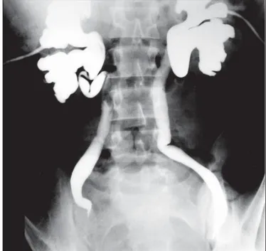
The patient underwent hemodialysis urgently. There-after, percutaneous nephrostomy catheters were inserted to the both kidneys by ultrasound guidence. Following a polyuric phase patients serum urea and creatinin levels sta-bilized (70 mg/ml and 1.3 mg/dl). Antegrade pyelography showed lateral displacement of both ureters and bilateral complete ureteral obstruction (Fig. 2).
We performed laparatomy using midline suprapubic incision. Pelvic hydatic cyst was observered arising from the mesentery and it was removed completelly. There was no adhesion to bladder. After the operation nephrostomy catheters were turned off and patient began to void nor-mally. Urine volume and density returned to normal levels. BUN and creatinine levels stayed in the stable and then nephrostomy catheters were removed.
The histopathological evaluation confirmed the hy-datid disease. The patient discharged from the hospital with albendazole treatment 10 mg/kg/day in 2 equal doses for 6 months duration.
Discussion
Hydatid disease is endemic in Turkey, especially in central and eastern Anatolia region. The hydatid cycts are mostly located in the liver and lungs. Pelvic hydatid disease is a rare form of hydatid disease.
Pelvic hydatid cycsts are usually secondary to the rup-ture (either spontaneously or accidentally during surgery). In our case patient had a history of hydatid disease surgery, we believe that the pelvic cyst was secondary to the previ-ous surgery.
Pelvic hydatid disease mostly present itself with ob-structive symptoms and pathologies of genitourinary tract due to its space occupying nature. In reported cases, the presenting features were obstruction of labor, compression to the fallopian tubes causing hydrosalpinx, obstructive azoospermia, and voiding disfunction (2,3,4). Rarely the voiding disfunction might ended with renal failure (5). Unilateral ureteral obstruction case has been reported but obstruction of the ureter was due to intraluminal Hydatid disease, not because of external compression (6). Horcha-ni reported 27 retrovesical HD but in only 1 patient had advanced renal failure and dilated both ureters related to external compression to ureters (7). In our case the pelvic huge mass compresed the ureter and cause hydroureter-onephrosis without any symptom of voiding disfunction or compression of the bladder neck. The progression of the cysts are usually slow 1cm/year, but might be faster in some cases, in our case the growth rate was fast because only 3 years passed from the liver hydatid disease (5). There is a large expansion space offered by the peritoneal cavity be-cause of this fact the patient didn’t suffer from abdominal pain until the end stage renal failure developed. And also patient didn’t have any voiding difficulty, because of the lo-cation of the cyst. We advise physicians in endemic areas to rule out hydatid disease in cases of obstructive pathologies of the urogenital tract also to give maximum care during surgery to decrease implantation to other sides.
Figure 1. Non-contrast CT image of huge pelvic hytadid cyst.
Figure 2. Antegrade pyelography showing lateral displacement of both ureters and bilateral complete ureteral obstruction.
Ankara Üniversitesi Tıp Fakültesi Mecmuası 2006; 59(2)
82 Pelvic hydatid disease causing renal failure
References
1. Von Sinner W. Advanced medical imaging and treatment of human cystic echinococcosis. Semin Roentgenol 1997; 32:276-290.
2. Emir L, Karabulut A, Balci U et al. An unusual cause of urinary retention: A primary retrovesical echinococcal cyst. Urology 2000; Nov 1; 56:856i-856.
3. Dede S, Dede H, Çalıskan E et al: Recurrent pelvic hydatid cyst obstructing labor, with a concomitant hepatic primary. A case report. Journal of Reproductive Medicine 2002; 47:164-166.
4. Laghzaoui M, Aderdour M, Bouhya S et al. Hydatic cycts of the fallopian tube: a case report. J Gynecol Obstet Biol Reprod 2002; 31:390-392.
5. Emir L, Germiyanoglu C, Lokumcu A ve Ark.Retrovesical Echinococal Cyst Causing Renal Failure an Nokturnal enuresis in a Child: Journal of Pediatric Surgery 2001; 36:1-3.
6. Merchant SA, Patel VH, Relekar R et al.Ureteral Obstruction in Hydatid Disease. J Pediatr Surg 2001; 36:385-387.
7. Horchani A, Nuira T, Chtourou M, et al. Retrovesical Hydatid Disease: A Clinical Study of 27 Cases Eur Urol 2001; 40:655-660.