Muhittin Yilmaz
1*, H. Ramazan Yilmaz2, Ali Alas
31Department of Biology, Faculty of Science and Art, Kafkas University, 36100 Kars-Turkey
2Department of Medical Biology and Genetics, Faculty of Medical, Suleyman Demirel University, 32100 Isparta-Turkey
3Department of Natural Sciences, Faculty of Education, Aksaray University, 68100 Aksaray-Turkey
*Corresponding author: [email protected]
An electrophoretic taxonomic study on serum
proteins of Acanthobrama marmid,
Leuciscus cephalus, and Chondrostoma regium
Abstract
In this study, native polyacrylamide gel electrophoresis (Native-PAGE) and sodium dodecyl sulfate-polyacrylamide gel electrophoresis (SDS-PAGE) were applied to the serum proteins of
Leuciscus cephalus, Acanthobrama marmid and Chondrostoma regium (Cyprinidae) fish taken
from) the Karakaya Dam (Malatya, Turkey). The electrophoregrams showed that there were similarities and differences in the molecular weight (MW) of the serum proteins among the three species. In the Native-PAGE, the total number of serum protein bands of L. cephalus, A.
marmid and C. regium were 5, 8, 7, respectively. In the SDS-PAGE, 13 protein bands were
obtained in L. cephalus, 11 in A. marmid and 11 in C. regium. In the electrophoregrams obtained from native-PAGE, the MW of the protein bands were found to be different in the fish species except the 80.3 kD protein band which was detected in the three fish species. In the SDS-PAGE, the protein band of 21.7 kD was observed in the three fish species. Protein bands of 63.4, 52.3 and 49.5 kD were present only in L. cephalus and C. regium. On the other hand, it was seen that other protein bands had similar molecular weights.
Keywords: Fish, native-PAGE, SDS-PAGE, serum proteins, taxonomy.
Yilmaz M, Yilmaz HR, Alas A (2007) An electrophoretic taxonomic study on serum proteins of
Acanthobrama marmid, Leuciscus cephalus, and Chondrostoma regium. EurAsia J BioSci 1, 3,
22-27.
www.ejobios.com/content/1/3/22-27
In the past, the identification of fish species was carried out mainly by examining the external morphological characteristics. In the present day, electrophoresis of sarcoplasmic proteins, serum proteins, liver proteins and a number of enzymes often have been used by some researchers as an aid in the species identification of fish (Focant et al. 1981, Miyazaki et al. 1998, Pinerio et al. 2001). Recent studies indicated that serum proteins can present taxonomic values, when the serum proteins of different fish species are examined electrophoretically (Khan and Gadru 1988). We previously showed that
serum protein bands of Capoeta trutta and
Capoeta capoeta umbla were significantly
different (Yilmaz et al. 2000). Unfortunately, there has been no taxonomical study with these species by serum protein electrophoresis in Turkey.
In the present investigation, the serum proteins of Leuciscus cephalus,
Acanthobrama marmid and Chondrostoma regium have been analyzed using the native
and SDS-PAGE technique and the resemblances and differences between the species was established.
Received: June 2007 Accepted: August 2007 Printed: November 2007
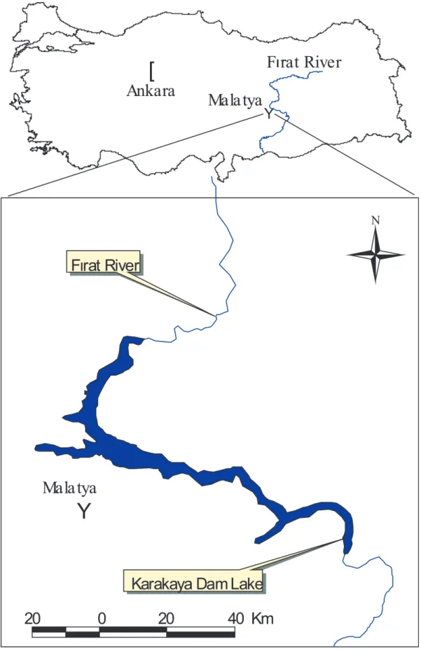
In the study, Leuciscus cephalus,
Acanthobrama marmid and Chondrostoma regium belonging to the Cyprinidae family
were used and the fish were obtained in the summer season from Karakaya Lake (Malatya, Turkey) (Fig. 1).
The fish were transported alive to the laboratory. A 2-3 ml blood sample was taken from the dorsal aorta of the fishes and injected into tubes, which contain EDTA. The blood samples were centrifuged at 1500 Rpm for 10 min at +4°C and to separate the serum. The obtained serum was used for the analysis of proteins and electrophoresis. Protein content was determined by using the method of Lowry et al. (1952) with bovine serum albumin as a standard.
SDS-PAGE was performed according to the Laemmli (1970) and O'Farrell (1975) methods. Proteins were separated on ft-16x10 cm dimension and a 1 mm thick slab gel. Slab gel consist of stacking gel [which proteins (stocked)] and a running gel part on which proteins seperate. Running gel containing 10% acrylamide was polymerized 12 hrs before electrophoresis and stacking gel containing 4% acrylamide was poured and polymerized 2 hrs before sample application. Each sample was mixed with a sample buffer which contained 10% glycerol, 2% mercaptoethanol, 2% SDS and 0.01 brom phenol blue. Protein concentrations were adjusted to 2 µg/µl and 0.2 µg/µl, then heat-denatured and run on the SDS-PAGE. For SDS-PAGE, 20µl sample were loaded on the stacking gel. And 200 Volts appilied the brom phenol blue was present on the lowest side of the gel. Following electrophoresis, the proteins were stained with 0.125% Commassie Brilliant Blue R-250 in 40% ethanol and 7% acetic acid, and then destained in acetic acid. Bovine serum albumine (66 kD) and pepsin (34.7 kD) were used as the protein standard in the SDS-PAGE electrophoresis and pepsin (34.7 kD) and ß-lactoglobulin (18.4) were used as protein standart for the native PAGE. Molecular weight of the proteins were calculated according to Weber et al. (1972).
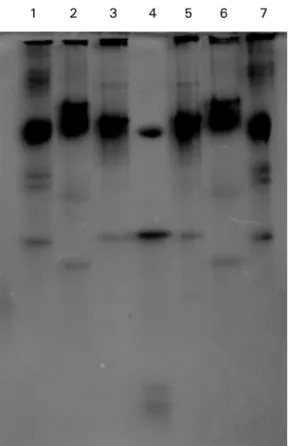
In the native-PAGE, the protein bands of
Leuciscus cephalus, Acanthobrama marmid
and Chondrostoma regium were 5, 8, 7, respectively (Fig. 2). Molecular weights of the protein bands were 80.3, 61.2, 53.8, 41.5 and 30.4 kD in Leuciscus cephalus, 80.3, 76.4, 70.6, 68.8, 52.4, 45.5, 43.1, and 34.1 kD in Acanthobrama marmid, 80.3, 66.1, 56.6, 51.8, 44.2, 34.7, and 27 kD in
Chondrostoma regium. In the SDS-PAGE,
protein bands of Leuciscus cephalus,
Acanthobrama marmid and Chondrostoma regium were 13, 11, 11, respectively (Fig. 3).
Molecular weights of the protein bands were found to be 98.3, 89.2, 68.8, 65.1, 63.4, 52.3, 49.5, 43.1, 31.9, 31, 27, 21.7, and 9 kD in Leuciscus cephalus, 101, 76.8, 66.7, 61.7, 51, 38.2, 33.7, 31.1, 27.4, 21.7, and 9.2 kD in Acanthobrama marmid, 95.6, 63.4, 56.8, 52.3, 49.5, 36.6, 31.4, 28.8, 21.7, and 9.5 kD in Chondrostoma regium.
In general, taxonomic studies are based on morphometric measurements and anatomical characteristics. Electrophoresis of serum proteins have been widely used in the classification of fish. These kinds of studies brought about a new look to taxonomical evaluation. Discrimination of related taxa can be easily made according to their electrophoretic results of serum proteins (Theophilus and Rao 1998). In a previous study by using SDS-PAGE, the serum protein bands of Capoeta trutta and Capoeta capoeta
umbla were separated and analyzed by using
SDS-PAGE. The serum protein band numbers of these fishes were 16 and 11, respectively (Yilmaz et al. 2000). In the other research, the sarcoplasmic proteins of Orthrias insignis
euphyraticus and Cyprinion macrostomus
were separated by using SDS-PAGE and, the electrophoregram showed that there were differences between the two species in both the number of bands and the molecular weight of the sarcoplasmic proteins (Yilmaz et al. 2005).
MATERIAL AND METHODS RESULTS
Similarly, the liver proteins of six species belonging to the Cyprinidae family, Acheilognathinae, Leuciscinae and Gobioninae subfamilies were separated using SDS-PAGE and Cyprinus carpio and Pseudogobius
esocinus esocinus gave the smallest genetic
distance. Nevertheless, Tribolodon hakonensis had almost equal genetic distances to the three other species (Miyazaki et al. 1998). In an other investigation, the serum proteins of the female Cyprinus carpio. And male
Ctenopharyngodon idella were analysed using
SDS-PAGE and it was stated that there were differences in the electrophoregrams of each species (Li 1991). Sexual dimorphism in electrophoretic patterns of blood serum proteins of a smelt [Hypomesus nipponensis] was investigated (Komagata et al. 1991). They found that in the electrophoretic
analyses of blood serum proteins were different in the female and male Hypomesus
nipponensis. In other research, the serum
protein profiles of parr and smolt in masu salmon (Oncorhynchus masou) have been analysed by two-dimensional SDS-PAGE and they have identified two proteins (43.80 kD) as possible smolt-specific serum proteins, these proteins were not detected in parr. The presence of differences in serum protein profiles between parr and smolt were confirmed using two-dimensional SDS-PAGE (Ura et al. 1994).
The serum proteins of five species of freshwater fish (Sarotheredon galilaeus,
Tilapia zillii, Orecohromis niloticus, Clarias lazera, and Barbus bynni) were studied,
electrophoretically. Eight fractions of serum
Fig 2. Native-PAGE of the serum proteins of
Leuciscus cephalus, Acanthobrama marmid
and Chondrostoma regium (1stlane belongs
to Acanthobrama marmid, 2ndlane Leuciscus cephalus 3rdlane Chondrostoma regium, 4th
lane the standard proteins, 5thlane Chondrostoma regium, 6thlane Leuciscus cephalus and 7thlane Acanthobrama marmid).
Fig 3. SDS-PAGE of the serum proteins of Leuciscus
cephalus, Acanthobrama marmid and
Chondrostoma regium (1stlane belongs to Acanthobrama marmid, 2ndlane Leuciscus cephalus 3rdand 4thlane Chondrostoma regium, 5thlane the standard proteins, 6th
lane Chondrostoma regium, 7thlane Leuciscus cephalus and 8thlane Acanthobrama marmid).
proteins in S. galilaeus, T. zillii and O. niloticus were reported. In the same research, seven and five fractions of serum proteins were found in C. lazera and B. bynni, respectively (Zowail and Baker 1998). In other investigation, genetic variations about the relationship among the characid fish Alestes
dentex and cyprinid fishes Barbus bynni, and Labeo niloticus common in the River Nile at El
Minia, Egypt, and the Chinese grass carp,
Ctenopharyngodon idella were studied.
Variations are found in fourteen enzymatic and two non-enzymatic proteins encoded by twenty-one structural loci. The characid fish
A. dentex demonstrated the minimum levels
of polymorphism. According to genetic similarity and distance between characid and cyprinid fishes, they closely related to each other (Shahin 1999).
In the study of mentioned, serum protein band numbers of Acanthobrama marmid and
Chondrostoma regium have shown a similarity
to serum protein band numbers of Capoeta
capoeta umbla mentioned above. However,
serum protein band patterns of the Leuciscus
cephalus studied were found to be different.
In conclusion, these fishes are easily distinguished by native and SDS-PAGE, taxonomically.
Focant B, Jacob MF, Huriaux F (1981) Electrophoretic comparison of the proteins of some perch (Perca fluviatilis L.) head muscles. J. Muscle Res. Cell Motil. 2, 3, 295-305.
Khan AR, Gadru M (1988) Electrophoretic patterns of blood serum protein of some fish of Kashmir. Trop. Freshwater Biol. 11, 62-70.
Komagata K, Kawarabayashi S, Kuwabara R (1991) Sexual dimorphism in electrophoretic patterns of blood serum proteins in a smelt Hypomesus nipponensis. Suisan Gakkaishi Bull. Jap. Soc. Sci. Fish 57, 8, 1599.
Laemmli UK (1970) Cleavage of structural proteins during the assemble, of the head of bacteriophage T4. Nature 227, 680.
Li C (1991) Electrophoretic analysis on the serum proteins of xinggua red carp, grass carp and their hybrid F sub (1). Freshwater Fish Danshui Yuye 6, 12-14.
Lowry OH, Rosebrough NJ, Farr AL, Randall RJ (1952) Protein measurement with the folin phenol reagent. J. Biol. Chem. 193, 265-271.
Miyazaki JI, Hirabayashi T, Hosoya K, Iwami TA (1998) Study of the systematics of cyprinid fishes by two dimensional gel electrophoresis. Environ. Biol. Fish. 52, 173-179.
O'Farrell PH (1975) High resolution two-dimensional electrophoresis of biological properties and significance. Comp. Biochem. Physiol. 88, 497-501.
Pineiro C, Vazquez J, Marina AI, Barros-Velazquez J, Gallardo JM (2001) Characterization and partial sequencing of species-specific sarcoplasmic polypeptides from commercial hake species by mass spectrometry following two-dimensional electrophoresis. Electrophoresis 22, 8, 1545-1552.
Shahin AAB (1999) Phylogenetic relationship between the characid nile fish Alestes dentex, cyprinid Barbus bynni, Labeo niloticus, and the introduced grass carp, Ctenopharyngodon idella elucidated by protein electrophoresis. J. Egypt Ger. Soc. Zool. 29, 21-42.
Theophilus J, Rao PR (1998) Electrophoretic studies on the serum proteins of the three species of genus channa. Indian J. Fish 35(4), 294-297.
Ura K, Hara A, Kawamura H, Yamauchi, K (1994) Immunochemical studies on serum proteins in juvenile masu salmon, Oncorhynchus masou. Comp. Biochem. Physiol. 1078, 2, 225-229. Weber K, Pringle J, Osborn M (1972) Measurement of molecular weights by electrophoresis on
sds-acrylamide gel. Meth. Enzymol. 26, 3.
Yilmaz M, Cigremis Y, Turkoz Y, Gaffaroglu, M (2000) An electrophoretic taxonomic study on blood serum proteins of some fish in Karakaya Dam Lake. J. of Selcuk Univ. Veterinary Sci. 16, 1, 89-92.
Yilmaz M, Cigremis Y, Turkoz Y, Gaffruoglu M (2005) A taxonomic study on Orthrias insignis
euphraticus (Banarescu and Nalbant, 1964) and Cyprinion macrostomus (Heckel, 1843) by
sarcoplasmic protein electrophoresis. G.U. Journal of Science 18, 1, 61-68.
Zowail MEM, Baker SME (1998) Genetic, biochemical polymorphism and similarity coefficient of five species of freshwater fish. J. Egypt Ger. Soc. Zool. 25, 75-88.
Acanthobrama marmid, Leuciscus cephalus ve Chondrostoma regium'un Serum
Proteinleri Uzerine Taksonomik Bir Calisma
Ozet
Bu calismada, Karakaya Baraj Golu (Malatya)'nden yakalanan Leuciscus cephalus, Acanthobrama marmid and
Chondrostoma regium ( Cyprinidae)'nin serum proteinleri dogal poliakrilamid gel elektroforez (Native-PAGE)
ve Sodyum dodeosil sulfat-poliakrilamid jel elektroforezine (SDS-PAGE) uygulandi. Elektroforegramlar uc turun serum proteinlerinin molekuler agirliklari arasinda benzerlik ve farkliliklarin varligini gostermistir. L.
cephalus, A. marmid and C. regium'un total serum protein bandlari Native-PAGE'de sirasiyla 5, 8, 7 ' dir.
SDS-PAGE'de L. cephalus icin 13, A. marmid icin 11 ve C. regium icin 11 protein bandi elde edildi. Native-PAGE'den elde edilen elektroforegramlarda protein bandlarinin molekuler agirliklari 80.3 kD protein bandi haric, uc balik turunde farkli bulundu. Uc balik turunde protein bandlarinin molekuler agirliklari SDS-PAGE'de 21.7 kD olarak belirlendi. 63.4, 52.3 and 49.5 kD protein bandlari sadece L. cephalus ve C. regium'da mevcuttu. Diger taraftan, diger protein bandlarinin molekuler agirliklarina yakin oldugu goruldu.