A. O. Vet. Fak. Derg. 36 (2): 413-418. 1989
THE NASAL CARTILAGES OF THE OONKEY (EQl:US ASI!\"l1Sq.
R. Merih Hazıroğlu 1 Merkebin (Equus asinus L.) burun kıkırdakları
Özet: Burun kık ırdak lan, makro anatomik olarak on adet (5 er-kek, 5 dişi) ergin merk ep başı üzerinde incelendi. Burun deliklerinin is-keletini cartilago alaris'lerin şekillendirdiği görüldü. Cartilago alaris'in, cartilago septi nasi ve cartilago ııasi lateralis dorsalis'in uzantısı olduğu saptandı. Yapdan diseksiyonlarda cartilago nasi lateralis ventralis'in yassı ince bir kıkırdak olduğu ve fissura palatina'yı doldurduğu gözlendi. Cartilago nasalis accessoria medialis'in oral kısmm//ı plica alaris'in içi-ne yer/eşll'ği ve cartilago nasi lateralis ventralis ile conclw nasalis vent-ralis' ten arjin aldığı teshit edildi. Gözlenen bulgulann atm burun kık ır-daklanyla olan farkhlıkları tartışi/dı.
Summary: The nasal cartilages were described in the adult donkey (5 male, 5 female). Anatomical ji"ndings observed were discussed with those of the horse.
Introduction
Aıı the veterinary anatomy textbooks considered both the horse and donkey has the same anatomical structures (I, 4,5,6). But during the last years it was noticed that same anatamical variations were found between the horse and donkey (2, 3, 8).
The nasal cartilages of the domestic animals studied by same aut-hors (i, 4, 5, 6, 9). The aim of this study was to elucidate the nasal cartilages of donkey and to discuss differentiation wİth the horse's na-sal cartilages.
R. MERİH HAZTROGLU
Materials and Methods
This study was performed on 10 heads of the adult donkey (5 male, 5 female) preserved in 10
%
formalin. The nasal cartilages were carefully dissected and terminated according to the N.A.V. 1983 (7).Results and Discussion
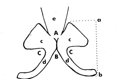
it was observed that the skeleton of the nostrils in the donkey inc-luded by two comma~shaped alar cartilages placed transversaııy back to back. The alar cartilages supportcd the nostrils dorsaııy, medially and ventrally, and the dorsal part of cach alar cartilages was prolonged and flattened to form a plate-like lamina, the ventral part caııed cornu was narrow. Thcsc anatomical variations were similar those of reported in the horse (I, 4,5.6). In addition to these, it was found that the ave-rage distance between the top of the lamina and the bottom of the cor-nu of alar cartilages was 5,i cm (Fig. 1, a-b) The average angles bet-wecn alar cartilages' lamina (A), and cornu (B), and each alar carti-lage's lamina and cornu (C) were 60, 70, 80 degrees, respectively (Fig. i)
Fig.ı. Alar cartilages of donkcy (schematic); c- Lamina of alar cartilage, d- Cornu of alar cartilage, e- Dorsal lateral nasal cartilagc (Merkebin cartilago alaris'i; c- Cartilago alarİs' in lamina'sı, d- Cartilago alaris'in cornu'su, c- Cartilago nasi lateral is dorsalis)
nONKEY NASAL CARTILAGES 4J5
Getty (4) has reported that alar cartilages attached to the rostral end of the cartilaginous nasal septum by connective tissue. In this study, it was found that alar cartilages were cranial extension of carti-laginous nasal septum and dorsal lateral nasal cartilages.
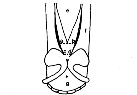
From the dorsal border of cartilaginous nasal septum, a thin, narrow plate, the dorsal lateral nasal cartilage curved outward for a short distance on either side was defined. The dorsal lateral nasal car-tilage was very narrowand not supported of the nostrils in any part. These findings are also rccorded in the literature related with the horse (J, 4, 5, 6). The average width of dorsal lateral cartilage at the rostral edge of nasal bone (Fig. 2, a-b) and the apex of this cartilage (Fig. 2, c-d) was 2,8 cm and 0,8 cm, respectively. Getty (4) has stated that the width of the dorsal lateral nasal cartilage is 3 to 4 cm in the horse.
Fig. 2. Nasal cartilages of donkey, dorsal view (scbernatic); e- Nasal bone, f- Nasal proccss of incisive bone, g- Body of incisivc bone (Merkebin burun kıkırdaklarının öoı'sal'den gö-rünüşü; c- Os nasale, f- Os incisivum'un proc. nasalis'i, g- Corpus ossis incisivi)
Getty (4) has announced that the ventral lateral nasal cartilage fill the palatine fissure in the horse. According to Nickel et al (6) the narrow ventral lateral nasal cartilage covers only palatine suture or may be
416 R. MERiH HAZJROGLU
absent, whereas Koch (5) noted that there is no vcntral lateral nasal cartilage or it can be seen rudimentary. Nonetheless, Ackerknecht (i)
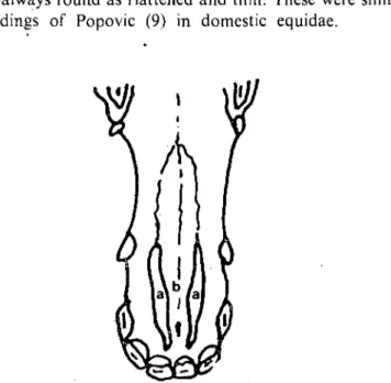
recorded that this cartilage is absent in the horse. The ventral lateral nasa! cartilage extended from the anterior part of ventral border of cartilaginous nasal septum into the palatine fissure in the donkey (Fig. 3, a). It was always found as flattened and thin. These were similar to reported findings of Popovic (9) in domestic equidae.
Fig. 3. Ventral lateral nasal carıilage of donkey, ventral view (schernatic); a) Ventral late-ral nasal cartilage, b- Palaıine process of lhe incisive bone (Merkebin carıilago nasi latera-Jis venıralis'i, venlral'den görünüş; a- Carıilago nasi laleralis venıralis, b- Os incisivurn'un
proc. palalinus'u)
The ventral lateral nasal cartilage's average length was 6 cm. The width in the narrowest and the largest parts of cartilage were O,i cm and 0,5 cm respectively. Although Popovic (9) has rccorded that its length is 8- 10 cm and, width is 2-4 mm in domestic equidae.
In the donkey, the yentral lateral nasal cartilage was initially 10-cated in the nasal surface of the maxilla. After fiııing to the palaıine fissure, it continued to the palatine surface of the body of incisive bone and, ended at i cm aboral of the first incisor teeth.
The oral portian of medial accessory nasal cartilage was found inside the alar fold. This cartilage was originated from the ventral
na-OONKEY NASAl CARTrlAGES 417
sal concha and the ventral lateral nasal cartilage, and it was large and "S" shaped. These are same with Popovic's (9) findings in domestic equidae. However, Koch (5) has stated that it is free and "S" shaped in the horse.
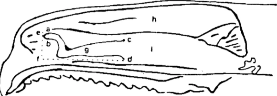
The average width at the oral (Fig. 4, a-b) and aboral (Fig. 4, c-d) parts of medial accessory nasal cartilage were 0,5 cm and 1,5 cm. Ac-carding to Fig. 4 the average distance between e and f, and f and d were 2,5 cm and 7 cm, respectively.
_____ h
•• e~
_:.~.~C
=====
-,: ... ~.d ~
~~
__
~t2
Fig. 4. Nasal cavity of donkey; sagittal section ""ith septum removed (schematic); g- Medi-al accessory nasal cartilage h- Oorsal concha İ-Ventral concha (Merkepte cavum nasi; septum nasi uzaklaştırılmış sagittal kesit, g- Cartilago nasalis accessoria medialis, h-
Conc-ha nasalis dorsalis, i- Concha nasalis ventralis)
The nasal cartilages in the donkey and its additional anatomical findings were described in this study, and were found some differences from registered findings associatcd with horse in classical veterinary anatomy textbooks.
Referenees
i. Ackerknecht, E. (1974): Das Eingell'eides system in Ellenberger-Baunı's Handbııch der
'erg/eichenden Anatol1lie der Haustiere. Auf. 18, Springer- Verlag-Berlin, Heidelberg,
New York.
2. Ahmed, M.A.A., Anis, H. and Moustafa, M.S.M. (1985): Arteria linguofacialis of the
donkey (Eqııus asimıs). Zbl. Vet. Med. C. 14: 47-53.
3. Ahmed, M.A.A., Anis, H. and Mou~tafa, M.S.M.(l985): Veins of the head and neck of the donkey (Equus asinus). Zbl. Vet. Med. C. 14: 149-157.
4. Getty, R.(ı975): Sisson and Grossman's The Anatonıy of the Donıes/ic Anima/s. 5 th ed. Vol. I-II, W.B. Saunders Company, Philadelphia London-ToroOlo.
41R R. MERIH HAZTROGLU
5. Koch. T. (1970): Lehrbııch der Veterinar-Anatomie, Auf. 2, Band II, Yeb Gustav Fischer Yerlag Jena.
6. Nickel, R., Schummer, A., Seiferle, E. (1979): The Viscem of the Domestic Mamma/s.
2nd ed., Yerlag Paul Parey Berlin-Hamburg.
7. Nomina, Anato~ica Yet~rinaria (1983): 3th ed. Ithaca, New york.
8. Öcal, M.K., Hazıroğll., R.M. (1988): Merkebin (Eqııııs asinııs L.) Medıılla spinalis'i üzerinde Komparatif-Morf%jik araştırmalar. 1. Segment/erin transI'ersal kesit/erinin incelenmesi. A.Ü. Yet. Fak. Derg. 3S (i) 55-68.
9. Popovic, S. (1964): Eine Dm'stellııng der morph%gischeıı Eigellttim/ichkeiten des knor-peligeıı Nasengen/stes bei Halıss(Jııgetiercn. Anat. An7. 114: 379-388.