SENSING AND SENSITIZER ACTIVATION BY
BIOLOGICAL THIOLS AND 1,2 -DIOXETANES BASED
CHEMILUMINESCENCE PROBES
A DISSERTATION SUBMITTED TO
MATERIALS SCIENCE AND NANOTECHNOLOGY PROGRAM OF THE GRADUATE SCHOOL OF ENGINEERING AND SCIENCE
OF BILKENT UNIVERSITY
IN PARTIAL FULFILLMENT OF THE REQUIREMENTS FOR THE DEGREE OF
DOCTOR OF PHILOSOPHY
By
İLKE ŞİMŞEK TURAN September, 2014
I certify that I have read this thesis and that in my opinion it is fully adequate, in scope and in quality, as a thesis of the degree of Doctor of Philosophy.
………. Prof. Dr. Engin U. Akkaya (Advisor)
I certify that I have read this thesis and that in my opinion it is fully adequate, in scope and in quality, as a thesis of the degree of Doctor of Philosophy.
………. Assoc. Prof. Dr. Tamer Uyar
I certify that I have read this thesis and that in my opinion it is fully adequate, in scope and in quality, as a thesis of the degree of Doctor of Philosophy.
………. Assoc. Prof. Dr. Özgür Altan Bozdemir
I certify that I have read this thesis and that in my opinion it is fully adequate, in scope and in quality, as a thesis of the degree of Doctor of Philosophy.
………. Assoc. Prof. Dr. Dönüş Tuncel
I certify that I have read this thesis and that in my opinion it is fully adequate, in scope and in quality, as a thesis of the degree of Doctor of Philosophy.
………. Assist. Prof. Dr. Salih Özçubukçu
Approved for the Graduate School of Engineering and Science
………. Prof. Dr. Levent Onural
Dedicated to
my husband and my mother,
for their continuous support and encouragement
i
ABSTRACT
SENSING AND SENSITIZER ACTIVATION BY BIOLOGICAL THIOLS AND 1,2-DIOXETANES BASED CHEMILUMINESCENCE PROBES
İlke Şimşek Turan
PhD in Materials Science and Nanotechnology Supervisor: Prof. Dr. Engin Umut Akkaya
September, 2014
Biologically important biothiols like Cystein (Cys), Homocystein (Hcy) and Glutathione (GSH) are vital for the maintenance of cellular redox status and alterations in their levels is linked to a number of severe diseases such as AIDS, cancer and Alzheimer‟s therefore the design and synthesis of nitroolefin functionalized bodipy dyes responding to biological thiols by both absorbance and emission changes have been accomplished. Through the incorporation of hydrophilic groups, bright signaling of biothiols in the longer wavelength region of the visible spectrum is deemed to operate in biological environment. With this knowledge, bioconjugation of the nitroolefin functionalized dyes with thiol groups like those belonging to cysteine residues on proteins has been proved via large spectral changes and targeted to visualize dynamics of proteins, cell-cell interactions, mechanisms of life cycles of proteins. Hence, the result suggests that nitroolefin functionalization of BODIPY dyes is a promising way to sense biological thiols and hence labeling proteins having thiol groups. Since GSH plays vital roles in the oxidative stress exists within the cells and thus, high concentration of it is the indication of cancer development, design and synthesis of cancer related parameter based activation of bodipy based photosensitizers have been achieved to enhance spatiotemporal selectivity in photonic sensitization of dissolved molecular oxygen and thus, improves the potential and practice of photodynamic therapy and their effectiveness are validated by cell culture studies. Chemiluminescence in principle can provide a rapid, qualitative and/or quantitative test for analytes of interest; because of that synthesis of novel probes for the sensing of biologically important (fluoride) anion have been devised to combine the power of chemiluminescence and self immolative amplifiers which offers a chemical avenue for enhancing the signal produced in response to a given analyte. Through the development of chemiluminogenic perspective for sensing of palladium ions, rapid and selective response of probe to
ii
palladium ions with regardless of their charge in aqueous environment have been accomplished. Considering the convenience of the methods and substantial results, we are confident that other probes combining the power of chemiluminescence will emerge.
Keywords: fluorescence, chemosensors, biological thiols, protein labeling,
iii
ÖZET
BİYOLOJİK TİYOLLERİN TANISI İLE FOTOSENSİTİZÖR
AKTİVASYONU VE 1,2-DİOKSETAN TEMELLİ
KEMİLÜMİNESAN PROBLAR
İlke Şimşek TuranMalzeme Bilimi ve Nanoteknoloji, Doktora Danışman: Prof. Dr. Engin Umut Akkaya
Eylül, 2014
Biyolojikçe aktif olan tiyollerden sistein (Cys), homosistein (Hcy) ve glutatiyon (GSH), hücresel redoks durumunun düzenlenmesinde hayati önem taşımaktadır ki hücresel değerlerindeki değişiklikler AIDS, kanser, Alzheimer gibi birçok rahatsızlıkla ilişkilendirilmektedir. Bu nedenle, biyolojikçe aktif tiyollere tepki veren nitroolefin grubu ile fonksiyonlandırılmış bodipy boyalarının dizayn ve sentezi hedeflenmektetir ve bu probların tiyollere olan seçiciliği de absorbans ve emisyondaki değişikiklerle kanıtlanmıştır. Hidrofilik gruplar ile modifiye edilen probların, biyolojikçe aktif tiyollere uzun dalga boyundaki tepkileri, bu moleküllerin biyoloik ortamlarda uygulanabilirliğini kanıtlamaktadır. Bu bilgiler ışığında, Nitroolefin ile fonksiyonlandırılmış bodipy boyalarının proteinlerin tiyol grupları ile biyokonjugasyonları, protein dinamiklerini gözlemlemek, hücrelerin birbirleriyle etkileşimleri belirlemek, proteinlerin yaşam döngülerinin mekanizmalarını anlamak amacıyla tasarlanmış olup, bu konjugasyon, boyar maddenin fotofiziksel özelliklerindeki değişikler ile kanıtlanmıştır. Elde edilen sonuçlar, BODIPY boyalarının nitroolefin fonksiyonlandırılmasının, hem biyolojik tiyollerin algılanması hemde tiyol grupları bulunduran proteinlerin etiketlenmesi açısından gelecek vaad eden bir yol olduğunu göstermektedir. Biyolojik öneme sahip tiyol içeren bu bileşiklerden olan GSH, hücre içi oksidatif stresin düzenlemesinde son derece büyük önem taşımaktadır ki hücre içerisinde yüksek konsantrasyonu kanser gelişimi ile ilişkilendirilmektedir. Hedef dışı hassasiyeti ortadan kaldırmak amacıyla (GSH) varlığında sönümlendirici modülün uzaklaştırılması ile aktiflendikten sonra singlet oksijen üretebilen sönümlendirilmiş seçici PDT fotoduyarlaştırıcılarının geliştirilmesi hedeflenmekte olup GSH‟ın aktif olmayan kromoforu, terapatik pencerede uyarıldığı zaman singlet oksijen üretebilen son derece etkili bir fotoduyarlaştırıcıya dönüştürebildiği hücre deneyleriyle kanıtlanmıştır. Elde edilen sonuçlar ışığında, PDT‟nin pratikte uygulanabilirliğine de yeni bir yön verilmektedir.
iv
Kemiluminesans prensipte analitlerin hızlı, kalitatif ve kantitatif belirlenmesinde kullanılan son derece etkili bir yöntem olduğu için kemilüminesan temelli kendini feda eden moleküler sistemleri florür iyonunu tayin edecek şekilde tetikleyici bir grup ile fonksionlandırmayı başardık. Dizayn ettiğimiz bu molekülün bir birim florür iyonuna karşılık çoklu kemiluminesans özellikte muhabir gruplarının salınımını ve bu molekülün sadece florür iyonuna seçicilik gösterdiğini analitik olarak kanıtladık. Paladyumun aşırı alınımının astım, bulantı, saç dökülmesinde artış ve dermatit gibi birçok önemli rahatsızlığın gelişmesine neden olduğudan kemiluminogenik perspektif kullanılarak hassas ve spesifik olarak paladyum iyonunu tayin edebilecek yeni probların dizayn ve sentezini hedefledik. Pd probu olarak kemilüminesan reaktiflerin geliştirilmesi planlanmış ve tasarlanan sensörlerin kemilüminesans yaparak kendi ışımalarını sağlaması paladyumun algılanmasını daha avantajlı kılmaktadır. Yaptığımız bu çalışmalardaki, kemilüminesansa dayalı metodların uygunluğu ve elde edilen sonuçlara dayanarak, kemilümenesans temelli yeni problar tasarlanacağına inanıyoruz.
Anahtar kelimeler: floresans, sensör, biyolojikçe aktif tiyoller, protein işaretleme,
v
ACKNOWLEDGEMENT
I would like to express my gratitude to my supervisor Prof. Dr. Engin U. Akkaya for giving me chance to be a member of his research group. I would like to express my sincere thanks for his guidance, support, and patience during the course of this research. I am also grateful to him for teaching us how to become a good scientist by looking everything from different point of views. Besides his scientific perspective, I admire his personality, view of life and I believe it is a privilege to know him as a person and as a scientist. I will never forget his support throughout my life.
I am sincerely grateful for my close friend Özlem Seven for her help, encouragement during the studies and especially for her endless support, understanding, caring throughout years that I have known her. It is good to know that there are people like her in this world. I would like to express my sincere thanks to my dear friend Seylan Ayan for her understanding, kind collaboration, joyful friendships and caring. I would like to express my special thanks to Özge Yılmaz, for her valuable help, we have confronted difficulties together. Additionally, I would like to thank to Deniz Yıldız for her valuable joyful friendship and I am very happy to know her. They have made tough events and days bearable. I feel lucky to have such a great friends.
I would like to thank to Ayşegül Gümüş, Fatma Pir Çakmak for their partnership and friendship.
I would like to thank TÜBİTAK (The Scientific and Technological Research
Council of Turkey) for financial support in the form of scholarship during PhD programme. Also, our experimental work was supported by TÜBİTAK in the form of a grant 112T480 and 113Z526.
I would like to thank to present and past members of Akkaya Lab. Nisa Yeşilgül, Hale Atılgan, Fazlı Sözmen, Ahmet Atılgan, Bilal Uyar, Ahmet Bekdemir, Elif Ertem, Hatice Turgut, Tuğçe Durgut, Jose Luis Bila, Ceren Çamur, Darika Okeava, Melek Baydar, Dilek Karışan, Ruslan Gluiyev, Onur Büyükçakır, Yusuf Çakmak,
vi
Sündüs Erbaş Çakmak, Safacan Kölemen, Tuğba Özdemir and rest of the SCL (Supramolecular Chemistry Laboratory) members. It was wonderful to work with them.
I would like to thank to Zeynep Aytaç, Aslı Çelebioğlu, Yelda Ertaş, Gözde Uzunallı for their endless help.
I also would like to thank all members of UNAM family for providing a multidisciplinary research atmosphere.
I would like to express my gratitude to my family and my husband, Tufan Turan for their endless love, support, and understanding.
vii
LIST OF ABBREVIATIONS
AcOH : Acetic Acid
Bodipy : Boradiazaindacene CHCl3 : Chloroform DDQ : Dichlorodicyanoquinone DMF : Dimethylformamide ET : Energy Transfer Et3N : Triethylamine
FRET : Förster Resonance Energy Transfer HOMO : Highest Occupied Molecular Orbital ICT : Internal Charge Transfer
IFE : Inner Filter Effect
LUMO : Lowest Unoccupied Molecular Orbital MALDI : Matrix-Assisted Laser Desorption/Ionization MS : Mass Spectroscopy
NMR : Nuclear Magnetic Resonance PET : Photoinduced Electron Transfer TFA : Trifluoroacetic Acid
THF : Tetrahydrofuran
TLC : Thin Layer Chromotography TOF : Time of Flight
viii
TABLE OF CONTENTS
ABSTRACT ... i ÖZET... iii 1. INTRODUCTION ... 1 2. BACKGROUND 1: ... 4 2.1 Photoluminescence ... 42.1.1 Principles and Characteristics of Luminescence ... 5
2.1.2 Phenomena of Fluorescence ... 8
2.1.3 Factors Affecting Fluorescence... 10
2.2 Fluorescent Probes ... 11
2.2.1 Photoinduced Electron Transfer (PET) ... 13
2.2.2 Photoinduced Charge Transfer (PCT) ... 15
2.2.3 Design Strategies for Probe Development ... 15
2.3 Protein Labeling ... 20
2.3.1 Fluorescence based labeling of proteins ... 21
2.3.1.1 Fluorescent- protein based labeling ... 22
2.3.1.2 Chemical Labeling of Strategies ... 23
2.4 Photodynamic Therapy ... 26
2.4.1 Photosensitizer and Singlet Oxygen as Key Elements of PDT ... 27
2.4.1.1 Photosensitizer ... 27
2.4.1.2 Singlet Oxygen ... 28
2.4.2 Mechanism of Action and Biological Response ... 29
ix
3. Chromogenic and Fluorogenic Sensing of Biological Thiols in Aqueous Solutions
using Bodipy Based Reagents ... 35
3.1 Objective ... 36
3.2 Introduction ... 36
3.3 Design of Probes ... 38
3.4 Results and Discussion ... 40
3.4.1 Synthetic Approach ... 40
3.4.2 Working Principle ... 41
3.4.3 Spectral Proofs for Sensing Process ... 42
3.5 Experimental Details ... 48
3.5.1 Synthesis ... 48
4. Nitroolefin Functionalized Bodipy Dyes for Protein Labeling ... 60
4.1 Objective ... 61
4.2 Introduction ... 61
4.3 Design of Labeling Agents... 62
4.4 Results and Discussion ... 64
4.4.1 Synthetic Approach ... 64
4.4.2 Working Principle ... 65
4.4.3 Spectral Proofs for Sensing and Labeling Process ... 66
4.5 Experimental Details ... 70
4.5.1 Protein Labeling Studies ... 70
4.5.2 Dialysis ... 70
4.5.3 Synthesis ... 71
5. Near IR Absorbing Bodipy Derivatives as Glutathione Activated Photosensitizers for Selective Photodynamic Action ... 77
x
5.1 Objective ... 78
5.2 Introduction ... 78
5.3 Design of Photosensitizers ... 80
5.4 Results and Discussion ... 82
5.4.1 Synthetic Approach ... 82
5.4.2 Working Principle ... 85
5.4.3 Spectral Proofs for Activation Process ... 86
5.5 Experimental Details ... 95
5.5.1 Singlet Oxygen Measurements ... 95
5.5.2 Singlet Oxygen Quantum Yield Studies ... 95
5.5.3 Live Cell Studies ... 97
5.5.4 Synthesis ... 99
6. BACKGROUND 2: ... 111
6.1 Historical Evolution ... 111
6.2 General Principles ... 112
6.3 Main Chemiluminescent Systems ... 116
6.3.1 Acyl Hydrazides ... 116
6.3.2 Acridinium Esters... 119
6.3.3 Peroxalate Derivatives ... 120
2.3.4 Dioxetane Derivatives ... 124
7. Chemiluminescence Sensing of Fluoride Ions Using a Self-Immolative Amplifier ... 131
7.1. Objective ... 132
7.2. Introduction ... 132
xi
7.2.1.1 Self Immolative Dendrimers ... 134
7.2.2 Anion Sensing ... 135
7.2.2.1 Fluoride Ion Sensing ... 136
7.3 Design of Chemiluminescent Probe ... 137
7.4 Results and Discussion ... 138
7.4.1 Synthetic Approach ... 138
7.4.2 Working Principle ... 141
7.4.3 Spectral Proofs for Chemiluminogenic Fragmentation... 142
7.5 Experimental Details ... 151
7.5.1 Preparation of Test Strips ... 151
7.5.2 Detection Limit Measurements ... 151
7.5.3 Synthesis ... 152
8. Chemiluminogen Probes for Palladium ion in Polar Organic Media ... 161
8.1. Objective ... 162
8.2. Introduction ... 162
8.2.1 Palladium Sensing ... 162
8.2.1.1 Fluorescent Detection of Palladium ions ... 164
8.3 Design of Chemiluminescent Probe ... 165
8.4 Results and Discussion ... 166
8.4.1 Synthetic Approach ... 166
8.4.2 Working Principle ... 168
8.4.3 Spectral Proofs for Chemiluminogenic Fragmentation... 170
8.5 Experimental Details ... 176
8.5.1 Detection Limit Measurements ... 176
xii
9. CONCLUSION ... 180
REFERENCES ... 183
xiii
LIST OF FIGURES
Figure 1: Electromagnetic Spectrum1 ... 5
Figure 2: (a) General representation of luminescence mechanism, exc represents excitation, em represents emission and heat represents the nonradiative transition (b) Representative Energy Level Diagram A*: Excited State of Activator, A: Ground State of activator, R: Radiative transition to the ground state or emission, NR: Non-radiative transition or heat. ... 6
Figure 3: Jablonski diagram representing the typical photophysical processes in molecules 2 ... 7
Figure 4: Representative scheme for the photophysical mechanism of reductive PET ... 14
Figure 5: Representative scheme for the photophysical mechanism of oxidative PET ... 14
Figure 6: Schematic Representation of PCT Mechanism ... 15
Figure 7: pH responsive fluorogenic probes ... 16
Figure 8: Fluorogenic probes based on complexation with metal ions... 17
Figure 9: Turn on fluorescent sensing of cyanide ... 18
Figure 10: Fluorescence turn-off (A) and turn-on (B) probes for the detection of thiols ... 18
Figure 11: Fluorescence based detection of thiols via conjugate addition. ... 19
Figure 12: Fluorescence based detection of thiols via nucleophilic substitution ... 19
Figure 13: Labeling of ACP proteins on cell surface... 24
Figure 14: Covalent labeling techniques for the hAGT system ... 24
Figure 15: Noncovalent labeling strategies based on tetracysteine/biarsenical system46 ... 25
xiv
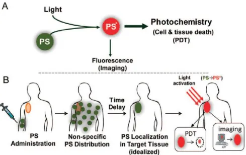
Figure 17: (A) Schematic representation of PDT (B) Schematic representation of
sequence of events in PDT59 ... 31
Figure 18: Schematic representation of FRET based quenching ... 33
Figure 19: Photosensitizer activation through cleavable activation mechanism. ... 33
Figure 20: Photoinduced electron transfer based control of PDT action. ... 33
Figure 21: pH activatable photosensitizer ... 34
Figure 22: Structure of thiol bearing biological compounds ... 37
Figure 23: Type of reactions based on Sulfhydryl containing biological thiols67 .... 38
Figure 24: Structures of probes proceeds based on conjugate addition of thiols. ... 38
Figure 25: Design elements of Thiol Probes ... 39
Figure 26: Pursued Synthetic route for thiol probe 1 ... 41
Figure 27: Design elements of the nitroolefin-BODIPY conjugate thiol probe 1. ... 42
Figure 28: Stacked partial 1 H-NMR spectra of thiol probe 2 (A) and conjugate addition product 2-ME adduct (B) in CDCl3 at 25 °C. ... 43
Figure 29: UV-vis absorption spectra (A) and fluorescence spectra (B) of the thiol probe 1 (5 μM) upon increased Cys concentrations (0-400 equiv.) in 50 mM HEPES: CH3CN (80:20, v/v, pH=7.20, λex: 500 nm at 25 °C). ... 44
Figure 30: UV-vis absorption spectra (A) and Fluorescence Spectra (B) of the Thiol Probe 1 (4 μM) upon increased Cys concentrations (0-400 equiv.) in 50 mM HEPES ( pH=7.2, λex:500 nm at 25 °C). ... 45
Figure 31: Absorption spectra (A) and Fluorescence spectra (B) of Thiol Probe 1 (4 μM) upon addition of 200 eqv. of Cys, Hcy, ME and GSH in 50 mM HEPES (pH=7.2, λex:500 nm at 25 °C). ... 45
Figure 32: Fluorescence response of the thiol probe 1 (2.4 μM) toward biothiols (Cys, Hcy and GSH; 200 equiv. each) and other natural amino acids (400 equiv.) in 50 mM HEPES: CH3CN (80:20, v/v, pH = 7.20, λex: 500 nm at 25 °C). ... 46
xv
Figure 33: UV-vis absorption spectra (A) of the Thiol Probe 3 (4 μM) upon increased Cys concentrations (0-1000 equiv.) in 50 mM HEPES: CH3CN (80:20, v/v,
pH=7.2, λex:600 nm at 25 °C). Fluorescence spectra of the red-emitting thiol probe 3
(5 μM) upon increased Cys concentrations (0-400 equiv.) in 30 mM 100% HEPES solution (pH=7.04, λex:600 nm at 25 °C). ... 47
Figure 34: Bodipy based Protein Labeling Agents ... 62 Figure 35: (A) Design elements of the nitroolefin-BODIPY conjugate for protein labelling (B) Chemical Structures of Bodipy based Protein Labeling Agents... 63
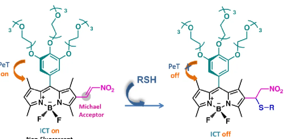
Figure 36: Pursued Route for the synthesis of PL1 and PL3 ... 65 Figure 37: Working Principle of the nitroolefin-BODIPY conjugates PL 3. ... 66 Figure 38: UV-vis absorption spectra (A) and Fluorescence Spectra (B) of the PL 1 (1 μM) upon increased Cys concentrations (0-100 equiv.) in 1X PBS: DMSO (5:95, v/v, pH=7.40, λex: 490 nm at 25 °C). ... 67
Figure 39: UV-vis absorption spectra (A) and Fluorescence Spectra (B) of the PL 3 (2.5 μM) upon increased Cys concentrations (0-200 equiv.) in 1X PBS:DMSO (5:95, v/v, pH=7.40, λex: 520 nm at 25 °C). ... 67
Figure 40: Comparison of aromatic regions of 1H-NMR of PL1 and PL2 and that of their adducts with mercaptoethanol ... 68
Figure 41: (A) Absorbance spectra (B) Fluorescence spectra of PL1 and BSA-PL 1 conjugate in 50 mM HEPES:ACN (80/20, v/v, pH:7.4 λex: 490 nm at 25 0C) ... 69
Figure 42: (A) Absorbance spectra (B) Fluorescence spectra of PL2 and BSA-PL 2 conjugate in 50 mM HEPES:ACN (80/20, v/v, pH:7.4 λex: 500 at 25 0C) ... 69
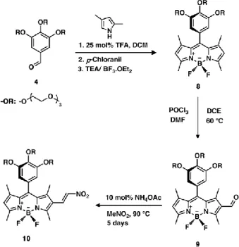
Figure 43: Structure of Designed Activatable Photosensitizer. ... 81 Figure 44: Target photosensitizers for GSH mediated activity control ... 82 Figure 45: Synthesis of target bodipy core via acid catalyzed condensation of 4- and 2-hydroxybenzaldehydes with 2,4-dimethylpyrrole ... 83
xvi
Figure 47: Chemical Structure of Control Photosensitizer 33. ... 85 Figure 48: GSH-mediated activation of photosensitizers for the cytotoxic singlet oxygen generation ... 86
Figure 49: Absorbance Spectra of GSH-mediated Activation of Photosensitizers 2-3 in in DMSO: 1X PBS (50:50, v/v, pH: 7.4, at 25 °C). ... 87 Figure 50: Fluorescence Emission Spectra of GSH-mediated Activation of Photosensitizers 2-3 in (4 µM) DMSO: 1X PBS (50:50, v/v, pH: 7.4, λex: 655 nm at
25 °C). ... 88 Figure 51: Absorbance spectrum of trap molecule (2,2'-(Anthracene-9,10-diyl)bis(methylene)dimalonic acid) with PS1-2 (4 µM) in DMSO: 1X PBS (50:50, v/v, pH: 7.4, 25 °C) LED applied at 660 nm. ... 89 Figure 52: Absorbance spectrum of trap molecule (2,2‟-(Anthracene-9,10-diyl)bis(methylene)dimalonic acid) with Bodipy 22, 26, 29 (4 µM) in DMSO: 1X PBS (50:50, v/v, pH: 7.4, 25 °C) LED applied at 660 nm. ... 90 Figure 53: Fluorescence microscope images of Annexin-V-FITC stained HCT116 cells in the presence of 20 nM sensitizer 1. Cells were either subjected to 4 hours of red LED irradiation at 660 nm for 4 hours, followed by 20 h incubation in the dark (A), or just 24 hours of dark incubation (B). Hoechst-33258 stains nuclear DNA in all conditions. Arrows point to the apoptotic cells with fragmented chromatin (bright blue) and Annexin V positive membrane (green). Images are captured at 40x. ... 91
Figure 54: Growth inhibition of HCT116 cells was determined by NCI-SRB assay. Various concentrations of sensitizer 1 (1-250 nM) were used to determine cell death. Red bars show cell growth inhibition under 4 h irradiation with red LED, followed by 20 h incubation in the dark, black bars indicate cell growth inhibition following 24 h incubation in the dark. ... 93
Figure 55: The decrease in absorbance spectrum of trap molecule (2,2'-(Anthracene-9,10-diyl)bis(methylene)dimalonic acid) in the presence of Bodipy 33 (4 µM) in DMSO: 1X PBS (50:50, v/v, pH: 7.4, 25 °C) LED applied at 660 nm for 180min. 94
xvii
Figure 56: 1O2 dependent degradation of trap molecule. ... 95
Figure 57: Schematic representation of direct and indirect CL. (*) indicates excited product... 113
Figure 58: Reaction Mechanism of Luminol. 84 ... 117
Figure 59: Structures of Luminol Derivatives. ... 118
Figure 60: Most probable mechanism responsible for the chemiluminescence of acridinium esters and the alternative routes.84 ... 119
Figure 61: Possible reaction pathway for the PO-CL system. ... 121
Figure 62: Structures of Peroxalate based Chemiluminogenic Compounds... 122
Figure 63: Literature examples for PO-CL system ... 123
Figure 64: Properties of dioxetane structure115 ... 124
Figure 65: Production of an electronically excited acetone for thermal decomposition of tetramethyldioxetane115 ... 125
Figure 66: The two modes of decomposition of 1,2-dioxetanes: (I) the diradical mechanism and (II) the chemically initiated electron exchange chemiluminescence (CIEEL). The diradical mechanism most often generates triplet excited states (T1) while CIEEL generally results in singlet states (S1).84 ... 126
Figure 67: Decomposition mechansim of adamantyl subtituted dioxetane derivatives121 ... 127
Figure 68: Design parameters of Chemiluminogenic Dioxetane Derivatives ... 127
Figure 69: Emission Spectra of Dioxetanes ... 129
Figure 70: Literature examples for Dioxetane Chemiluminescence ... 130
Figure 71: Schematic Representation of Non-Amplified and Amplified Eliminations126 ... 133
Figure 72: Self Immolative Dendrimer Fragmentation 129 ... 134
xviii
Figure 74: Literature Examples for Fluoride Anion Sensors ... 136 Figure 75: Pursued synthetic route for the synthesis of 1,2-dioxetane derivative. . 140 Figure 76: Synthesis of the self-immolative chemiluminogenic Fluoride sensor ... 141 Figure 77: Self-immolation mechanism and multivalent response ... 142 Figure 78: A) Chemiluminescence spectra of probe+F- in the presence of increasing F- concentrations. Probe concentration is 100 µM in DMSO. B) Chemiluminescence spectra of probe+F- in the presence of increasing F- concentrations. Probe concentration is 500 µM in DMSO/PBS (1X, 90/10, pH 7.2). ... 143 Figure 79: Chemiluminescence Intensity of Probe at different percentages of buffer (PBS 1X, pH 7.2) in DMSO. Probe concentration is 500 µM ... 144 Figure 80: pH-dependent chemiluminescence intensity of Probe in the presence of F -(5mM). Probe concentration is 500 µM in DMSO/Buffer (90/10 for pH 4-5.5, NaOAc buffer 50 mM, for pH 7.2, PBS 1X). ... 144
Figure 81: Chemiluminescence intensity of probe in the presence of AcOH (5 mM) and F- (5 mM). Probe concentration is 500 µM in DMSO/NaOAc (50 mM, 90/10, pH 4). ... 145
Figure 82: Chemiluminescence spectra of probe upon addition of 10 equiv. of I-, Br-, Cl-, CN-, AcO-, H2PO4-, HSO4-, NO3-, F- and Probe concentration is 500 µM in
DMSO/PBS (1X, 90/10, pH 7.2). ... 146
Figure 83: Selective chemiluminescent response of the fluoride probe 9 under ambient light. ... 146
Figure 84: PMMA on glass impregnated with probe, exposed to increasing concentrations of F- (12.5 mM to 100 mM) in THF (top, a-d). Same strips exposed to 250 mM fluoride and in varying concentrations of buffer (PBS, pH 7.2) DMSO (e-h, 40, 30, 20, 10 %). ... 147
Figure 85: Integrated luminescence from the PMMA strips showing the response to increasing fluoride (A) and water (B) concentrations. ... 147
xix
Figure 86: A) Chemiluminescence spectra of RC in the presence of increasing F- concentrations. Probe concentration is 100 µM in DMSO. B) Chemiluminescence spectra of RC in the presence of increasing F- concentrations. Probe concentration is 500 µM in DMSO/PBS (1X, 90/10, pH 7.2). ... 148 Figure 87: Comparison of chemiluminescence intensity of Probe and reference compound in the presence of increasing F- concentrations. Probe concentration is 100 µM in DMSO. ... 149 Figure 88: Comparison of chemiluminescence intensity of Probe and RC in the presence of increasing F- concentrations. Probe concentration is 500 µM in DMSO/PBS (1X, 90/10, pH 7.2). ... 149
Figure 89: Chemiluminescence decomposition spectra of RC and Probe. ... 150 Figure 90: Pd-catalyzed depropargylation reaction147 ... 165 Figure 91: Fluorescent detection of Pd ion via depropargylation reaction ... 165 Figure 92: Synthetic scheme for chemiluminescent palladium probes 46 and 48 .. 168 Figure 93: Proposed chemiluminescent depropargylation process catalyzed by Pd ions ... 169
Figure 94: Proposed chemiluminescent Pd0 catalyzed Tsuji-Trost reaction ... 170 Figure 95: Chemiluminescence Spectra of probe 46 (200 µM) in the presence of increasing concentrations of PdCl2 (concentrations: 0.1, 0.2, 0.3, 0.4, 0.5, 0.6 mM) in
DMSO-H2O (95:5, v/v) solution with Na2CO3-NaHCO3 Buffer (50 mM, pH: 9.0)
involving PPh3 (1 mM) at 70 °C. ... 171
Figure 96: Chemiluminescence Spectra of pH dependent deallylation of Dioxetane 46 (200 µM) in the presence of varying concentrations of PPH3 in DMSO-H2O (95:5,
v/v) solution with Na2CO3-NaHCO3 Buffer (50 mM, pH: 9.0) involving PdCl2 (0.4
mM), at 70 °C. ... 172 Figure 97: Chemiluminescence Intensity of Dioxetane 46 (200 µM) at different percentages of buffer (Na2CO3-NaHCO3 buffer, 50 mM, pH: 9.0) involving PdCl2
xx
Figure 98: Chemiluminescence Spectra of pH dependent deallylation of Dioxetane 46 (200 µM) in the presence of PdCl2 (0.4 mM), PPh3 (1 mM) in DMSO-H2O (95:5,
v/v) solution with Kpi Buffer (50 mM for pH: 7.0, 8.0) (Na2CO3-NaHCO3 Buffer (50
mM for pH: 9.0-10.8) involving PPh3 (1 mM) at 70 °C ... 173
Figure 99: Chemiluminogenic response of the probe toward various Pd species A= PdCl2, B= Na2PdCl4, C= Na2PdCl6, D=Pd(OAc)2, E=Pd(PPh3)4 ... 174
Figure 100: Chemiluminescence Emission Intensity of Dioxetane 46 (200 µM) upon addition of different metal ions in DMSO-H2O (95:5, v/v) solution with Na2CO3
-NaHCO3 Buffer (50 mM, pH: 9.0) involving PPh3 (1 mM) at 70 °C. Metals ions
xxi
LIST OF TABLES
Table 1. Optical properties of the probes 1 and 3. ... 47 Table 2: Photophysical properties of dyes.* relative quantum yields. Reference dye: Rhodamine 6G in water (f :0.95) ... 68
Table 3: IC50 values of sensitizers in HCT116 cell line ... 92
1
CHAPTER 1
1. INTRODUCTION
Science is always fascinated by the magnificence of the nature. Mankind discovers notable ideas by observing it with wide open eyes and the questions are clarified by the whispers of the nature. Nature reaches down mankind in several forms and “light” is one of the most indispensable one since one of the prerequisites required for the permanence of life is light. There would be no vegetation and thus, no food chain in the absence of light. We can explain several everyday phenomena like rainbows, growth of garden flowers by thinking about light. There is remarkable harmony between the light and matter or living organisms. When the light is appreciated in the electromagnetic spectrum, the term “Luminescence” takes part in the literature of the science.
In this thesis, “light” will be introduced to you in two parts as “let the light be there” and “let the light glow there”. As it is understood from the heading, first part covers the three glorious projects based on fluorescence phenomena that require the presence of the light. The second part however, covers the two wondrous projects at which light are produced (glow) there.
First part of the thesis is composed of three different projects which are based on the significance biological thiols. Biologically important biothiols like Cystein (Cys), Homocystein (Hcy) and Glutathione (GSH) are vital for the maintenance of cellular redox status and alterations in their levels is linked to a number of severe diseases such as AIDS, cancer and Alzheimer‟s. Judicious design of dyes carrying nitroolefin substituents in conjugation with the BODIPY core, yields dyes which respond to biological thiols by both absorbance and emission changes. The result is bright signaling of biologically relevant thiols in the longer wavelength region of the visible
2
spectrum and in aqueous solutions (Chapter 3). With this knowledge, nitroolefin functionalized dyes were targeted to result in conjugation of nitroolefin with thiol groups such as those belonging to cysteine residues on proteins in order to visualize dynamics of proteins, cell-cell interactions, mechanisms of life cycles of proteins, etc. To prove bioconjugation of the dyes with proteins, absorbance and emission changes were recorded after reaction with both L-cysteine and Bovine Serum Albumin (BSA) and large spectral changes were obtained (Chapter 4). From these biological thiols, Glutathione is related with the oxidative stress exits in cells. In consideration of high intracellular Glutathione concentrations in the cancer cells, we designed and synthesized a series of Bodipy based sensitizers which can generate cytotoxic singlet oxygen only after a glutathione mediated cleavage of the electron sink module because enhanced spatiotemporal selectivity in photonic sensitization of dissolved molecular oxygen is an important target for improving the potential and practice of photodynamic therapy. Cell culture studies not only validate our design, but also suggest an additional role for the relatively hydrophobic quencher module in the internalization of the photosensitizer (Chapter 5).
The other part of the thesis contains two different projects which are developed based on chemiluminescence. In the case of chemiluminogenic sensing of fluoride ions (Chapter 7), Enhanced chemiluminescence signal is obtained when electronically triggered dioxetane cleavage is initiated by fluoride mediated deprotection of the silyl-protecting group, followed by self-immolation via 1,4-quinone-methide rearrangement. The reaction takes place even when the probe is trapped within a PMMA layer on top of a glass plate. In that arrangement, fluoride in aqueous solutions can be detected selectively at low micromolar concentrations. Rapid assessment of fluoride concentrations in drinking water could be a possible application, and the bright chemiluminescence of the probe or structurally related derivatives could provide a promising alternative. New methodology has been developed for sensing of palladium ions (Chapter 8). We have used the power of chemiluminescence for rapid, qualitative and/or quantitative test for analytes of interest; we have analyzed the palladium ions regardless of their charge and source in
3
aqueous environment. Considering the convenience of the method and substantial results, we are confident that other chemiluminogenic probes will emerge.
4
CHAPTER 2
2. BACKGROUND 1:
“LET THE LIGHT BE THERE”
2.1 Photoluminescence
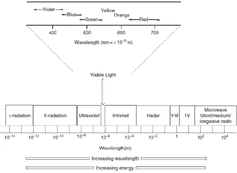
One of the prerequisites required for the permanence of life is “light”. There would be no vegetation and thus, no food chain in the absence of light. We can explain several everyday phenomena like rainbows, growth of garden flowers, etc., if we appreciate the visible light in electromagnetic spectrum that bring us to the substantial question: what happens when light hits to matter. This can be explained by considering the harmony between radiation and matter. For example, if there is harmony between radiation and matter, absorption of energy is possible otherwise, reflection or scattering occurs.1 Light can be characterized by electromagnetic radiation of a definite wavelength (λ). The wavelengths from 400nm to 750nm represent the visible part of the spectrum which corresponds to the region of shorter wavelength range of γ-rays to longer wavelength end of radio waves (figure 1). While the color of a chemical compound depends on which part of electromagnetic spectrum it absorbs, the transmitted spectral mixture of light determines the color that we see. Additionally when we are working with dilute solutions, by considering spectral characteristics of absorbed light, we can get information about the electronic structure and concentration of the sample.2
5
Figure 1: Electromagnetic Spectrum1
2.1.1 Principles and Characteristics of Luminescence
The umbrella term for light emission processes is “Luminescence” which can be defined as the emission of light resulted from the excitation of a chemical compound trough high energy radiations or electrons. When a chemical compound is exposed to the light which has energy equal to the energy of possible electronic transition, some of the light absorbed by compound leads to the excitation of an electron to a higher energy orbital which relaxes back to ground state by losing their excess energy through several possible pathways.3 There are actually two forms of radiation in competition which are radiative and nonradiative transitions. If electronically excited molecule relaxes back to ground state by emitting its excess energy radiatively through the emission of photon or nonradiatively vibrations which heats the lattice (figure 2). These deactivation processes can be visualized simply via The Perrin-Jablonski diagram (figure 3).
6
Figure 2: (a) General representation of luminescence mechanism, exc represents excitation, em represents emission and heat represents the nonradiative transition (b) Representative Energy Level Diagram A*: Excited State of Activator, A: Ground State of activator, R: Radiative transition to the ground state or emission, NR: Non-radiative transition or heat.
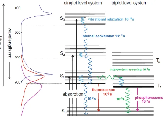
When light hits to a chemical compound, an electron from ground state (S0) is
jumped to an excited state depending on the energy of absorbed light. Since the excited state of a molecule by absorption occurs through conservation of the electron spin-paring, the excited state is called as singlet state (S1). According to Kaska‟s rule,
either luminescence emission or chemical reaction by excited molecules originates from ν=0 of S1 or T1 because relaxations from higher electronically excited states to
S1 (or T1) occurs swiftly. If the absorbed light has higher energy required for a simple
electronic transition, the excess energy is converted into vibrational and rotational energy which leads to excitation of upper vibronic levels of excited state. When absorbed light having excess vibrational energy leads to the excitation of an electron to a higher vibrationally excited state and vibrational relaxation occurs within the order of 10-12s between vibrationally excited state v>0 and v=0 of given state as a result of possible collisions with solvent molecules. On the other hand, internal
conversion occurs from upper excited electronic levels like S2, S3, etc. to the lowest
electronically excited state S1 by preserving the same spin multiplicity. Internal
conversion occurs within the order of 10-14-10-11s between excited states and the order of 10-9 -10-7s between S1 and S0.
7
Figure 3: Jablonski diagram representing the typical photophysical processes in molecules 2
Another nonradiative process is based on intramolecular transitions which occur between isoenergetic states of different multiplicities called intersystem crossing. This is a spin forbidden process and it can be obtained as a result of spin-orbit coupling which is the action of an effective magnetic field and it can originate while electrons orbit around the charged nuclei. This causes little mixing of singlet and triplet states thus in turn, electron can move from S1 to T1. In a molecule, spin-orbit
coupling can be enhanced with the introduction of heavy atoms like bromine and iodine.
After the excited state molecule release its excess energy through vibrational relaxation or interval conversion in order to reach S1 state, it can relaxes back to
ground state via the emission of light. This is called as fluorescence and it occurs between the states of same multiplicity and in the order of 10-9s. Although
8
fluorescence competes with other deactivation processes of same multiplicity when electrons are excited to higher vibrational levels of S1 state or higher vibrational
levels of upper energetic states, it always wins the race from S1 state.
When an electron is moved to T1 state, it can also relax back to ground state with
emission of light. This spin-forbidden radiative transition is called as
phosphorescence. Since transitions to S0 are forbidden, the rate of emission is slow
thus in turn, the phosphorescence lasts typically milliseconds to few hours so that emission can continue even after light source has been removed.4
2.1.2 Phenomena of Fluorescence
The world of fluorescence is a world of beautiful color.
In the darkness, all the ordinary colors of our daylight would disappear.
Only the intensely glowing hues of fluorescent substances touched by the
Ultraviolet beam shine out with striking clarity.
Sterling Gleason, 1960
Fluorescence can be defined as the radiative transition which can occur between the states of same multiplicity.5,6 Fluorescence can be used for the quantitative analysis of fluorescence compounds since emission intensity is proportional to the concentration of the compound. Due to the high sensitivity and selectivity of fluorescence, it has diverse spectrum of application areas. Because of that in order to comprehend and interpret the analysis, there are several parameters related with fluorescence which has to be learned deeply.
The energy of fluorescence is less than that of absorption because of that fluorescence occurs at longer wavelengths than absorbance. When a molecule is excited upon exposure to light, there are possibilities of collisions between the
9
molecule and solvents so that molecule loses some of its energy as heat. Therefore, since energy and wavelength are inversely proportional to each other, lower energy fluorescence emission occurs at longer wavelength which is spectrally red shifted. The difference between band maxima of absorption and emission spectra is called as
Stokes Shift. There are several causes of Stokes shift such that rapid decay to lowest
vibrational level of S1, solvent effects, excited state reactions, complex formation and
energy transfer.7
Quantum yield and fluorescence efficiency are one of the most important characteristics of a fluorophore. Quantum yield (Фf) can be defined as the number of
emitted photons by S1 relative to the number of absorbed photons by S0. The highest
value of quantum yield approaches to unity in molecules relatively rigid which have large and planar conjugated systems. In the case of more flexible molecules, quantum yield will be low due to the possibility of having high vibrational and rotational freedom compared to rigid ones. Quantum yield is specific for each fluorescent compound and it is independent of either excitation or emission wavelength.8 Fluorescence lifetime (τ) can be defined as the average value of time which is spent by molecules in the excited states before relaxing back to ground state. It is related with the decay rate of fluorescence intensity after a short excitation pulse. These two parameters are closely related by
Фf = τf/ τn equation 1
where τn is the natural lifetime of excited state which represents the lifetime that
fluorophore would have if fluorescence is the only way for a molecule deactivates from lowest excited singlet state.9 Lifetime becomes shorter in the case of quenching processes.
Fluorescence quenching is the decrease or suppression of the fluorescence intensity
via different mechanisms. The decrease or suppression in the fluorescence intensity has been observed due to formation of transition complex when excited fluorophore contacts with quencher like molecular oxygen, compounds bearing heavy atoms, halogen ions or another fluorophore. The possible quenching mechanisms are static
10
and collisional according to the type of contact between fluorophore and quencher. Static quenching occurs when a fluorophore and quencher takes place in ground state thus, forms a nonfluorescent complex whose efficiency depends on both the concentration of quencher and the formation constant of the complex. On the other hand, collisional quenching occurs when excited fluorophore is deactivated upon contact with the quencher. Oxygen, halogens, amines and electron deficient molecules act as quenchers but the quenching mechanism is different and depends on the fluorophore-quencher pair. For example, quenching resulted from halogens or heavy atoms occur via spin orbit coupling and inter system crossing.10 Another term is photobleaching which can be defined as photo-induced chemical destruction of fluorophore upon exposure to excitation that leads to the loss of ability of the fluorophore to fluoresce.11
2.1.3 Factors Affecting Fluorescence
There are several factors having considerable effects on fluorescence quantum yield that can be listed as molecular structure, substituent effects, solvent effects, temperature and viscosity. These factors are explained in order.
Molecular Structure: Fluorescence is mostly observed in compounds having rigid structures as a result of aromaticity or extended conjugation. Consequently, the higher the rigidity of the compound, the lesser the vibrational and rotational freedom, the higher the probability of fluorescence since the energy gap between S1 and S0
becomes large so that fluorescence predominate over nonradiative processes.4
Substituent Effect: The introduction of freely rotating substituents into aromatic compounds decreases both fluorescence quantum yield and intensity due to the enhanced probability of rotational and vibrational freedom. Additionally, modification of aromatic compounds with electron donating substituents increases fluorescence quantum yield due to the increased rate of radiative decay. On the other hand, introduction of electron withdrawing substituents having sp2 hybridized
11
nonbonding electrons and heavy atoms reduces the fluorescence quantum yield owing to the mixed spin-orbital electronic motions of aromatic system and enhanced probability of spin-triplet intersystem crossing.9
Solvent Effect: Solvent has profound effects on fluorescence emission in different ways. For instance, fluorescence intensity could be decreased in solvents containing heavy atoms due to the increase in probability of spin orbit coupling thus in turn, increase in phosphorescence. Hydrogen bonding between fluorophore and solvent has significant effect on fluorescence whose intensity has been affected by the stabilization or destabilization effect of generated hydrogen bond on the electronic excited state. When excited, organic molecules become more polar and thus, enhances the interaction between the dye and its molecular environment resulting in the energy loss.12
Temperature: Temperature and fluorescence is inversely proportional with each other since the increase in temperature leads to increase in either molecular motions or collisions via lowering viscosity of the solvent and thus, fluorescence decreases.
Viscosity: Fluorescence can also be affected by viscosity of the solvents which determines the collisions between solvent molecules and excited state molecules. In viscous solutions, the molecular collisions are reduced which reduces the energy transfer. Consequently, the lesser the molecular collisions, the lower the energy loss and thus in turn, the higher the fluorescence.7
2.2 Fluorescent Probes
Methods based on fluorescence like fluorescence spectroscopy, fluorescence imaging and fluorescence indicators become indispensible diagnostic, monitoring and analytical tools in biochemistry, material science, biotechnology, environmental and analytical chemistry since they provide worthless information for investigating the molecular interactions in chemical and biological systems.
12
Fluorogenic probes can be defined as the reagent which can convey the information upon interaction with the analyte as the changes in its photophysical characteristics and based on these spectroscopic changes, analyte of interests could be detected8,13. Fluorescent probes have wide range of application areas due to distinct advantages offered by fluorescence detection in terms of simplicity, sensitivity, selectivity, monitoring of dynamic changes in space and in time, having high spatial and temporal sampling capability and response time.14 Generally chromogenic and fluorogenic probes are composed of three vital parts which are recognition site, signaling unit and linker.13 While recognition site is designed in a way as to recognize the analyte of interest, signaling unit is designed in a way as to provide visible, fluorescent readouts as a result of a change in photophysical properties upon reaction of recognition site with the analyte. Linkers are responsible for the connection of recognition and signaling moiety however, they are mostly integrated directly. Although various types of fluorescent probes specific to different analytes exist, there is still growing need for the development of fluorescent probes with improved sensitivity and selectivity, minimum response time and minimum perturbation in biological environment. Because of that for the development of rational fluorescent probes, there are several significant parameters required to be considered extensively.15 (a) Designed probe should respond only to the analyte of interest and elicit fluorescent response (b) The fluorescence obtained upon interaction with the analyte should be bright which means that it should acquire large extinction coefficient at the excitation wavelength and high quantum yield. (c) Probe should have high stability against the chemicals and light. (d) Fluorescence emission by turn-on or shifts in excitation and /or emission wavelength is more favored than turn off response. Actually, applications of fluorescent probes for the identification of molecular interactions are very important in the sense of chemistry and biology. Therefore, fluorescent probes are designed to work in live cells, too and this brings the consideration of other parameters during the design of fluorogenic probes. (a) The probe should be selective to the analyte and it should not respond to other cellular analytes. (b) Fluorogenic probes should represent high brightness with low dye concentration in order to eliminate photo cytotoxicity to live cells. (c) Probes
13
designed to operate in near IR range is better for live cell studies since the possibility of photodamage is low and additionally, biological samples have low background emission in NIR region which enhances the signal to noise ratio.
There are several photophysical processes responsible for the changes in fluorescence like quenching via collision, photoinduced electron transfer, exciplex formation, photoinduced charge transfer, energy transfer, etc. In this thesis, analyte detection by fluorogenic probes proceeds over mostly two different photophysical processes which are photoinduced electron transfer (PeT) and photoinduced charge transfer (PCT).
2.2.1 Photoinduced Electron Transfer (PeT)
Probes designed to operate via PeT processes contain acceptor (fluorophore), linker and donor (chelator constructs). The use of linkers is necessary to prevent the full conjugation between acceptor and donor molecule. In case of Bodipy dye, introduction of donor group in the meso position of Bodipy fluorophore enables the disconnection between the acceptor and donor units due to the perpendicular arrangement of donor group. PeT process can be explained better by using simple representative molecular orbital (MO) diagrams.16 When fluorophore is exposed to the light, an electron from highest occupied molecular orbital (HOMO) is promoted to the lowest molecular orbital (LUMO). Since HOMO level of donor is higher than HOMO level of the acceptor, electron from HOMO of donor is transferred to the HOMO of acceptor which enables the PeT process and thus, fluorescence is quenched (i.e. probe is in fluorescence off state). When the analyte is added to the medium, it interacts with the chelator unit and energy of HOMO level becomes lower than HOMO level of fluorophore; consequently PeT becomes inactive (i.e. probe is fluorescence on state). As a result of this, excited electron relaxes back to ground state and fluorescence emission can be observed (figure 4).17
14
Figure 4: Representative scheme for the photophysical mechanism of reductive PeT
In the case of oxidative PeT which is shown schematically in figure 5, electron donation occurs from fluorophore (donor) to the chelator (acceptor). When fluorophore is exposed to light, an electron from HOMO level of donor is promoted to LUMO level. Since LUMO level of donor is higher in energy than that of acceptor, excited electron is transferred from LUMO of donor to the LUMO of acceptor; consequently fluorescence is in off state. Upon reaction with the analyte, LUMO level is higher than that of donor. Therefore, PeT becomes inactive (i.e. probes is fluorescent on state)15. In the literature there are several examples which operate through oxidative PeT mechanism based on Bodipy dyes.18,19
15
2.2.2 Photoinduced Charge Transfer (PCT)
PCT operates in probes when electron donating moiety is in full conjugation with electron withdrawing moiety and intramolecular charge transfer from donor to the acceptor occurs upon excitation; consequently, changes in dipole moment leads to the stokes shift which is observed as red or blue shifts in the fluorescence emission spectra. When the analyte like a cation interacts with electron donor moiety of the probe, the ability to donate electrons is reduced. Therefore, due to the presence of full conjugation, a blue shift is observed (figure 6a). On the other hand, when analyte like a cation interacts with acceptor moiety, it enhances electron withdrawing ability of the acceptor group thus in turn, a red shift is observed (figure 6b). Beside these shifts, there will be changes in either quantum yield or lifetimes of the probes.16
Figure 6: Schematic Representation of PCT Mechanism
2.2.3 Design Strategies for Probe Development
In the literature, there are several classification types for the design strategies required for probe development. Analyte detection can be achieved via three different reaction mechanisms based on (a) protonation-deprotonation (b) complexation (c) carbon-carbon bond forming and breaking reactions.13 Additionally, analyte recognition mechanisms based on carbon-carbon bond forming
16
and bond breaking reactions can be diversified by nucleophilic addition/substitution type reactions.20 Studies presented in the thesis are developed based on the conjugate addition and nucleophilic substitution reactions of analytes so that these reactions are explained deeply in next chapters.
Protonation-deprotonation mechanism has been used to develop pH responsive
fluorogenic probes. They are mostly designed to work in neutral pH however modification via OH, COOH and amino groups enable the development of probes with wide pH ranges. In order to enhance the response range, probes should be modified with the introduction of multiple H+ responsive units in different positions of the molecule.21 Since pH sensitive groups like OH, COOH and amino have affinity for the metal ions, there is a possibility of complexation reaction which can be eliminated by the arrangement of electronegative groups such that they cannot enable the formation of suitable cavities for metal ions. Large number of pH probes has been reported in the literature based on xanthene22-24, bodipy25-28 and cyanine
29-31
(figure 7).
Figure 7: pH responsive fluorogenic probes
Complexation based probes could be developed based on the attachment of receptor
and reporter units either covalently or noncovalently. In this type of probes, construction of specific receptor units for the analyte is important because probes respond via the combination of different photophysical processes. Additionally, since several electronegative groups have been introduced for the development of complexation probes, the effect of pH should be examined. Furthermore, the great
17
challenge in the design and development of this type of fluorogenic probes is the high selectivity for a specific metal ion because most metal ions may interfere with each other due to having similar reactivities (e.g., Mg2+ vs Ca2+, Ag+ vs Hg2+, and Cd2+ vs Zn2+). Complexation based probe in figure 8 works based on the PeT process. Due to electron transfer from donor (receptor) to the acceptor (fluorophore), PeT is on state and consequently, fluorescence of the probe is weak. However, in the presence Zn+2 ion, receptor unit makes complexation with the ion and thus, fluorescence emission is enhanced due to the inactivation of PeT.32
Figure 8: Fluorogenic probes based on complexation with metal ions
Probes based on carbon-carbon bond forming and breaking reactions could be accepted as reaction based probes which can be classified into different groups based on the reaction types. In general, since these probes recognize the analyte through specific reactions, the spectroscopic changes in the fluorophore will be irreversible which enhances the sensitivity of the probe whereas reduces the applicability of it for dynamic range studies. Probes designed based on the nucleophilic addition to carbonyl group can be applied for the detection of anions, neutral species. When considering the nature of the reaction in terms of chemistry behind it, analytes with high nucleophilicity can be detected by using this type of reaction based probes. For example, cyanide, bisulfite, carboxylates and amines are the common analytes for this type of reaction. Although photophysical changes can be adjusted accordingly, these probes work based on the PeT process. Cyanide ion
18
can be detected via cyanohydrin formation (figure 9). Probe A is non-fluorescent since PeT operates. When cyanide is added to reaction medium, it attacks to carbonyl carbon of the fluorophore forming cyanohydrin adduct; consequently PeT becomes inactive thus in turn, fluorescent is turned on.33
Figure 9: Turn on fluorescent sensing of cyanide
Carbonyl addition based probes can be designed for the sensing of biological thiols
which can be detected upon the formation of thiazoline ring through the nucleophilic addition of thiols to aldehyde moiety. In the case of formaldehyde derived fluorescein (figure 10a), selective detection of thiols has been achieved via turn-off fluorescence response.34 When coumarin has been used for the detection of thiols, turn-on fluorogenic response has been observed.35
Figure 10: Fluorescence turn-off (A) and turn-on (B) probes for the detection of thiols
19
Since thiols are soft nucleophiles, they prefer to attack to soft electrophiles. Because of that biological thiol detection operates through the Michael addition reaction which can also be named as 1, 4-conjugate addition. Many fluorescent probes have been developed based on the thiol addition to maleimide unit modified fluorophore like bodipy as in figure 11a.18 Beside maleimide unit, quinone derived fluorophores can be used for the conjugate addition of thiols as in figure 11b.36
Figure 11: Fluorescence based detection of thiols via conjugate addition.
Thiols can also be detected by nucleophilic substitution reaction by use of electron sink 2,4-dinitrobenzenesulfonyl moiety37 or by the use of disulfide bridges.38
20
2.3 Protein Labeling
Proteins are one of the major biological macromolecules which are composed of different combinations of amino acids. Proteins can be accepted as worker biological molecules existing in all living organisms whose functions like catalysis of metabolic reactions, DNA replication, molecular recognition, visualization of cell-cell interactions, protein dynamics, mechanisms of life cycles of proteins, etc. affect whole organization of life. Because of that it is important to figure out how they function normally, how they interact with other molecules or how some diseases are caused by the abnormal shapes of them.39 Protein labeling can be depicted as tagging of proteins in order to figure out their functions, interactions and movements either in
vitro or in vivo. In the literature, there are several approaches which enable the
introduction of small probes bearing wide range of photophysical properties to purify target proteins in vitro. Labeled purified proteins can be applied to identify protein-protein interactions but they have differences in behavior compared to those in live cells. Proteins have at least two functional groups which can be used for the labeling procedures are amino functionality (N-terminus) and carboxyl functionality (C-terminus). In order to label the proteins, chemical methods like covalent bonding can be used to interact with functional groups on aminoacids specifically. Additionally, nonspecific attachment methods to N or C terminus of amino acids are also possible. Other than these, methods involving enzymes requiring related polymerases, ATP and labeled amino acids or nucleotides can also be used to labels proteins and amino acids. The expression of tagged proteins via in vitro translation is difficult since this requires proper protein length, folding and post-translational modifications which cannot be provided by some of the kits.
21
2.3.1 Fluorescence based labeling of proteins
The need for chemical protein labels has led the researchers to develop new strategies for labeling of proteins. Traditional methods used for labeling studies are not good enough for in vivo studies since purification of protein, chemical labeling, repurification and reintroduction into cells by invasive methods like microinjection are hardly necessary.40 Spectroscopic measurement based methods are more prosperous since they are minimally invasive; allow real time monitoring of cellular events without any perturbation. For this reason, spectroscopic tags must be employed for labeling of the target proteins. Among spectroscopic methods, use of fluorescence based methods is more promising because fluorescent response is obtained swiftly in a short time. Beside fast signal acquisition, sensitivity of
detection has been developed which enable single molecule detection due to the
development of current technologies.41 Additionally, dyes can be prepared in multiple colors which can be applied for multiplex assays. Furthermore, since the size of the tag is small, this reduces the possibility of perturbation in the behavior of the tagged protein. Different from enzymatic reactions in which the signals amplify and diffuse, fluorescent signals is localized with high spatial resolution. Moreover, fluorescent labeling reagents are stable and robust in biological environment. Lastly, the labeling process is very straightforward.41,42
Characteristics for a good labeling tag and method: First of all, after the determination of labeling strategy, either the nature of suitable tag or the type of conjugation reaction has to be considered extensively. As labeling strategy, choice of tag is very important for effectiveness of the method. Small-sized tags should be preferred in order to eliminate the possibility of perturbation in natural behavior of labeled protein. Preferred tag should show performance equally in cell surface, in cytosol and also in an organelle. Having multiple choices of tags are beneficial in order to introduce multiple probes with wide spectrum ranges. The methods have to be chosen in a way as to ensure specific and selective labeling of the target protein in
22
a cell which contain other biomolecules. The interaction between label and protein must be stable enough to enable the stable interaction between two molecules so that label remains securely attached to the protein during the assay. Lastly, labeling reaction should be designed to be orthogonal to any reactions.42
2.3.1.1 Fluorescent- protein based labeling
Fluorescent labeling of proteins has started with the use of green fluorescent protein (GFP) or one of its variants when their genetic sequence becomes available. After the expression of gene fusion, autofluorescent GFP fusion could be detected microscopically. Due to its small size (ca.27kDa), compactness, its single domain structure enable GFP to be fused to a variety of proteins without causing any interferences in native protein functionality.40 Modified versions of GFP like cyan fluorescent protein (CFP), yellow fluorescent protein (YFP) and blue variants have been generated to offer various emission wavelengths with improved photo-stability. Availability of multicolored variants of fluorescent proteins (FP) contributes to the development of cell biology studies by consenting the multicolor labeling of multiple proteins in a single cell. FP based labeling of proteins is an extremely sensitive method since FP fused proteins are only fluorescent species in the target cells. Beside, fluorescence intensity can be used for the determination of amount of protein since there is a direct correlation between them. This method can be used for live cell monitoring of either dynamic distributions or localizations of proteins, protein-protein interactions and quantifying the expression levels of protein-proteins in real time.42 Moreover, since fluorescent proteins are highly stable and it cannot be sheltered by proteins barrel structure, they can be used to track the localization and movement of fusion proteins in cells.
On the other hand, this approach brings several intrinsic limitations. GFP is a large protein consisting of 238 amino acid proteins which can interfere with localization,