Ameliorative Effect of Omega-3 in Carbon Tetrachloride Toxicity
Musa KARAMAN
1
Hasan ÖZEN
2Serpil DAĞ
2Onur ATAKİŞİ
3Gülşen ÇIĞŞAR
4Oktay KAYA
5[1] This study was supported by the Scientific Research Fund of Kafkas University (Project No: BAP VF 07-2014) 1 Department of Pathology, Faculty of Veterinary Medicine, Balikesir University, TR-10145 Balikesir - TURKEY 2 Department of Pathology, Faculty of Veterinary Medicine, Kafkas University, TR-36100 Kars - TURKEY 3 Department of Biochemistry, Faculty of Art and Science, Kafkas University, TR-36100 Kars - TURKEY 4 Department of Emergency, Faculty of Medicine, Kafkas University, TR-36100 Kars - TURKEY
5 Department of Physiology, Faculty of Medicine, Trakya University, TR-22030 Edirne - TURKEY
Article Code: KVFD-2016-15862 Received: 11.04.2016 Accepted: 11.10.2016 Published Online: 12.10.2016
Citation of This Article
Karaman M, Özen H, Dağ S, Atakişi O, Çığşar G, Kaya O: Ameliorative effect of omega-3 in carbon tetrachloride toxicity. Kafkas Univ Vet Fak Derg,
23, 77-85, 2017. DOI: 10.9775/kvfd.2016.15862
Abstract
Omega-3 is a polyunsaturated fatty acid known to have immunomodulatory functions. In the present study, ameliorative potential of omega-3 in experimental carbon tetrachloride (CCl4) toxicity was investigated. Total of 40 adult male Wistar albino rats were allocated into five groups and were
subcutaneously given once every two days for 6 weeks the followings: Group 1 (Control): 0.5 ml/kg serum physiologic, Group 2 (Omega): 0.5 g/kg omega-3, Group 3 (Vehicle): 0.5 ml/kg pure olive oil, Group 4 (CCl4): 0.5 ml/kg CCl4, Group 5 (CCl4 + Omega): 0.5 ml/kg CCl4 plus 0.5 g/kg omega-3. At
the end of the treatments, blood samples were collected and necropsy was performed for collection of liver tissues. Serum AST, AlT, GGT, TAC, TOC, triglyceride, and visfatin levels were detected. liver morphology and immunoreactivities against TGF-α, TGF-β, PPAR-α, and PPAR-γ were assessed. Serum AST, AlT, GGT, and TOC levels significantly increased while TAC level decreased in CCl4 given animals as compared to the control group. No significant
changes were observed in triglyceride and visfatin levels. Immunohistochemical staining revealed increased TGF-α and TGF-β expressions and decreased PPAR-α and PPAR-γ expressions in liver of CCl4 given animals. Omega-3 supplementation has prominent effects in correcting the biochemical and
immunohistochemical parameters studied as well as the tissue morphology. The results of the investigation indicated that omega-3 has ameliorative effects on the oxidative tissue degeneration and inflammatory processes induced by CCl4 treatment in rats.
Keywords: CCl4, Omega-3, TGF-α, TGF-β, PPAR-α, PPAR-γ
Karbon Tetraklorür Toksikasyonunda Omega-3’ün Koruyucu Etkisi
Özet
Omega-3 bağışıklığı düzenleyici fonksiyonları olduğu bilinen çoklu doymamış bir yağ asididir. Bu çalışmada, deneysel karbon tetraklorür (CCl4)
toksikasyonunda omega-3’ün koruyucu potansiyel etkisi araştırılmıştır. Toplam 40 adet ergin erkek Wistar albino rat eşit beş gruba ayrıldı ve 6 hafta süresince her iki günde bir olmak üzere subkutan yolla deneklere tarif edilen uygulamalar yapıldı; Grup 1 (kontrol): 0.5 ml/kg serum fizyolojik, Grup 2 (Omega): 0.5 g/kg omega-3, Grup 3 (Taşıt): 0.5 ml/kg saf zeytin yağı, Grup 4 (CCl4): 0.5 ml/kg CCl4, Grup 5 (CCl4 + Omega): 0.5 ml/kg CCl4 artı 0.5 g/kg
omega-3. Araştırma süresinin sonunda, kan örnekleri alınan deneklere karaciğer doku örneklerinin toplanması amacıyla nekropsi uygulandı. Serum AST, AlT, GGT, TAC, TOC, trigliserid ve visfatin seviyeleri belirlendi. Karaciğer morfolojisi incelendi ve TGF-α, TGF-β, PPAR-α ve PPAR-γ immunreaktiviteleri tespit edildi. Kontrol grubu ile karşılaştırıldığında CCl4 verilen hayvanlarda serum AST, AlT, GGT ve TOC seviyelerinin anlamlı derecede arttığı, TAC seviyesinin
ise azaldığı gözlemlendi. Trigliserid ve visfatin seviyelerinde anlamlı bir değişikliğin olmadığı belirlendi. İmmunohistokimyasal boyamalarda CCl4 verilen
hayvanlarda TGF-α ve TGF-β immunreaktiviteleri artarken PPAR-α ve PPAR-γ immunreaktivitelerinde azalma dikkati çekti. Omega-3 takviyesinin incelenen biyokimyasal ve immunohistokimyasal parametreler üzerine olumlu etki gösterdiği belirlendi. Çalışma sonuçları omega-3’ün CCl4 uygulanan ratlarda
oluşan oksidatif doku hasarı ve yangısal süreci azaltmada etkili olduğunu göstermektedir. Anahtar sözcükler: CCl4, Omega-3, TGF-α, TGF-β, PPAR-α, PPAR- γ
INTRODUCTION
liver is the primary organ in various metabolic activities. However, countless physiological and biochemical functions
as well as its anatomic localization subject the organ in development of infectious and toxic degenerations [1,2].
liver may recover after acute degeneration, if not chronic inflammation and/or death may develop. The worst result
İletişim (Correspondence)
+90 543 8708684of the chronic inflammation is cirrhosis. liver cirrhosis may cause portal hypertension, hepatic encephalopathy, and organ failure that may result in death of the patient [3,4].
Carbon tetrachloride (CCl4) is a chemical compound
frequently used in experimental studies to induce liver cirrhosis in laboratory animals as a model for human. It is a transparent, non-flammable, easily vaporizable, and colorless liquid [5,6]. In mammalian body, CCl
4 is first
metabolized to trichlormethyl (CCl3) by the enzymatic
reduction of cytochrome p-450 and then converted to trichlormethylperoxy radical (OOCCl3)in the presence
of oxygen [5]. These reactive free radical products of CCl
4
may react with polyunsaturated fatty acids and cause the production of reactive oxygen derivatives, which triggers lipid peroxidation in biological membranes [7-9]. Oxygen
radicals can induce a cascade of events that result in cellular degeneration, increased Kupffer cell activation in liver, and increased expression of some pro-fibrogenic and pro-inflammatory factors such as Tumor necrosis factor alpha (TNF-α) and Transforming growth factor beta 1 (TGF-β1) [10]. TGF-β1 functions in conversion of quiescent
hepatic stellate cells into myofibroblast-like cells and suppresses degradation and stimulates production of extracellular matrix proteins [10], thereby is important in the
development of hepatic fibrosis [11].
Omega-3 fatty acids are unsaturated fatty acids that have double bounds at the third carbon atom from the end of the carbon chain. Omega-3 fatty acids, which include dokozaheksanoic asit (DHA), eikozapentaenoic acid (EPA) and α-linolenic acid (AlA), are the part of cellular membranes and required for normal biological functioning. DHA and EPA are members of long chained polyunsaturated fatty acids and found in fish oil in large amounts [12,13]. Studies on omega-3 fatty acids showed
that they have anti-oxidant, anti-inflammatory, anti-hyper-tensive, and anti-apoptotic effects[14,15].
Free radicals are known to involve in degeneration of cellular molecules and hence play role in aging, cancer, arteriosclerosis, and cirrhosis. Omega-3 fatty acids were shown to partially protect against these conditions by inhibiting development of ischemia, inflammation and production of free radicals [2,16-18].
Peroxisome proliferated activated receptors (PPARs) are nuclear receptors and found in 3 isoforms, namely PPAR-α, -β, and -γ [19,20]. PPAR-α is most frequently expressed
in organs rich in fat tissue such as liver, heart, skeletal muscle, brown adipose tissue and kidney. Monocytes, macrophages, lymphocytes, vascular endothelial and smooth muscle cells show expression of PPAR-α the most. While PPAR-β is expressed mostly in fat tissue, skin and brain, PPAR-γ is seen in fat tissue, large intestine, heart, kidney, pancreas and spleen [21-23]. PPARs are activated by
fatty acids and their derivatives. PPAR-α is activated by leukotriene B4 while PPAR-β with prostaglandin J2 [24,25].
Visfatin is an adipokin having insulin like functions and excreted from adipose tissue. Its expression is affected by TNF-α and interleukin 6 [26]. It was shown that visfatin
play roles in lipid metabolism and inflammation [27]. It
was shown that PPAR-γ ligands could increase the gene expression of visfatin in macrophages [28].
In this study, the roles of TGF-α, TGF-β, PPARs and recently discovered molecule, visfatin, were investigated in an experimental hepatic fibrosis model in rats induced by CCl4 and also the potential protective effect of omega-3.
Oxidant and antioxidant capacities as well as enzymatic changes in liver tissue in relation to the tissue degeneration were investigated by immune-histopathological and bio-chemical means.
MATERIAL and METHODS
Animal and Treatments
The ethical approval for the research was confirmed by Kafkas University Animal Care and Use Committee (Registration Number: 2012-71). All procedures were conducted in accordance with the ‘Guide for Care and Use of laboratory Animals’ published by the National Institutes of Health and the ethical guidelines of the International Association for the Study of Pain.
Forty male Wistar-Albino rats weighing 270-300 g at 3 months of age were used in the study. The rats were housed at 20±2°C and 12 h/12 h light/dark cycle through the study. Standard pellet diet and tap water were provided ad libitum. The animals were divided into five groups each containing 8 animals and treated once every two days during a 6-week period with subcutaneous injections of the followings: Group 1 (Control): 0.5 ml/kg serum physiologic, Group 2 (Omega): 0.5 g/kg omega-3 (18% eicosapentaenoic acid and 12% docosahexaenoic acid), Group 3 (Vehicle): 0.5 ml/kg pure olive oil, Group 4 (CCl4): 0.5 ml/kg CCl4 mixed 1:1 volume with olive oil, and
Group 5 (CCl4 + Omega): 0.5 ml/kg CCl4 mixed 1:1 volume
with olive oil plus 0.5 g/kg omega-3. Biochemical Analysis
At the end of the 6-week treatment period, intra- cardiac blood samples were collected into the serum tubes under ether anesthesia. Serum was separated by centrifugation of the blood samples at 3000 rpm for 10 minutes. The samples were maintained at -20ºC until further analyses. Serum aspartate aminotransferase (AST) (ERBA DDS), alanine aminotransferase (AlT) (ERBA DDS), gamma glutamyl transferase (GGT) (TMl), tri-glyceride (IBl), TAC and TOC (REl Assay Diagnostics, Gaziantep-Turkey) were measured colorimetrically using a spectrophotometer (Eon Biotex, USA), serum visfatin (SUN RED CAT NO: 201-11-0472) levels were determined using an ElISA kit.
Histopathology
Animals were sacrificed by decapitation under ether anesthesia. At necropsy, liver samples were collected and fixed in 10% buffered formaldehyde solution. After routine procedures, paraffin blocks were prepared and cut at 5 μ thickness for hematoxylin and eosin staining. The liver sections were viewed under light microscope for evaluation of pathological changes.
Immunohistochemistry
Avidin biotin peroxidase method with diaminobenzidine substrate color development was used for immunohisto-chemical staining for PPAR-α, PPAR-γ, TGF-α, and TGF-β in liver tissue sections. Antigen retrieval was accomplished by 0.01% trypsin treatment at 37°C for 30 min. Antibody dilutions were as follow: PPAR-α (abcam ab8934) 2 μg/ ml, PPAR-γ (abcam ab19481) 4 μg/ml, TGF-α (abcam ab112030) 5 μg/ml, and TGF-β (abcam ab66043) 5 μg/ml. All antibody incubations were done at room temperature for 1 h. liver sections were finally counterstained with Mayer’s hematoxylin and examined under light microscope. Immunoreactivity was evaluated based on the density of the immunostain as weak, moderate, and strong.
in situ TUNEL Method for Apoptosis
Apoptotic cell death in liver was studied by DeadEndTM
Colorimetric TUNEl System (Promega, Madison, WI, USA). Tissue sections cut at 4 μ thickness were processed through xylene and alcohol series. After several rinses in phosphate buffered saline (PBS), the sections were treated with Proteinase K solution for 30 min. Then, the sections were placed in an equilibration buffer and incubated with the reaction buffer composed of biotinylated nucleotide mix and terminal deoxynucleotidyl transferase at 37ºC for 1 h. After incubation with sodium citrate solutions, endogenous peroxidase activity was blocked by 3% H2O2. The sections
were incubated with streptavidin horseradish peroxidase solution, and color development was accomplished by 3,3-diaminobenzidine/H2O2. The sections were rinsed in
distilled H2O, counterstained with 0.1% methyl green,
rinsed in distilled H2O2, dehydrated in butanol and xylene
and finally coverslipped with Permount. Statistical Analysis
Statistical analysis were performed by the statistical package SPSS, version 10.0 (SPSS Inc., Chicago, Il, USA). Statistical analysis of data was carried out using one-way analysis of variance (ANOVA) followed by Duncan test. Results were expressed as mean ± standard error (mean ± SE). P values less than 0.05 were considered significant.
RESULTS
Biochemical Findings
The findings of biochemical investigations are summarized in Table 1. There were significant changes at activities of serum AlT, AST, GGT, TAC, TOC and triglycerides in the only CCl4-treated group as compared to the control
group, indicating that CCl4 caused liver damage (P<0.05).
No significant changes in visfatin were recorded in any of the groups studied.
Histopathological Findings
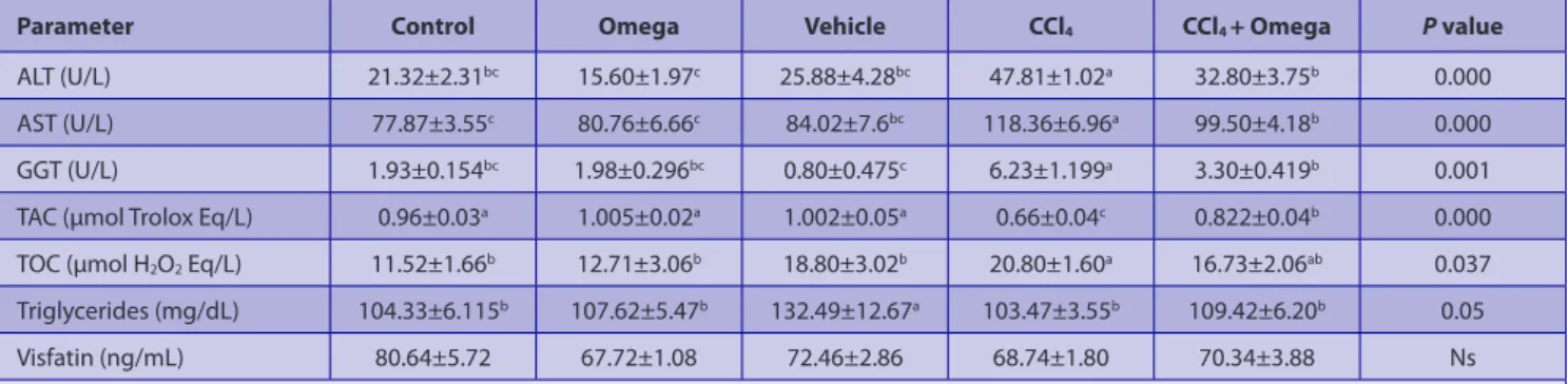
Normal liver histomorphology was observed in groups of control (Fig. 1a), omega, and vehicle (Figures not shown). In only CCl4 given group, hydropic degeneration,
occasional coagulation necrosis, and sinusoidal dilatation were observed. Formation of fibrosis starting from the portal regions through the parenchyma with presence of occasional necrotic hepatocytes and mononuclear cellular infiltration was evident. Interlobular areas were widened due to development of fibrosis. Pseudolobule formations were also recognizable in severely affected regions (Fig. 1b). The severity of the histopathological changes decreased in CCl4 + Omega given group as compared to the only CCl4 given group. The most recognizable findings in CCl4 +
Omega group were the fatty and hydropic degeneration of hepatocytes located close to the portal region. No fibrotic
Table 1. Serum levels of AST: Aspartate aminotransferase, ALT: Alanine aminotransferase, GGT: Gamma glutamyl transferase, TAC: Total antioxidant capacity, TOC: Total oxidant capacity, triglycerides, and visfatin in groups
Tablo 1. AST: Aspartat aminotranferaz, ALT: Alanin aminotransferaz, GGT: Gama glutamil transferaz, TAC: Total antioksidan kapasite, TOC: Total oksidan kapasite, trigliserit ve visfatin serum seviyeleri
Parameter Control Omega Vehicle CCl4 CCl4 + Omega P value
AlT (U/l) 21.32±2.31bc 15.60±1.97c 25.88±4.28bc 47.81±1.02a 32.80±3.75b 0.000
AST (U/l) 77.87±3.55c 80.76±6.66c 84.02±7.6bc 118.36±6.96a 99.50±4.18b 0.000
GGT (U/l) 1.93±0.154bc 1.98±0.296bc 0.80±0.475c 6.23±1.199a 3.30±0.419b 0.001
TAC (μmol Trolox Eq/l) 0.96±0.03a 1.005±0.02a 1.002±0.05a 0.66±0.04c 0.822±0.04b 0.000
TOC (μmol H2O2 Eq/l) 11.52±1.66b 12.71±3.06b 18.80±3.02b 20.80±1.60a 16.73±2.06ab 0.037
Triglycerides (mg/dl) 104.33±6.115b 107.62±5.47b 132.49±12.67a 103.47±3.55b 109.42±6.20b 0.05
Visfatin (ng/ml) 80.64±5.72 67.72±1.08 72.46±2.86 68.74±1.80 70.34±3.88 Ns
changes were observed in any of the animals in this group. Hepatocytes located in midzonal and central areas were normal in histomorphology (Fig. 1c).
Immunohistochemistry
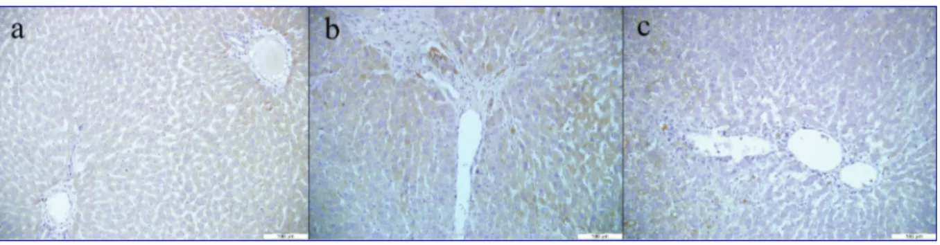
- TGF-α: In the control group, mainly weak immunoreactivity against TGF-α antibody was seen throughout the hepatic regions (Fig. 2a). Compared to the control, increased immunoreactivity was noted in liver of rats given CCl4 only
(Fig. 2b). The degree of immunoreactivity against TGF-α
antibody in CCl4 + Omega group was near to (Fig. 2c) that observed in the control group. Immunoreactivity in omega and vehicle groups was also similar to that of the control group (Figures not shown).
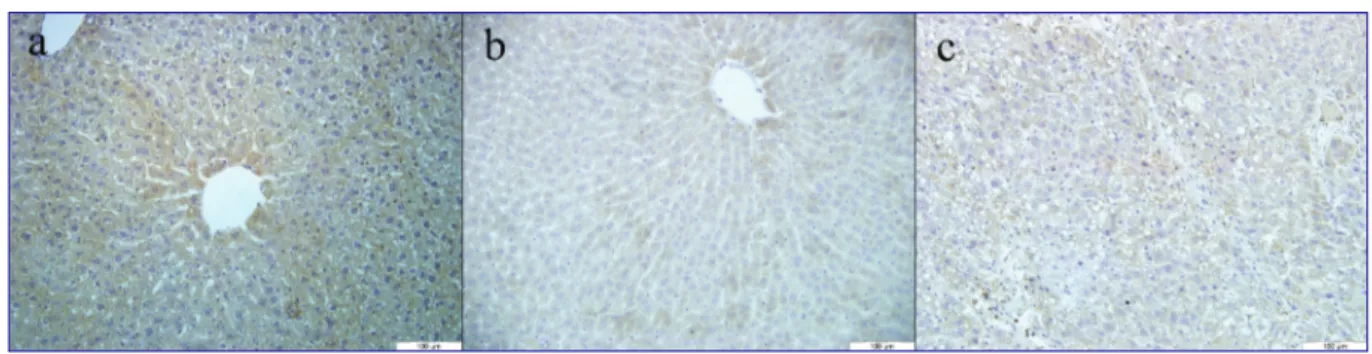
- TGF-β: Very weak immunoreactivity against TGF-β was observed in the control group (Fig. 3a). In CCl4 only given
group, cytoplasmic immunoreactivity was observed in the hepatocytes throughout the liver sections (Fig. 3b). This immunoreactivity was stronger in hepatocytes located close to the portal regions. TGF-β immunoreactivity in CCl4 +
Fig 1. a) Control: Normal liver histomorphology, H&E, b) CCl4: Hepatic fibrosis developing from the portal region through the hepatic
lobules. Degenerated and occasional necrotic hepatocytes, accumulation of mononuclear cells, and some attempt for ill-formation of hepatic lobules, H&E, c) CCl4 + Omega: Periportal fatty and hydropic degeneration, no fibrotic changes, H&E
Şekil 1. a) Kontrol: Karaciğerin normal histomorfolojisi, H&E, b) CCl4: Portal bölgeden hepatik lobüllere doğru gelişen hepatik fibrozis.
Dejenere ve nekrotik hepatositler, mononüklear hücrelerin toplanması ve bazı hatalı hepatik lobüllerin oluşma çabası, H&E, c) CCl4 +
Omega: Periportal alanda yağ ve hidropik dejenerasyonu, fibrotik değişiklikler bulunmamaktadır, H&E
Fig 2. TGF-α immunohistochemistry; a) Control: Weak immunoreactivity, b) CCl4: Moderate to strong immunoreactivity, c) CCl4 + Omega:
Weak immunoreactivity
Şekil 2. TGF-α immunohistokimyası; a) Kontrol: Zayıf immunoreaktivite, b) CCl4: Ortadan güçlüye değişen derecede immunoreaktivite,
c) CCl4 + Omega: Zayıf immunoreaktivite
Fig 3. TGF-β immunohistochemistry; a) Control: Weak immunoreactivity, b) CCl4: Moderate to strong immunoreactivity, c) CCl4 + Omega:
Weak immunoreactivity
Şekil 3. TGF-β immunohistokimyası; a) Kontrol: Zayıf immunoreaktivite, b) CCl4: Ortadan güçlüye değişen derecede immunoreaktivite,
Omega group was prominently reduced compared to the CCl4 only given group (Fig. 3c). The pattern and density for
TGF-β immunoreactivity in group 2 and 3 were similar to that of the control group (Figures not shown).
- PPAR-α: Strong immunoreactivity against PPAR-α was observed in the control group (Fig. 4a). Immunoreactivity was stronger in the periportal region as compared to the central region. In CCl4 only given group, mostly no
immunoreactivity was seen except in few scattered cells
(Fig. 4b). Weak to moderate immunoreactivity against
PPAR-α was observed in CCl4 + Omega group (Fig. 4c). PPAR-α immunoreactivity in group 2 and 3 were similar to that of the control group (Figures not shown).
- PPAR-γ: Moderate to strong immunoreactivity against PPAR-γ was noted in the control group (Fig. 5a). In CCl4
only given group, weak immunoreactivity was observed. Immunoreactivity in this group was mostly observed in the Kupffer cells though hepatocytes also showed weak immunoreactivity (Fig. 5b). Moderate immunoreaction was recognized in CCl4 + Omega group (Fig. 5c). PPAR-γ
immunoreactivity in group 2 and 3 were similar to that of the control group (Figures not shown).
Apoptosis
No apoptotic cell death was detected by in situ TUNEl
method in any of the groups including CCl4 (Figures not
shown).
DISCUSSION
liver acts as the center of many important biological activities such as synthesis and/or storage of substances in the body. Hence, it might be considered that it is the most important organ in many metabolic activities. During the metabolic activation and/or detoxification of substances degenerative changes may take place resulting acute or chronic toxicity. Fatty accumulation and cirrhosis can also develop as a result of long lasting activities [1,29,30].
Despite the efforts of medical treatments, the medical management of the cases with liver degenerations might be insufficient and require other regimens [31]. The use
of herbal substances and/or other non-classical medical treatments might be considered as a supplemental application [32,33]. In this study, the effect of omega-3
supplementation was assessed in the CCl4-mediated
experimental liver degeneration model in rats.
CCl4-induced liver injury is a well-known animal model
in hepatic toxicity studies [6,15,33,34]. The mode of toxicity
induced by CCl4 is that it is metabolically converted to
CCl3 by cytochrome P450, and CCl3, under molecular oxygen
rich environment, reacts with cellular proteins and
poly-Fig 4. PPAR-α immunohistochemistry; a) Control: Strong immunoreactivity, b) CCl4: Very weak immunoreactivity, c) CCl4 + Omega: Weak
to moderate immunoreactivity
Şekil 4. PPAR-α immunohistokimyası; a) Kontrol: Güçlü immunoreaktivite, b) CCl4: Oldukça zayıf immunoreaktivite, c) CCl4 + Omega:
Zayıftan ortaya değişen derecede immunoreaktivite
Fig 5. PPAR-γ immunohistochemistry; a) Control: Moderate to strong immunoreactivity, b) CCl4: Very weak immunoreactivity, c) CCl4 +
Omega: Moderate immunoreactivity
Şekil 5. PPAR-γ immunohistokimyası; a) Kontrol: Oratadan güçlüye değişen derecede immunoreaktivite, b) CCl4: Oldukça zayıf
unsaturated fatty acids to form more toxic trichlormethyl peroxy radicals and other oxygen radicals such as O2−, OH,
and H2O2. Adduct formation with membrane phospholipids
are primarily important in cellular degeneration through development of lipid peroxidation [5]. Disrupted membrane
integrity is the main cause of cellular degeneration and resulting necrosis observed microscopically. CCl4-induced
liver degeneration and necrosis of hepatocytes were previously described by others [5,6,35-37]. In the present study,
severe necrotic changes were as well observed due to CCl4
treatment. Direct damage of lipid membranes as a result of free radicals is responsible for cellular degeneration. Increased TOC and decreased TAC levels support the notation that oxidative cellular degeneration is the cause of liver degeneration and necrosis.
Mononuclear cell infiltration and fibrosis was described in experimental CCl4 toxicity studies [5,7,15,35,36]. Same findings
were also noted in the present study. Additionally, fatty degeneration observed in many hepatocytes in CCl4
given animals in the current study can be explained by interruption of triglyceride passage to blood and as a result their accumulation inside the cells resulting cellular vacuolation. Some studies also described apoptotic cellular death in experimental CCl4 toxicities [38,39]. Such
a cellular death was not observed in the present study. Severe cellular degeneration probably did not allow the development of apoptotic pathways as necrotic changes was the dominant cellular change in the CCl4 given rats.
In evaluation of liver degeneration, liver function tests are commonly applied. Biochemical markers such as AlT, AST, GGT, albumin etc, can therefore provide priceless clinical clues about the development and degree of liver injury [40,41]. Upon hepatocellular degeneration release of
these substances into the circulatory system increases as a result of the loss of membrane integrity. CCl4 has been
previously shown to cause increased serum levels of AST and AlT [1,32,36,42,43]. In the present study, serum AlT and
AST levels were significantly higher in CCl4 given rats as
compared to the control. AlT is normally found higher in liver and kidney tissues whereas AST is higher in heart and skeletal muscles. Therefore, significant increase in serum AlT level correlates with liver injury. It has also been reported that AST level might increase in the early phase of liver degeneration and then the level could decrease with time [40,41]. In the present study increased levels of both AlT
and AST clearly shows the presence of liver injury.
GGT is expressed in liver hepatocytes located close to the bile ducts, as well as renal proximal tubules, pancreas and small intestines. Serum GGT level mostly reflects the liver GGT level, and hence increased serum GGT level is usually an indicator of liver injury [40,41]. In the present study,
increased serum GGT level may indicate the presence of degeneration in those hepatocytes located close to the portal regions. In histopathological observations, fibrotic changes and periportal degenerations in hepatocytes
correlates the findings of biochemical analysis. Therefore, increased serum AlT, AST, and GGT levels as well as histopathological observations indicate the presence of liver injury in CCl4 given rats.
In estimating the presence of cellular degeneration in general, detection of reactive oxygen species is used commonly[15,33,36,43]. However, detection of individual oxidant
and/or antioxidant products can be very difficult as well as time and money consuming. Besides, analyzing the individual results from different oxidant and antioxidant products can be very complicated. Therefore, measuring total oxidant and antioxidant capacities and making comparisons are much in use and practical today. Even TAC and TOC can be used in monitoring the treatment in use for a given disease or experimental study [44,45]. In the present
study, TOC increased while TAC decreased in CCl4 given
animals as compared to the control. The role of oxidative stress in CCl4-induced hepatic toxicity has been previously
shown as well [1,46,47]. On the other hand, in a CCl
4 toxicity
study, unchanged TAC level has also been recorded though presence of increased TOC [48]. Omega-3 supplementation
in the current investigation significantly increased the level of TAC as compared to the CCl4 only given group. In
addition, TOC level reduced, though not significantly, in the omega-3 supplemented group. These findings, together with the results of serum AlT, AST, and GGT levels, clearly indicated that omega-3 supplementation reduced the oxidative degenerative effects that are induced by CCl4.
liver is a key organ in lipid metabolism and plays important roles in fatty acid uptake, conversion, oxidation, and synthesis. It has been shown that CCl4 administration
increases cholesterol and triglycerides levels [1,49-51]. In the
current investigation, an increase in triglyceride level was observed only in the vehicle group in that olive oil was given to the rats. However, no significant changes were observed among the other groups. Therefore, the result of this investigation contradicts the previous studies. It has been suggested that the increase in hepatic triglyceride content in olive oil given mice might be due to the disrupted mitochondrial fatty acid oxidation [52]. This might
be the case in the present study.
In regulation of lipid metabolism, adipocytokines, which are released from the adipose tissue, may be critical. They have been also shown to be responsible for differentiation of stem cells to myofibroblast-like cells, which produces large amount of extracellular matrix substances [4,31,53]. Therefore,
some of the adipocytokines have been suggested to be responsible in development of hepatic fibrosis [53,54].
Visfatin, which is a newly discovered adipocytokine found in adipocytes, hepatocytes, lymphocytes, monocytes and neutrophils, was studied to investigate its relationship with liver injury in the present study. The role visfatin in acute and chronic liver degenerations and fibrosis has not been well studied before and there are some controversial results. While it has been shown that visfatin concentration
is lower in cirrhotic patients [55], in other studies no
correlation was found between fibrosis and serum visfatin level [56,57]. We also did not detect any significant changes in
serum visfatin level on any of the groups studied indicating that visfatin has no apparent role on the omega-3 supplementation or the liver injury induced by CCl4.
TGF-α and TGF-β expression in liver were investigated immunohistochemically to estimate the roles of these cytokines in liver injury [15,43]. TGF-α is known to be
present in healthy liver as well as many other organs. It is a strong mitogen and thought to play role in hepatocyte regeneration after hepatic injury. Harada et al.[58] stated
that TGF- α expression is closely related to severity of liver dysfunction. TGF-α is able to activate hepatic stellate cells, and that triggers the development of hepatic fibrosis [59]. It
has been shown that following CCl4 treatment expression
of TGF-α rapidly increases and after few days prominently decline indicating that TGF-α play role early in the liver degeneration [38]. Similar finding and continously increased
expression of TGF-β1 was also described by Tian et al.[60].
We have detected increased TGF-α immunoreactivity in rats given only CCl4, and the immunoreactivity was near
to control group in omega-3 supplemented animals indicating that omega-3 has beneficial effects in inhibiting the degenerative effects of CCl4.
TGF-β has been described as an important cytokine in development of hepatic fibrosis. Sources of TGF-β in liver has been described primarily to be Kupffer cells and stellate cells [61]. TGF-β is known to be the most profibrotic
cytokine and its expression significantly increases in hepatic cirrhosis [57]. It has been shown in a rat model of
CCl4 toxicity that TGF-β mRNA expression significantly
increases as the Kupffer cell number increase indicating that these cells are primarily responsibly in development of hepatic fibrosis [14]. In the present investigation, TGF-β
immunoreactivity increased in CCl4 given animals, and
omega-3 supplementation lowered the immunoreactivity to the level that was observed in the control group.
In the present investigated, we have studied the expression of PPARs in liver tissue. All three isoforms of PPAR, -α, -β, and -γ, are known to have important regulatory roles in not only inflammatory processes but also lipid biosynthesis, glucose metabolism, cellular proliferation and differentiation [19,62]. It has been suggested that PPAR-α
plays a role in hepatic steatosis, and ciprofibrate, which is a potent ligand for PPAR-α, has a potential to prevent the negative effects, indicating that PPAR-α overexpression or activation is important in amelioration of degenerative changes in liver [63]. PPAR-γ overexpression in liver has also
been suggested to be important and inhibits the fibrotic changes in liver [34,64]. The number of PPAR-γ expressing cells
has also been shown to decrease by CCl4 treatment [34,42].
In the current investigation, immunohistochemical reactivity of both PPAR-α and PPAR-γ, increased in omega-3 supple-mented rats compared to the only CCl4 injected animals.
Therefore, our results correlate the findings of previous studies [42,65,66] indicating CCl
4 treatment prominently
reduce the expression of PPAR-γ, and that omega-3 supplementation has beneficial effects in reducing the development of degenerative changes induced by CCl4.
In conclusion, we have observed that intraperitoneal injection of CCl4 at a dose of 0.5 mg/kg in rats causes fatty
degeneration and necrosis as well as fibrosis especially in the periportal region in liver. Biochemical analysis for AST, AlT and GGT confirmed the hepatic injury. Decreased TAC and increased TOC levels indicated that the degenerative changes in liver were mediated by oxidative pathways. Visfatin, a recently discovered adipocytokine known to play role in fat metabolism, was shown to have no significant effect in hepatic fibrosis in the given experiment. On the other hand, increased TGF-β and decreased PPAR-α and PPAR-γ expressions were recognized to be associated with development of hepatic fibrosis in CCl4-treated animals.
Omega-3 supplementation, on the other hand was shown to normalize the above-mentioned parameters studied, indicating clearly that it has beneficial effect in amelioration of the degenerative changes induced by CCl4. It seems that
the protective effect of omega-3 is mediated primarily by inhibiting the lipid peroxidation.
REFERENCES
1. Kober H,Tatsch E, Torbitz VD, Cargnin LP, Sangoi MB, Bochi GV, da Silva AR, Barbisan F, Ribeiro EE, da Cruz IB, Moresco RN: Genoprotective
and hepatoprotective effects of Guarana (Paullinia cupana Mart. var. sorbilis) on CCl4-induced liver damage in rats. Drug Chem Toxicol, 39, 48-
52, 2016. DOI: 10.3109/01480545.2015.1020546
2. Lee S, Kim S, Le HD, Meisel J, Strijbosch RAM, Nose V, Puder M:
Reduction of hepatocellular injury after common bile duct ligation using omega-3 fatty acids. J Pediatr Surg, 43, 2010-2015, 2008. DOI: 10.1016/j. jpedsurg.2008.05.030
3. Berardis SI, Dawisthi Sattwika P, Najimi M, Sokal EM: Use of
mesenchymal stem cells to treat liver fibrosis: Current situation and future prospects. World J Gastroenterol, 21, 742-758, 2015.
4. Raafat N, Abdel Aal SM, Abdo FK, El Ghonaimy NM: Mesenchymal
stem cells: In vivo therapeutic application ameliorates carbon tetrachloride induced liver fibrosis in rats. Int J Biochem Cell Biol, 68, 109-118, 2015. DOI: 10.1016/j.biocel.2015.09.003
5. Manibusan MK, Odin M, Eastmond DA: Postulated carbon tetrachloride
mode of action: A review. J Environ Sci Health C Environ Carcinog Ecotoxicol
Rev, 25, 185-209, 2007. DOI: 10.1080/10590500701569398
6. Hong RT, Xu JM, Mei Q: Melatonin ameliorates experimental hepatic
fibrosis induced by carbon tetrachloride in rats. World J Gastroenterol, 15, 1452-1458, 2009.
7. Muriel P, Alba N, Pérez-Alvarez VM, Shibayama M, Tsutsumi VK:
Kupffer cells inhibition prevents hepatic lipid peroxidation and damage induced by carbon tetrachloride. Comp Biochem Physiol C Toxicol
Pharmacol, 130, 219-226, 2001. DOI: 10.1016/S1532-0456(01)00237-X 8. Sheweita SA, Abd El-Gabar M, Bastawy M: Carbon
tetrachloride-induced changes in the activity of phase II drug-metabolizing enzyme in the liver of male rats: Role of antioxidants. Toxicology, 165, 217-224, 2001. DOI: 10.1016/S0300-483X(01)00429-2
9. Basu S: Carbon tetrachloride-induced lipid peroxidation: Eicosanoid
formation and their regulation by antioxidant nutrients. Toxicology, 189, 113-127, 2003. DOI: 10.1016/S0300-483X(03)00157-4
Zhang YG, Wu WJ, Di HL, Li Y, Yu J: Activation of peroxisome proliferator
activated receptor alpha ameliorates ethanol mediated liver fibrosis in mice. Lipids Health Dis, 12, 1-10, 2013. DOI: 10.1186/1476-511X-12-11
11. Cheng K, Yang N, Mahato RI: TGF-beta1 gene silencing for treating
liver fibrosis. Mol Pharm, 6, 772-779, 2009. DOI: 10.1021/mp9000469
12. Simopoulos AP: The importance of the ratio of omega-6/omega-3
essential fatty acids. Biomed Pharmacother, 56, 365-279, 2002. 10.1016/ S0753-3322(02)00253-6
13. de Lorgeril M, Salen P: New insights into the health effects of dietary
saturated and omega-6 and omega-3 polyunsaturated fatty acids. BMC
Med, 10, 1-5, 2012, DOI: 10.1186/1741-7015-10-50
14. Pastor-Clerigues A,Marti-Bonmati E, Milara J, Almudever P, Cortijo J: Anti-inflammatory and anti-fibrotic profile of fish oil emulsions
used in parenteral nutrition-associated liver disease. PLoS One, 9, 1-25, 2014. DOI: 10.1371/journal.pone.0115404
15. Shaaban AA, Shaker ME, Zalata KR, El-kashef HA, Ibrahim TM:
Modulation of carbon tetrachloride-induced hepatic oxidative stress, injury and fibrosis by olmesartan and omega-3. Chem Biol Interact, 25, 81-91, 2014. DOI: 10.1016/j.cbi.2013.10.008
16. Aguilera CM, Ramirez-Tortosa CL, Quiles JL, Yago MD, Martínez-Burgos MA, Martínez-Victoria E, Gil A, Ramirez-Tortosa MC:
Monounsaturated and omega-3 but not omega-6 polyunsaturated fatty acids improve hepatic fibrosis in hypercholesterolemic rabbits. Nutrition, 21, 63-71, 2005. DOI: 10.1016/j.nut.2004.06.029
17. Calder PC: Polyunsaturated fatty acids and inflammation. Prostaglandins Leukot Essent Fatty Acids, 75, 197-202, 2006. DOI: 10.1016
/j.plefa.2006.05.012
18. Raptis DA, Limani P, Jang JH, Ungethüm U, Tschuor C, Graf R, Humar B, Clavien PA: GRP120 on Kupffer celss mediates
hepato-protective effects of ω-3 fatty acids. J Hepatol, 60, 625-632, 2014. DOI: 10.1016/j.jhep.2013.11.006
19. Issemann I, Green S: Activation of a member of the steroid hormone
receptor superfamily by peroxisome proliferators. Nature, 347, 645-650, 1990. DOI: 10.1038/347645a0
20. Pawlak M, Lefebvre P, Staels B: Molecular mechamism of PPAR-α
action and its impact on lipid metabolism, inflammation and fibrosis in non-alcoholic fatty liver disease. J Hepatol, 62, 720-733, 2015. DOI: 10.1016/j.jhep.2014.10.039
21. Wolfrum C, Borrmann CM, Borchers T, Spener F: Fatty acids
and hypolipidemic drugs regulate peroxisome proliferator-activated receptors alpha - and gamma-mediated gene expression via liver fatty acid binding protein: A signaling path to the nucleus. Proc Natl Acad, 98, 2323-2328, 2001. DOI: 10.1073/pnas.051619898
22. Akbiyik F, Ray DM, Bozkaya H, Demirpence E: ligand- and
species-dependent activation of PPAR alpha. Cell Physiol Biochem, 14, 269-276, 2004. DOI: 10.1159/000080336
23. Tunca R, Devrim AK, Sözmen M, Dağ S, Güngör O, İpek E: The
effects of gemfibrozil and ovariectomy on the peroxisome proliferator activated receptors (PPARs) in mice with experimentally induced obesity.
Kafkas Univ Vet Fak Derg, 19, 783-792, 2013. DOI: 10.9775/kvfd.2013.8788 24. Devchand PR, Keller H, Peters JM, Vazquez M, Gonzalez FJ, Wahli W: The PPAR alpha-leukotriene B4 pathway to inflammation control. Nature, 384, 39-43, 1996. DOI: 10.1038/384039a0
25. Xin X, Yang S, Kowalski J, Gerritsen ME: Peroxisome
proliferator-activated receptor gamma ligands are potent inhibitors of angiogenesis in vitro and in vivo. J Biol Chem, 74, 9116-9121, 1999. DOI: 10.1074/ jbc.274.13.9116
26. Hutcheson J: Adipokines influence the inflammatory balance in
autoimmunity. Cytokine, 75, 272-279, 2015. DOI: 10.1016/j.cyto.2015.04.004
27. Al-Suhaimi EA, Shehzad A: leptin, resistin and visfatin: The missing
link between endocrine metabolic disorders and immunity. Eur J Med
Res, 18, 1-13, 2013. DOI: 10.1186/2047-783X-18-12
28. Mayi TH, Rigamonti E, Pattou F, Staels B, Chinetti-Gbaguidi G:
liver x Receptor (lXR) activation negatively regulates visfatin expression in macrophages. Biochem Biophys Res Commun, 404, 458-462, 2011.
DOI: 10.1016/j.bbrc.2010.12.002
29. Lee SH, Heo SI, Li L, Lee MJ, Wang MH: Antioxidant and
hepatoprotective activities of Cirsium setidens NAKAI against CCl4
-induced liver damage. Am J Chin Med, 36,107-114, 2008. DOI: 10.1142/ S0192415X0800562X
30. Kopec AK, Joshi N, Luyendyk JP: Role of hemostatic factors in
hepatic injury and disease: Animal models de-liver. J Thromb Haemost, 14,1337-1349, 2016. DOI: 10.1111/jth.13327
31. Thompson AJ, Patel K: Antifibrotic therapies: Will we ever get there? Curr Gastroenterol Rep, 12, 23-29, 2010. DOI: 10.1007/s11894-009-0080-9 32. Chang BY, Lee DS, Lee JK, Kim YC, Cho HK, Kim SY: Protective
activity of kudzu (Pueraria thunbergiana) vine on chemically-induced hepatotoxicity: in vitro and in vivo studies. BMC Complement Altern Med, 16, 1-8, 2016. DOI: 10.1186/s12906-016-1023-2
33. Lu Y, Hu D, Ma S, Zhao X, Wang S, Wei G, Wang X, Wen A, Wang J: Protective effect of wedelolactone against CCl4-induced acute liver injury in mice. Int Immunopharmacol, 34, 44-52, 2016. DOI: 10.1016/j. intimp.2016.02.003
34. Wang Z, Xu JP, Zheng YC, Chen W, Sun YW, Wu ZY, Luo M: Peroxisome
proliferator-activated receptor gamma inhibits hepatic fibrosis in rats.
Hepatobiliary Pancreat Dis Int, 10, 64-71, 2011. DOI:
10.1016/S1499-3872(11)60009-X
35. Jadhav VB, Thakare VN, Suralkar AA, Deshpande AD, Naik SR:
Hepatoprotective activity of luffa acutangula against CCl4 and rifampicin
induced liver toxicity in rats: A biochemical and histopathological evaluation. Indian J Exp Biol, 48, 822-829, 2010.
36. Suzek H, Celik I, Dogan A, Yildirim S: Protective effect and
antioxidant role of sweetgum (Liquidambar orientalis) oil against carbon tetrachloride-induced hepatotoxicity and oxidative stress in rats. Pharm
Biol. 54, 451-457, 2015. DOI: 10.3109/13880209.2015.1045086
37. Sen I, Turgut K, Ok M, Kıran MM, Güzelbekteş H, Ortatatlı M, Birdane FM, Altunok V: Effects of nutritional therapy or n-acetyl-cysteine
(NAC) treatment on biochemical markers and liver histology in dogs with ccl4-induced hepatic necrosis. Revue Med Vet, 156, 483-490, 2005.
38. Liu J, Zhang QY, Yu LM, Liu B, Li MY, Zhu RZ: Phycocyanobilin
accelerates liver regeneration and reduces mortality rate in carbon tetrachloride-induced liverinjury mice. World J Gastroenterol, 21, 5465-5472, 2015. DOI: 10.3748/wjg.v21.i18.5465
39. Özsoy ŞY: The protective effect of kefir on carbon
tetrachloride-induced histopathological changes in the livers of rats. Kafkas Univ Vet
Fak Derg, 22, 403-408, 2016. DOI: 10.9775/kvfd.2015.14825
40. Limdi JK, Hyde GM: Evaluation of abnormal liver function tests. Postgrad Med J, 9, 307-312, 2003. DOI: 10.1136/pmj.79.932.307
41. Giannini EG, Testa R, Savarino V: liver enzyme alteration: A guide
for clinicians. CMAJ, 172, 367-379, 2005. DOI: 10.1503/cmaj.1040752
42. Guo C, Xu L, He Q, Liang T, Duan X, Li R: Anti-fibrotic effects of
puerarin on CCl4-induced hepatic fibrosis in rats possibly through the
regulation of PPAR-γ expression and inhibition of PI3K/Akt pathway. Food
Chem Toxicol, 56, 436-442, 2013. DOI: 10.1016/j.fct.2013.02.051
43. Chu X, Wang H, Jiang YM, Zhang YY, Bao YF, Zhang X, Zhang JP, Guo H, Yang F, Luan YC, Dong YS: Ameliorative effects of tannic acid on
carbon tetrachloride-induced liver fibrosis in vivo and in vitro. J Pharmacol
Sci, 130, 15-23, 2016. DOI: 10.1016/j.jphs.2015.12.002
44. Aydin M, Oktar S, Yonden Z, Ozturk OH, Yilmaz B: Direct and
indirect effects of kisspeptin on liver oxidant and antioxidant systems in young male rats. Cell Biochem Funct, 28, 293-299, 2010. DOI: 10.1002/ cbf.1656
45. Sözen S, Kisakürek M, Yildiz F, Gönültaş M, Dinçel AS: The effects
of glutamine on hepatic ischemia reperfusion injury in rats. Hippokratia, 15, 161-166, 2011.
46. Recknagel RO, Glende EAJ, Dolak JA, Waller RL: Mechanisms
of carbon tetrachloride toxicity. Pharmacol Ther, 43,139-154, 1989. DOI: 10.1016/0163-7258(89)90050-8
47. Weber LW, Boll M, Stampfl A: Hepatotoxicity and mechanism
Crit Rev Toxicol, 33, 105-136, 2003. DOI: 10.1080/713611034
48. Demiroren K , Dogan Y, Kocamaz H , Ozercan IH , Ilhan S, Ustundag B, Bahcecioglu IH: Protective effects of l-carnitine, N-acetylcysteine and
genistein in an experimental model of liver fibrosis. Clin Res Hepatol
Gastroenterol, 38, 63-72, 2014. DOI: 10.1016/j.clinre.2013.08.014 49. Ince S, Keles H, Erdogan M, Hazman O, Kucukkurt I: Protective
effect of boric acid against carbon tetrachloride-induced hepato- toxicity in mice. Drug Chem Toxicol, 35, 285-292, 2012. DOI: 10.3109/ 01480545.2011.607825
50. Li G, Wang XY, Suo YR, Wang HL: Protective effect of seed oil of
Herpetospermum pedunculosum against carbon tetrachloride-induced liver injury in rats. Saudi Med J, 35,981-987, 2014.
51. Yuan C, Li Z, Yi M, Wang X, Peng F, Xiao F, Chen T, Wang C, Mushtaq G, Kamal MA: Effects of polysaccharides from selenium-enriched
Pyracantha fortuneana on mice liver injury. Med Chem, 11, 780-788, 2015.
52. Ferramosca A, Savy V, Zara V: Olive oil increases the hepatic
triacylglycerol content in mice by a distinct influence on the synthesis and oxidation of fatty acids. Biosci Biotechnol Biochem, 72, 62-69, 2008. DOI: 10.1271/bbb.70369
53. Anty R, Lemoine M: liver fibrogenesis and metabolic factors. Clin Res Hepatol Gastroenterol,1, 10-20, 2011. DOI: 10.1016/S2210-7401(11)
70003-1
54. Kukla M, Mazur W, Bułdak RJ, Zwirska-Korczala K: Potential role
of leptin, adiponectin and three novel adipokines-visfatin, chemerin and vaspin-in chronic hepatitis. Mol Med, 17, 1397-1410, 2011. DOI: 10.2119/ molmed.2010.00105
55. de Boer JF, Bahr MJ, Böker KH, Manns MP, Tietge UJ: Plasma levels
of PBEF/Nampt/visfatin are decreased in patients with liver cirrhosis.
Am J Physiol Gastrointest Liver Physiol, 296, G196-201, 2009. DOI: 10.1152/
ajpgi.00029.2008
56. Baranova A,Jarrar MH, Stepanova M, Johnson A, Rafiq N, Gramlich T, Chandhoke V, Younossi ZM: Association of serum adipocytokines
with hepatic steatosis and fibrosis in patients with chronic hepatitis C.
Digestion, 83, 32-40, 2011.
57. Waluga M, Kukla M, Żorniak M, Kochel-Jankowska A, Kajor M, Krzemiński T, Kotulski R: Visfatin and TGF-β1 in primary biliary
cirrhosis and two other common liver diseases. Folia Med Cracov, 55,
59-70, 2015.
58. Harada K, Shiota G, Kawasaki H. Transforming growth
factor-alpha and epidermal growth factor receptor in chronic liver disease and hepatocellular carcinoma. Liver, 19, 318-325, 1999. DOI: 10.1111/j.1478-3231.1999.tb00056.x
59. Ohyama T, Yamazaki Y, Sato K, Horiguchi N, Ichikawa T, Kakizaki S, Takagi H, Mori M: Transforming growth factor-α attenuates hepatic
fibrosis: Possible involvement of matrix metalloproteinase-1. Liver Int, 31, 572-584, 2011. DOI: 10.1111/j.1478-3231.2011.02475.x
60. Tian XF, Ji FJ, Zang HL, Cao H: Activation of the miR-34a/SIRT1/
p53 signaling pathway contributes to the progress of liver fibrosis via inducing apoptosis in hepatocytes but not in HSCs. PLoS One, 11, 1-14, 2016. DOI: 10.1371/journal.pone.0158657
61. Seki E, Brenner DA: Recent advancement of molecular mechanisms
of liver fibrosis. J Hepatobiliary Pancreat Sci, 22, 512-518, 2015. DOI: 10.1002/jhbp.245
62. Abbott BD: Review of the expression of peroxisome
proliferator-activated receptors alpha (PPAR alpha), beta (PPAR beta), and gamma (PPAR gamma) in rodent and human development. Reprod Toxicol, 27, 246-257, 2009. DOI: 10.1016/j.reprotox.2008.10.001
63. Rao MS, Papreddy K, Musunuri S, Okonkwo A: Prevention/reversal
of choline deficiency-induced steatohepatitis by a peroxisome proliferator- activated receptor alpha ligand in rats. In Vivo, 16, 145-152, 2002.
64. Yang L, Chan CC, Kwon OS, Liu S, McGhee J, Stimpson SA, Chen LZ, Harrington WW, Symonds WT, Rockey DC: Regulation of
peroxisome proliferator-activated receptor-gamma in liver fibrosis. Am
J Physiol Gastrointest Liver Physiol, 291, G902-G911, 2006. DOI: 10.1152/
ajpgi.00124.2006
65. Liu Q, Wang CY, Liu Z, Ma XS, He YH, Chen SS, Bai XY: Hydroxysafflor
yellow A suppresses liver fibrosis induced by carbon tetrachloride with high-fat diet by regulating PPAR-γ/p38 MAPK signaling. Pharm Biol, 52, 1085-1093, 2014. DOI: 10.3109/13880209.2013.877491
66. Orfila C, Lepert JC, Alric L, Carrera G, Béraud M, Pipy B:
Immunohistochemical distribution of activated nuclear factor kappa B and peroxisome proliferator-activated receptors in carbon tetrachloride-induced chronic liver injury in rats. Histochem Cell Biol, 123, 585-593, 205. DOI: 10.1007/s00418-005-0785-2