INTRODUCTION
The major drawback of the resin-based restorations is their limited long-term durability despite the continuous evolution in adhesive technology over the past sixty years1,2). Over the years, in response to the demands of the clinicians, increasingly user-friendly and simplified adhesive systems that are less technique-sensitive have been developed. This trend recently led to the introduction of an increasing number of universal adhesive systems into the market.
Despite the continuing advances, in vivo as well as in vitro studies in the past decades have widely reported the progressive degradation of resin-dentin bonds3-5). The limited durability of contemporary adhesives has been shown to be caused by two main reasons including the increased hydrophilicity of the bonding agents that leads to water sorption and subsequent degradation of the infiltrated polymer matrices during physical and chemical challenges in the oral cavity, and degradation of the collagen matrix itself from the adhesive interfaces.
Two distinct groups of endogenous proteases; matrix metalloproteinase (MMPs) and cysteine cathepsins (CC) were shown to be involved in degradation of the collagen component of the bonding interface6-9). These endogenous enzymes become bound to the collagen matrix in dentin during development in their active preforms and could be uncovered and activated during routine adhesive restorative procedures such as etching with phosphoric acid or application of acidic monomers in the self-etch adhesives4-7,10).
Although endogenous protease activity following acidic treatments has been clearly reported in previous studies, most of the evidence is based on the evaluation
of the activity of proteases extracted from the dentin matrix or the addition of recombinant enzymes. This does not necessarily reflect the activity of matrix-bound proteases in situ in dentin. Additionally, the specific effects of universal bonding agents in comparison with self-etch adhesives or phosphoric acid on MMP- or CC-mediated degradation are not well known.
The objectives of the present study were 1) to evaluate the total MMP-activity of ethylenediaminetetraacetic acid (EDTA)-demineralized human dentin following the application of two commercial universal adhesives and two self-etch adhesives in comparison with phosphoric acid treatment; 2) to evaluate the release of collagen telopeptide fragments, C-terminal-telopeptide of type I collagen (ICTP) and C-terminal telopeptide (CTX), as specific markers identifying MMP- and CC-mediated degradation, respectively. The null hypothesis tested was that there will be no difference in the total MMP activity between the tested adhesives, and no difference will be observed between the MMP-mediated or CC-mediated degradation of EDTA-demineralized dentin matrix after application of the different self-etch adhesives.
MATERIALS AND METHODS
Seventy-eight human third molars were obtained from 18–22-year-old patients using a protocol approved by The Dental College of Georgia at Augusta University. The teeth were frozen and stored until required. The enamel and superficial dentin of the molars were removed below the deepest central fissure by horizontal sectioning and dentin discs of 1 mm thickness from the mid-coronal dentin were obtained using an Isomet saw (Buehler, Lake Bluff, IL, USA) under water cooling. Each dentin
Activation of matrix-bound endogenous proteases by self-etch adhesives
Bebek Serra OGUZ AHMET1, Roda SESEOGULLARI-DIRIHAN2 and Arzu TEZVERGIL-MUTLUAY2,3 1 Department of Prosthodontics, Istanbul Medipol University, School of Dentistry, Bagcilar, 34214, Istanbul, Turkey
2 Department of Restorative Dentistry and Cariology, Adhesive Dentistry Research Group, Institute of Dentistry, University of Turku, FI-20014, Turku,
Finland
3 Turku University Hospital, TYKS, University of Turku, FI-20014, Turku, Finland
Corresponding author, Bebek Serra OGUZ AHMET; E-mail: [email protected]
The study evaluated changes in total enzymatic activity and degradation of demineralized dentin following the application of universal or self-etch adhesives. The universal adhesives —Scotchbond Universal (SU) and All-Bond Universal (ABU) and self-etch adhesives —Adper Easy Bond (EB) and G-aenial Bond (GB) were used for 2 min pretreatment of the dentin beams. Phosphoric acid (PA) treatment as well as no treatment served as controls. Total enzymatic activity was analyzed before and after treatment, collagen degradation was assessed using mass loss, C-terminal telopeptide (CTX) and C-terminal-telopeptide of type I collagen (ICTP) release (24 h, 3-day, 3-week). Over three weeks of incubation, ICTP release of ABU treated beams was significantly higher than other groups (p<0.05), except for SU treated beams (p>0.05) and CTX release of GB treated beams was the highest among the groups with statistically significant difference (p<0.05). The results confirm that the universal adhesives tested have also potential to increase the enzymatic activity in dentin.
Keywords: Cycteine cathepsin, C-terminal telopeptide, C-terminal-telopeptide of type I collagen, Matrix metalloproteinase, Self-etch adhesive
Received Sep 12, 2019: Accepted Dec 25, 2019
Table 1 Commercial adhesive systems tested
Brand name Abbreviation Company Composition pH Lot No AdperTM Easy
Bond EB
3M, ESPE, St. Paul, MN, USA
MHP, HEMA, dimethacrylate resins, vitrebond copolymer (VCP), water, ethanol, filler, inititators,
2.4–2.7 426992
Scotchbond
Universal SU 3M, ESPE
MDP, HEMA, dimetharcylate resins, vitrebond copolymer (VCP), water,
ethanol, silane, inititators, 2.7 4676661 All-bond
Universal ABU
Bisco, Schaumburg, IL, USA
MDP, Bis-GMA, HEMA, water,
ethanol, inititators, 2.5–3.5 1200006790 G-aenial
Bond GB GC, Tokyo, Japan
4-MET, phosphoric acid ester monomer, dimetharcylate monomers, acetone,
water, silicone dioxide, inititators, 1.5 1102241 Bis-GMA: 2,2-bis[4-(2-hydroxy-3-methacrylyloxy-propoxy)-phenyl] propane, HEMA: 2-hydroxyethyl methacrylate, MHP: methacryloxyhexyl phosphate monomer, MDP: 10-methacryloyloxydecyl dihydrogen phosphate monomer, 4-MET: 4-methacryloxyethyl trimellitic acid, VCP: methacrylate functionalized polyalkenoic acid
disc was further sectioned to obtain dentin beams, 6×2×1 mm in dimension. All beams were completely demineralized in 0.5 M EDTA (pH 7.4) for 30 days at 4°C with constant stirring. Three-point flexure was used to confirm the absence of residual mineral. Mineralized dentin has a modulus of elasticity between 16 and 19 GPa. Dentin beams completely demineralized in EDTA have a modulus of elasticity of 2–2.5 MPa11). Thirty teeth were used for Generic MMP assay (n=5) and 48 teeth were used for the other tests (n=8). Commercially available universal bonding agents, self-etch adhesives, and phosphoric acid used in this study are listed in Table 1.
Measurement of total MMP activity of demineralized dentin matrices
Generic colorimetric MMP assay (Sensolyte Generic MMP assay, Anaspec, San Jose, CA, USA) was used to determine the baseline activity of EDTA-demineralized dentin beams. After EDTA-demineralization and rinsing, the beams were incubated in 300 µL of chromogenic thiopeptide substrate and assay buffer in a 96-well plate for 60 min at 25°C. Following incubation, the beams were removed from the wells and the 96-well plate was placed in a microplate reader (Synergy HT, BioTek Instruments, Winooski, VT, USA) to measure the baseline total MMP activity of each beam at 412 nm12).The beams were then distributed to different groups such that the mean baseline activity of each group was not statistically significant. The groups were: 1) Controls (CM) pretreated with distilled water, 2) 37% PA, 3) Easy Bond (EB), 4) Scotchbond Universal (SU), 5) All-Bond Universal (ABU), and 6) G-aenial Bond (GB) (n=5/group). The beams in each group were dipped in the respective bonding agents or acidic solution for 2 min (Table 1), rinsed, and incubated in fresh chromogenic substrate and assay buffer in the 96-well plate for 60 min at 25°C. After 60 min of incubation, the activity
was reassessed as previously described. The total MMP activity was expressed as a percentage of the untreated baseline level to determine the percent activation. Loss of dry mass
After demineralization, a set of beams (n=8/group) was thoroughly washed 4 times with distilled water for 2 h at room temperature. The beams were then placed in a sealed desiccator containing anhydrous calcium sulfate (Drierite, W.A. Hammond Company, Xenia, OH, USA) in order to desiccate the beams to a constant weight within 72 h. The initial dry mass of each beam was measured to the nearest 0.001 mg on an analytical balance (Mettler XP6 Microbalance, Mettler Toledo, Hightstown, NJ, USA). Dried dentin beams were assigned to the six groups, each consisting of 8 beams (n=8). The groups were arranged so that the mean initial dry mass of each group was similar in all groups. Following the pretreatment of rehydrated (in water at 4°C for 1 h) dentin beams with 37% PA, EB, SU, ABU or GB for 2 min in a dark environment, all the beams were placed in polypropylene tubes containing acetone and washed for 1 min using an ultrasonic device to remove possible remnants of adhesive resins. Demineralized beams with no adhesive pretreatment were used as control. After this procedure, all beams were placed into separate tubes and rinsed sequentially for 30 min, 10 min, and 10 min at room temperature by refreshing the acetone between each of the washes, and finally rinsed with distilled water for 10 min at room temperature before further incubation. After each incubation period, the beams were separately washed with distilled water for 12-4-4-4 h sequentially at 4°C with constant stirring and dried again as previously described. The loss of dry mass after each incubation period was calculated as percentage dry mass change with respect to the baseline dry mass of each sample. Dehydrated beams were rehydrated in water at 4°C for 1 h prior to each incubation period. Previously it
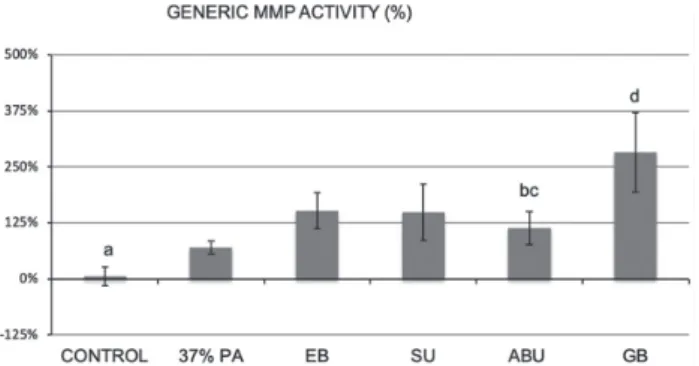
Fig. 1 Percent change in total MMP activity compared with untreated control group, using the Sensolyte Generic MMP assay kit.
Groups identified with different letters are significantly different (p<0.05).
Fig. 2 Loss of dry mass of demineralized dentin beams expressed as percent loss.
For each time period, the groups with the same lowercase letters (among a, b, c; α, β, γ, δ and w, x, y, z) are not statistically significant (p>0.05). For each adhesive tested as well as the control, the time period columns connected by a solid black bar are not statistically different (p>0.05).
was shown that 1 h was sufficient time for complete re-expansion of the dried beams13). The incubation medium contained 5 mM HEPES, 2.5 mM CaCl2.H2O, 0.05 mM ZnCl2, and 120 mM NaCl adjusted to pH 7.2.
Solubilized telopeptides of collagen
To evaluate collagen breakdown, enzyme linked immune-sorbent assays (ELISA) were used to measure enzyme-specific degradation products of the collagen. We analyzed the incubation media for pyridinoline-crosslink-containing degradation fragment of the ICTP as a determinant for MMP-mediated matrix degradation, and deoxypyridinoline-containing degradation fragment of the CTX as a determinant for cathepsin-mediated matrix degradation over the 24 h, 3 days and 3 weeks incubation periods. The amount of liberated telopeptide fragments due to MMP activity was measured using an ICTP elisa kit (UniQ EIA, Orion Diagnostica, Turku, Finland) while the liberated fragments due to cathepsin-K activity was measured using the Serum CrossLaps ELISA (Immunodiagnostic System, Farmington, UK) for the degradation by cathepsin K.
Briefly, aliquots of the incubation medium from dentin beams (n=8/group) were diluted 1:10 in saline and pipetted (50 µL per well) in dublicate into a 96-well plate. Procedures were performed following manufacturer’s instructions and absorbance was measured at 450 nm using a plate reader (Synergy HT, BioTek Instruments). The amount of CTX/ICTP release was calculated with a standard curve using standards with known concentrations provided in the kits. The assays were performed with 8 samples/incubation in duplicates. Statistical analyses
Normality of the data for dry mass loss, total MMP-activity, and the ICTP and CTX analyses were evaluated with the Shapiro-Wilk test. The data was found to be normal, (p>0.05) thus parametrical statistical tests were performed. The differences at each time-point were determined using ANOVA and Tukey tests. For the pairwise comparisons, repeated measures ANOVA was applied. For the statistical tests, SigmaPlot Version 13 software (Systat Software Inc., San Jose, CA, USA) was used at p=0.05.
RESULTS Total MMP activity assay
When demineralized dentin beams were used as a source of MMP, after 60 min incubation the baseline activity of all dentin beams was not significantly different among the groups (p>0.05). However, when the demineralized dentin beams were treated with 37% PA and the respective universal and self-etch adhesives, significant differences were observed among the groups (p<0.05, Fig 1). The control beams exhibited a 6% increase in total MMP activity during the second incubation period which was significantly lower than the beams treated with EB, SU, ABU, and GB (p<0.05). The group that was treated with 37% PA showed a 70% increase in
MMP activity which was significantly different from the control (p<0.05) but was significantly lower than EB, SU, and GB groups (p<0.05). The GB treatment group showed a 282% MMP activity which was significantly higher than all the other groups (p<0.05).
Loss of dry mass
Thirty-seven percent PA-treated beams showed a dry mass loss of 2.77±0.76 % over 24 h of incubation, which was slightly higher than control but significantly lower than the SU-treated beams, which showed the highest amount of dry mass loss (p<0.05), and significantly higher than the ABU- and GB- treated beams (p<0.05, Fig 2). After the 3-day and 3-week incubation periods, dry mass loss in the 37% PA group showed an increase resulting in significantly higher dry mass loss than all the other groups (p<0.05), except for the SU group,
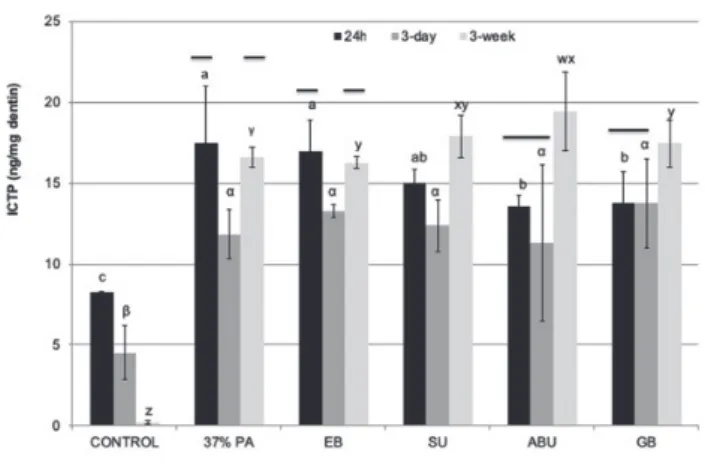
Fig. 3 The rate of ICTP telopeptide release from demineralized dentin beams.
Groups identified with different letters are significantly different (p<0.05). For each adhesive tested as well as the control, the time period columns connected by a solid black bar are not statistically different (p>0.05).
Fig. 4 The rate of CTX telopeptide release from demineralized dentin beams.
Groups identified with different letters are significantly different (p<0.05). For each adhesive tested as well as the control, the time period columns connected by a solid black bar are not statistically different (p>0.05).
which showed similar amount of mass loss (p>0.05). Comparison of the effect of incubation times showed a statistically significant increase in the dry mass loss during the 3-weeks incubation period compared to 24 h and 3 days (p<0.05) among all the groups, except for SU group which showed no significant differences over the mentioned time periods (p>0.05).
Release of solubilized telopeptides
The control group with no pretreatment showed the lowest ICTP release after 24 h incubation period, which was significantly different from all the other groups (p<0.05, Fig 3). On the other hand, ICTP release of the 37% PA-treated beams was significantly higher than
the ABU- and GB-treated beams (p<0.05). After 3-days of incubation, no statistically significant differences were observed in the ICTP release among the groups (p>0.05), except for the control group which showed the lowest release of ICTP (p<0.05). However, after three weeks incubation, the ICTP release of all groups showed an increase, and among the adhesive groups the ABU-treated beams was significantly higher than the other groups (p<0.05), except for SU-treated beams (p>0.05). Additionally, the amount of ICTP release of the SU, ABU and GB-treated beams at the end of the 3-week incubation period increased significantly compared to the 24 h and 3-days of incubation periods (p<0.05).
CTX release for the demineralized beams treated with SU, ABU, and GB at 24 h was significantly higher than the control, 37% PA, and EB groups (p<0.05), and the CTX release for beams treated with GB was almost 60-fold higher than the control (Fig. 4). At 3 days of incubation, the ABU-treated beams showed the highest amount of CTX release which was significantly higher than the PA-treated group only (p<0.05). Over 3 weeks of incubation, CTX release for the GB-treated beams was highest among the various groups, which was statistically significant (p<0.05). For the SU, ABU and GB groups, the amount of CTX release was significantly decreased over the 3-week incubation period compared to 24 h (p<0.05).
DISCUSSION
Recently, “universal” or “multimode” adhesives which represent the last generation of adhesive systems were introduced in the market and can be used as either an etch-and-rinse or self-etch technique14). In addition, this adhesive system allows practitioners to selectively etch the enamel margins with phosphoric acid prior to application of the self-etch adhesive as indicated.
The ability of etch and rinse or self-etch adhesives to increase the host-derived endogenous enzymatic activity in dentin matrices has been well documented previously15-17). In an effort to compare the universal adhesives with self-etch ones, in the present study, the total MMP activity of EDTA-demineralized dentin was measured immediately after application of the two universal and etch adhesives. Once activated by self-etch adhesives, the amounts of ICTP and CTX release by endogenous dentin MMPs and CCs, respectively, was also evaluated. We also assessed the loss of dry mass over time that indicates solubilization of the dentin matrix by endogenous MMP and CC activities8,18).
Demineralization of dentin was completed using 0.5 M EDTA. Despite being relatively slow, EDTA demineralization was previously shown to successfully preserve most of the matrix-bound proteases19). This would allow to expose matrix-bound MMPs and CCs to PA or other acidic monomers, without the buffering effects of apatite minerals in dentin15). In this study, we analyzed the total activity of EDTA-demineralized beams by using Generic MMP activity assay kit at baseline and after the pretreatment. In line with previously published
results, our results confirmed significant increases in the total MMP activity after acid treatment. These findings negated the null hypothesis of this study that there is no difference in the total MMP activity among tested adhesives. The control group with no acidic pretreatment showed only 6% increase compared to baseline, whereas 37% PA treated group showed a 70% increase in total MMP activity. A recent study reported that, acids were not necessary for the activation of dentin proteases due to the presence of some other glycoproteins, which could activate the MMP proforms20).Therefore, the slight increase in activity with demineralized control groups are expected, and in line with the previous literature15,17). On contrary, increase in PA treated group might seem in conflict with the results of previous studies claiming that 37% PA could denature the MMP activity during demineralization due to rapid exposure of the collagen matrix and its matrix-bound MMPs21,22).However, other reports also confirmed that the treatment of dentin with 37% PA did not denature the endogenous proteases in the dentin matrix15,17). Continual MMP or cathepsin activity of the previously EDTA-demineralized dentin powder was observed for 30 days before exposure to PA, where all of the calcium was removed, thus eliminating the formation of CaHPO4 precipitates over the demineralized collagen fibrils15). Furthermore, a direct evidence of gelatinase activity was also observed both for MMP-2 and MMP-919).Thus, previous methods might have underestimated the MMP activity21,22), and as confirmed also by the results of the present study, matrix-bound proteases could be far more resistant to denaturation compared to proteases in soluble form15). The increase in total MMP activity after treatment with universal or self-etch adhesives were adhesive dependent and, ranged between 113–282% (Fig. 1). Previous reports also confirmed an adhesive-dependent increase in specifically MMP-2 and-9 activities when two-step etch-and-rinse, as well as self-etch adhesive systems were used11,17). It is well known that MMPs can be activated in low pH environments by inducing the cysteine switch4), therefore the pH of self-etch or universal adhesives can contribute to the activation process. Among the tested adhesives, EB, ABU and SU are classified as ultra-mild adhesives with a pH range of 2.7–3.1 , whereas GB is classified as intermediate strong adhesive with a lower pH of 1.523). Moreover, 2-hydroxyethyl methylmethacrylate (HEMA)-free composition of GB compared to the other tested adhesives might also contribute to the initial activation results observed at this study. In previous studies, the presence of HEMA monomer was reported to have an inhibitory effect on MMPs17,24). Thus, the lower MMP activity of EB, SU, and ABU-treated beams compared to GB might be partly related to the HEMA content of these adhesive systems.
The dry mass loss as well as quantification of solubilized telopeptides were used as evaluate the degradation over three weeks of incubation period. The only source of ICTP telopeptide fragments in mineralized matrices is attributed to the telopeptidase activity of MMPs which optimally function at neutral Ph25-27). On
the other hand, the activity of Cathepsin-K generates a linear eight amino acid sequence on the CTX end, which is recognized as CTX fragments25). By measuring the amounts of ICTP and CTX release in the incubation medium, the relative MMP activity vs. cathepsin-K activity were followed. The results of telopeptidase activity require the rejection of the null hypothesis, since both CTX and ICTP activity showed differences between the adhesives. Furthermore, the CTX release of the beams were 10–20 fold lower than ICTP release in this study. Similar trend was observed in previous studies and attributed to the incubation medium pH2,15). The optimum pH for cathepsin-K activity is around 5.5, and a pH dependent activity reduction was observed at previous studies showing only 11% of the activity at neutral pH28).
After 24 h of incubation, all groups showed significantly higher ICTP release compared to no-treatment control group. The lowest ICTP release was observed for the control group, whereas the highest ICTP release was observed for the 37% PA group. Mass loss of PA and SU group correlated well with ICTP results confirming a higher mass loss with higher ICTP release. Over 3 days, the ICTP release was almost the same among the groups except for control, which was the lowest (p<0.05). After 3 weeks of incubation, cumulative ICTP release (data not shown) was similar among the adhesive or PA groups. This is in line with a previous report showing that relatively weak self-etching adhesives (i.e. pH=2.4) activated MMPs without any denaturation21). In a study by Mazzoni et al. acid-etched dentin powder was treated with five simplified etch-and-rinse adhesives for 1 min, and concluded that the endogenous enzymes present in dentin could be reactivated by the self-etching adhesives, which corroborated our findings17).In yet another study by Apolonio et al. it was shown that after the application of Easy Bond, the dentin-derived MMP-2 and MMP-9 activities were significantly increased within the hybrid layer29).
On the other hand, CTX release did not follow exactly the same trend. Similar to ICTP release control group showed significantly lower cumulative CTX release (203.3 pg/mg dentin) followed by PA group which only showed 359.1 pg/mg dentin CTX release. SU, ABU and GB ranged between 854.1–1983.8 pg/mg dentin, between 4–10 fold higher compared to control group. The lower CTX release observed with 37% PA brings the question if strong acids can denature cathepsin-K resulting in the lower amount of CTX fragments. The controlled effect of pH on matrix bound cathepsin activity requires further investigation.
CONCLUSIONS
Within the limitations of this in vitro study, it may be concluded that the universal and self-etching adhesives can activate matrix-bound dentin proteinases in the demineralized dentin matrices without denaturing the enzymes. The universal adhesives tested in the current
study showed similar amount of total ICTP release compared to the self-etch adhesives. The release rate of ICTP and CTX, as well as the percentage loss of dry mass following the activation of these endogenous enzymes, may vary as a result of the different chemical compositions and pH values of the adhesive systems used.
ACKNOWLEDGMENTS
The authors declare no conflict of interest. This study was funded by the Grant #296653 from the Academy of Finland to AT-M (PI) and EVO funding #13140 of Turku University Hospital to AT-M (PI). The authors do not have any financial interest in the companies whose materials are included in this article.
REFERENCES
1) De Munck J, Van Landuyt K, Peumans M, Poitevin A, Lambrechts P, Braem M, et al. A critical review of the durability of adhesion to tooth tissue: methods and results. J Dent Res 2005; 84: 118-132.
2) Tezvergil-Mutluay A, Agee KA, Mazzoni A, Carvalho RM, Carrilho M, Tersariol IL, et al. Can quaternary ammonium methacrylates inhibit matrix MMPs and cathepsins? Dent Mater 2015; 31: 25-32.
3) Bourd-Bouttin K, Fridman R, Fanchon S, Septier D, Goldberg M, Menashi S. Matrix metalloproteinase inhibition impairs the processing, formation and mineralization of dental tissues during Mouse molar development. Exp Cell Res 2005; 304: 493-505.
4) Chaussain-Miller C, Fioretti F, Goldberg M, Menashi S. The role of matrix metalloproteinases (MMPs) in human caries. J Dent Res 2006; 85: 22-32.
5) Tjäderhane L, Larjava H, Sorsa T, Uitto VJ, Larmas M, Salo T. The activation and function of host-derived matrix metalloproteinases in dentin matrix breakdown in caries lesions. J Dent Res 1998; 77: 1622-1629.
6) Hanna AR, Pereira JC, Granjeiro JM, Tjaderhane L. The role of matrix metalloproteinases in the oral enviroment. Acta Odontol Scand 2007; 65: 1-13.
7) Sulkala M, Tervahartiala T, Sorsa T, Larmas M, Salo T, Tjaderhane L. Matrix-metalloproteinase-8 (MMP-8) is the major collagenase in human dentin. Arch Oral Biol 2007; 52: 121-127.
8) Nascimento FD, Miciotti CL, Geraldeli S, Carrilho MR, Pashley DH, Tay FR, et al. Cysteine cathepsins in human carious dentin. J Dent Res 2011; 90: 506-511.
9) Tjaderhane L, Nascimento FD, Breschi L, Mazzoni A, Tersariol IL, Geraldeli S, et al. Optimizing dentin bond durability: control of collagen degradation by matrix metalloproteinases and cysteine cathepsins. Dent Mater 2013; 29: 116-135. 10) Mazzoni A, Nascimento FD, Carrilho M, Tersariol I, Papa V,
Tjaderhane L, et al. MMP activity in the hybrid layer detected with in situ zymography. J Dent Res 2012; 91: 476-472. 11) Carrilho MR, Tay FR, Donnelly AM, Agee KA, Tjaderhane L,
Mazzoni A, et al. Host-derived loss of dentin matrix stiffness associated with solubilization of collagen. J Biomed Mater Res B Appl Biomater 2009; 90: 373-380.
12) Tezvergil-Mutluay A, Mutluay MM, Gu L, Zhang K, Agee KA, Carvalho RM, et al. The anti-MMP activity of benzalkonium
chloride. J Dent 2011; 39: 57-64.
13) Agee KA, Becker TD, Joyce AP, Rueggeberg FA, Borke JL, Waller JR, et al. Net expansion of dried demineralized dentin matrix produced by monomer/alcohol saturation and solvent evaporation. J Biomed Mater Res 2006; 79: 349-358. 14) Hanabusa M, Mine A, Kubochi T, Momoi Y, Van Ende A,
Van Meerbeek B. Bonding effectiveness of a new multi-mode adhesive to enamel and dentine. J Dent 2012; 40: 475-484. 15) Tezvergil-Mutluay A, Mutluay M, Seseogullari-Dirihan R,
Agee KA, Key WO, Scheffel DLS, at al. Effect of phosphoric acid on the degradation of human dentin matrix. J Dent Res 2013; 92: 87-91.
16) Mazzoni A, Pashley DH, Nishitani Y, Breschi L, Mannello F, Tjäderhane L, et al. Reactivation of inactivated endogenous proteolytic activities in phosphoric acid-etched dentine by etch-and-rinse adhesives. Biomaterials 2006; 27: 4470-4476. 17) Mazzoni A, Scaffa P, Carrilho M, Tjaderhane L, Di Lenarda
R, Polimeni A, et al. Effects of etch-and-rinse and self-etch adhesives on dentin MMP-2 and MMP-9. J Dent Res 2013; 92: 82-86.
18) Tezvergil-Mutluay A, Agee KA, Hoshika T, Carrilho M, Breschi L, Tjäderhane L, et al. The requirement of zinc and calcium ions for functional MMP activity in demineralized dentin matrices. Dent Mater 2010; 26: 1059-1067.
19) Martin-DeLas Heras S, Valenzula A, Overall CM. The matrix metalloproteinase gelatinase A in human dentine. Arch Oral Biol 2000; 45: 757-765.
20) Fedarko NS, Jain A, Karadag A, Fisher LW. Three small integrin-binding ligand N-linked glycoproteins (SIBLINGS) bind and activate specific matrix metalloproteinases. Faseb J 2004; 18: 743-746.
21) Nishitani Y, Yoshiyama M, Wadgaonkar B, Breschi L, Mannello F, Mazzoni A, et al. Activation of gelatinolytic/ collagenolytic activity in dentin by self-etching adhesives. Eur J Oral Sci 2006; 114: 160-166.
22) Pashley DH, Tay FR, Yiu C, Hashimoto M, Breschi L, Carvalho RM, et al. Collagen degredation by host-derived enzymes during ageing. J Dent Res 2004; 83: 216-221. 23) Nagarkar S, Theis-Mahon N, Perdigão J. Universal dental
adhesives: Current status, laboratory testing, and clinical performance. J Biomed Mater Res B Appl Biomater 2019; 107: 2121-2131.
24) Tezvergil-Mutluay A, Agee KA, Hoshika T, Uchiyama T, Tjaderhane L, Breschi L, et al. Inhibition of MMPs by alcohols. Dent Mater 2011; 27: 926-933.
25) Garnero P, Borel O, Byrjalsen I, Ferreras M, Drake FH, McQueney MS, et al. The collagenolytic activity of cathepsin K is unique among mammalian proteases. J Biol Chem 1998; 273: 32347-32352.
26) Garnero P, Ferras M, Karsdal MA, Nicamhlaoibh R, Risteli J, Borel O, et al. The type I collagen fragments ICTP and CTX reveal distinct enzymatic fragments of bone collagen degradation. J Bone Miner Res 2003; 18: 859-867.
27) Osorio R, Yamauti M, Osorio E, Ruiz-Requena ME, Pashley D, Tay F, et al. Effect of dentin etching and chlorhexidine application on metalloproteinase-mediated collagen degradation. Eur J Oral Sci 2011; 119: 79-85.
28) Kometani M, Nonomura K, Tomoo T, Niwa S. Hurdles in the drug discovery of cathepsin K inhibitors. Curr Top Med Chem 2010; 10: 733-744.
29) Apolonio FM, Mazzoni A, Angeloni V, Scaffa PMC, Santi S, Saboia VPA, et al. Effect of a one-step self-etch adhesive on endogenous dentin matrix metalloproteinases. Eur J Oral Sci 2017; 125: 168-172.