ORIGINAL PAPER
Pin and plate fixation in complex distal humerus fractures:
surgical technique and results
Reza Shariar Kamrani&Saeed Reza Mehrpour&
Mohamad Reza Aghamirsalim&Reza Sorbi&
Ramin Zargar Bashi&Alper Kaya
Received: 28 July 2011 / Accepted: 14 August 2011 / Published online: 1 September 2011 # Springer-Verlag 2011
Abstract
Purpose Complex distal humerus fractures are difficult to fix by conventional methods, especially in comminuted low distal humerus fractures. We propose a technique using small diameter K-wires and a plate on the humeral shaft. Methods Between May 2007 and March 2009, 19 patients with poor bone quality showing comminuted or low distal humerus fractures involving the articular surface were referred to our institution and were primarily treated by this technique that we called“pin and plate fixation”. We have reviewed all the cases treated by this method. Results The average age was 46 years. All of the patients were followed up for a mean of 12 months and had a good range of motion (the average total arc of flexion-extension was 99°); the average Disabilities of the Arm, Shoulder and Hand (DASH) score was 18 points. The Mayo Elbow Performance Index was measured and the mean score was 88 in our patients.
Conclusions In this study, a technique has been evaluated for the treatment of complex distal humerus fractures. We recommend this technique in comminuted, osteoporotic or low distal humerus fractures in which other fixation methods seem difficult or even impossible.
Introduction
Distal humerus fractures are relatively uncommon and account for about 30% of elbow fractures [1]. They may occur in complex patterns which make operative treatment challenging and also prone to complications. Low fracture line, comminution and poor bone quality make the fixation more difficult [2,3].
Reconstruction of the articular surface of the distal humerus and rigid internal fixation for early range of motion (ROM), is the main prerequisite to improve functional results and avoid joint arthrosis. There are a number of techniques in the literature for complex fractures such as bilateral plate fixation [4], ring fixator [5], double tension band wiring (TBW) [8] and distal humeral plate [11], but there is no unique method of fixation for all complex fractures.
Although the preliminary results of the Locking Compression Plate (LCP) in supra-intercondylar frac-tures are promising with good outcome [4], and recently open reduction and internal fixation (ORIF) with the Distal Humeral Plate (DHP) system has been evaluated [5], these studies were not especially focused on very low supracondylar fractures. Some low supracondylar fractures contain small distal fragments, which are difficult to stabilise even with low twisted, multiple or anatomically pre-shaped LCP plates because there may not be enough space, even for a small number of screws in the distal fragment.
The purpose of this study is to present a novel technique of fixation for complex distal humerus fractures and to evaluate the functional outcome of patients with this type of fixation.
R. S. Kamrani
:
S. R. Mehrpour (*):
M. R. Aghamirsalim:
R. Sorbi
:
R. Zargar BashiDepartment of Orthopedic and Trauma Surgery, Tehran University of Medical Sciences, Tehran, Iran
e-mail: [email protected] A. Kaya
Department of Orthopaedics and Traumatology, Ufuk University, Ankara, Turkey
Materials and methods
Between May 2007 and March 2009, patients with low distal humerus fracture, comminuted articular surface fracture or fractures with poor bone quality were referred to our institute and were primarily treated using our technique using pin and plate fixation. The technique was used in the fractures confirmed to be difficult to treat, if not impossible, (pre- or intraoperatively) by surgeons and recommended to be reconstructed by other techniques.
The study reviewed 19 patients treated by this method (pin and plating) within three weeks of their initial injury. The patients were followed up at six-week intervals and at the last follow-up, assessment was performed by recording the elbow ROM and forearm supination and pronation, stability of the elbow and by reviewing the radiographs for displacement, union and heterotopic ossification. All of the patients completed the validated Disabilities of the Arm, Shoulder and Hand (DASH) questionnaire [8] and the Mayo Elbow Performance Index [7] at the last visit.
Surgical technique
The surgery was performed using a novel technique at a mean of 6.5 days after injury (1–20 days). In some cases, the technique was used in combination with other methods. At operation, all the patients were placed in a semi-lateral position with the injured extremity on the chest. All of the elbows were opened through a posterior midline skin incision curving medial to the olecranon tip. The ulnar nerve was explored at the elbow and anteriorly transposed, if necessary. All of the fractures were approached through a chevron olecranon osteot-omy. The fracture fragments, including articular surface fragments, were repositioned anatomically to reconstruct the articular surface and secured with smooth thin K-wires (1.2–1.5 mm). Then, fixation of the fracture was attempted using a distal humerus locking plate or 3.5-mm reconstruction plate. If the entire or some parts of the fracture could not be stabilised by standard techniques, we used our technique for unsecured part (s) (Fig. 1a–d). K-wires, which were used to provide
provisional assembly of the articular surface and tempo-rary fixation, were bent at the ulnar and/or radial side to contour the distal part of the humerus. Proximally, far from the fracture site, a small contoured three- or four-hole reconstruction plate was placed transversely to secure all of the K-wires on the shaft. The plate was fixed to the bone with two screws in a divergent direction to reduce the danger of iatrogenic fracture of the humerus intra- or postoperatively. Fixing the wires with the contoured plate
will avoid pulling the wires out in the early phase of elbow motion. All of the olecranon osteotomies were fixed with TBW. Early active ROM was started the day after the operation.
Results
In our study, 19 patients (ten male and nine female), with an average age of 46 years (range 17–73), were evaluated (Table 1). All of the patients had a low distal humerus fracture including articular surface fractures and fractures with poor bone quality. The fractures were categorised according to the AO classification (Table 1). The dominant hand was involved in nine patients (47.36%). The mechanism of injury was a motor vehicle accident in seven patients and falling from a height in the other 11. Only one patient was injured in sporting activities. One patient had an ipsilateral brachial plexus injury before the operation but had spontaneous recov-ery after several months. The fractures were fixed with only pin and plating in ten patients, and in the others a combination of pin and plating and other techniques was used (Table 1).
All of the patients were followed up for a mean of 12 months, with a range of five to 22 months. One patient (aged 73) died after five months and no other patient was lost to follow-up. The average total arc of flexion-extension on the injured side was 99°, with a range of 51–130°. We noted a mean of 115° flexion (range 90–130°) and 16° extension (range 0–39°). The forearm rotation was normal in all of the patients except for one (case 5) who had 40° pronation loss because of pin protrusion, and she regained her full pronation after pin removal.
Radiographs revealed that no fracture was fixed with greater than a 2-mm step or gap in the joint. There was no evidence of nonunion and the healing of the fracture was good in all of the patients.
All of the patients except for two filled out the DASH questionnaire and the Mayo score was noted at their last visit. One patient aged 73 died after five months (case 8), and the other had rheumatoid arthritis with multiple joint deformities (case 3) for whom DASH and Mayo scores could not be evaluated.
The average DASH score (ranges between 0 for normal function and 100 for severe disability) in our patients was 18 points (range 0–35 points). The Mayo Elbow Performance Index was measured and the mean score was 88 within the range of 60–100. In this index, greater scores show better outcome, and if the total score is between 75 and 89 points, the result can be considered good but not excellent.
All the patients achieved the maximal stability score except for one who had valgus instability in both of her elbows due to rheumatoid arthritis.
Complications
Two patients had an unfavourable arc of motion (52 and 70°) after physical therapy. One patient (case 2) had a concomitant ipsilateral brachial plexus injury, and the other (case 11) had congenital radial head dislocation. The rotational arc of motion was normal in all but one who had collapse of the capitulum and pin protrusion into the radiocapitular joint. Near full rotational arc of motion was regained after implant removal. Implants used for our technique were required to be removed only in two patients. One in a patient who complained of prominent pin and skin irritation, and the other had the pin protrusion into the joint. On the other hand, reoperation for the removal of prominent hardware of TBW that had been used to fix the fracture or the osteotomy site in the olecranon was performed in six patients. One patient had ulnar nerve palsy postoperatively, but symptoms decreased with time and resolved in six months with no objective evidence of deficit in the follow-up evaluation. None of the patients required re-operation for nonunion either at the fracture site or osteotomy site; however, one patient had fibrous union of the olecranon with no clinical symptoms. There was one superficial wound infection that resolved with oral
anti-biotics. Only two patients had heterotopic ossifications Brooker types I and II [7]
Discussion
Fractures involving the distal humerus are difficult to manage because of the complex anatomy. Many techni-ques have been described in the fixation of distal humerus fractures, such as double TBW [8], Y-plating [9], orthogonal plating, parallel position of the plates [10] and the DHP system [5].
Among distal humerus fractures, there are some types of low supracondylar fracture, which are more challeng-ing to fix and are believed to have a poor outcome regardless of technique. Thus, stable fracture fixation in these cases still remains a problem and from inves-tigations on treating this problem new techniques of fixation have emerged. Some authors have reported their best results by parallel precontoured plate in low supracondylar fractures [11]. O’Driscoll reported more
stability between shaft and distal fragment by parallel placement of two plates in the sagittal plane and linking the plates together through the bone, creating an arch. He noted that failure of fixation typically occurs in the distal fragment [14] which is more likely in low fractures. Alternatively, some reported placing the plates as distal as possible or adding an intermediate third or fourth plate if reconstruction with two plates is inadequate despite good
Fig. 1 a Comminuted distal humerus intra-articular fracture. b Fragments were fixed with thin K-wires. c K-wires are bent and fixed with a reconstruction plate. d Postoperative radiographs of the patient
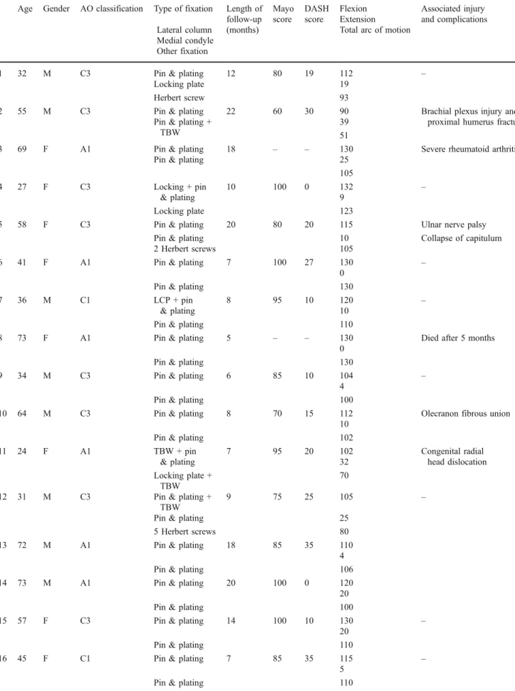
Table 1 Patient characteristics and results
Age Gender AO classification Type of fixation Length of
follow-up (months) Mayo score DASH score
Flexion Associated injury
and complications Extension
Total arc of motion Lateral column
Medial condyle Other fixation
1 32 M C3 Pin & plating 12 80 19 112 –
Locking plate 19
Herbert screw 93
2 55 M C3 Pin & plating 22 60 30 90 Brachial plexus injury and
proximal humerus fracture Pin & plating +
TBW
39 51
3 69 F A1 Pin & plating 18 – – 130 Severe rheumatoid arthritis
Pin & plating 25
105 4 27 F C3 Locking + pin & plating 10 100 0 132 – 9 123 Locking plate
5 58 F C3 Pin & plating 20 80 20 115 Ulnar nerve palsy
Pin & plating 10 Collapse of capitulum
2 Herbert screws 105
6 41 F A1 Pin & plating 7 100 27 130 –
0
Pin & plating 130
7 36 M C1 LCP + pin
& plating
8 95 10 120 –
10
Pin & plating 110
8 73 F A1 Pin & plating 5 – – 130 Died after 5 months
0
Pin & plating 130
9 34 M C3 Pin & plating 6 85 10 104 –
4
Pin & plating 100
10 64 M C3 Pin & plating 8 70 15 112 Olecranon fibrous union
10 102 Pin & plating
11 24 F A1 TBW + pin & plating 7 95 20 102 Congenital radial head dislocation 32 Locking plate + TBW 70
12 31 M C3 Pin & plating +
TBW
9 75 25 105 –
Pin & plating 25
5 Herbert screws 80
13 72 M A1 Pin & plating 18 85 35 110
4
Pin & plating 106
14 73 M A1 Pin & plating 20 100 0 120
20
Pin & plating 100
15 57 F C3 Pin & plating 14 100 10 130 –
20
Pin & plating 110
16 45 F C1 Pin & plating 7 85 35 115 –
5
bone stock [5]. Greiner et al. reported preliminary results of the LCP in supra-intercondylar fractures and noted that clinical and radiological results are promising with good outcome [6]. ORIF with the DHP system was evaluated by Arnander et al., and they reported a good outcome in the majority of the patients [12], but their study was not specifically focused on low fractures. Some low supra-condylar fractures contain small distal fragments, which are difficult to stabilise even with low twisted, multiple or anatomically pre-shaped LCP plates because there may not be enough space, even for a small number of screws in the distal fragment. Another option is TBW, which is used in low supracondylar humerus fractures [8], but this method of osteosynthesis has some disadvantages and limitations. It cannot be used in comminuted or very low oblique fractures (lambda type). Clinically, in our experi-ence TBW implants generally need to be removed, in contrast to our technique.
Our early results in low supracondylar fractures are reasonably acceptable, and we have found that stability achieved with our technique is secure enough to begin early active ROM.
Frankle et al. compared total elbow arthroplasty (TER) and ORIF for intra-articular distal humerus fractures in women older than 65 years of age and believe TER to be an option for this group [15]. However, the patient with TER will have severe limitation use of the limb and should be restricted to low demand patients, and good long-term outcome is dependent on patient compliance in avoiding lifting greater than 5 lb [14].
This type of fixation is much more stable for fragments that can only be secured with K-wires, especially in the osteoporotic setting, and this may be an absolute indication for our technique of osteosynthesis.
Relative indications for this technique are as follows: (1) Low supracondylar fractures, severe osteoporosis and severe comminution which are difficult or impossi-ble to fix by standard techniques. (2) The lack of facilities for the surgeon in complex fractures. (3) As an alternative to TBW in comminuted or low distal humerus fractures.
On the basis of our findings, this technique also has a role in the treatment of comminuted articular surface fractures, particularly when there is limited subchondral bone attached to these fragments. Some have suggested the Herbert screw for large articular fragments with nearby bony support and buried small threaded K-wires connecting subchondral bone in limited opposing bone [15–17]. The latter is what we have considered as a good indication for our method.
There are some advantages offered by this method. It is an easy and generally fast procedure to perform, when compared with traditional methods, as only K-wires suffice, which are mostly used in other methods as preliminary fixation.
With respect to cost, this procedure can reduce the patient’s expenses for orthopaedic implants compared with the expenses of other techniques since this procedure needs only a K-wire and a short plate for fixation.
Another advantage of this technique is that reoperation for implant removal, which is generally required after TBW, is not common.
The major potential specific problem of this method is loss of distal fragment reduction during K-wire contouring on the humeral shaft. Thus, we recommend using K-wires with appropriate thickness to avoid this problem.
Our study has several potential weaknesses related primarily to the short duration of follow-up.
Degenera-Table 1 (continued)
Age Gender AO classification Type of fixation Length of
follow-up (months) Mayo score DASH score
Flexion Associated injury
and complications Extension
Total arc of motion Lateral column
Medial condyle Other fixation
17 17 M C2 Pin & plating 13 95 20 110 –
15
Pin & plating 95
18 36 F C3 Pin & plating +
TBW
11 95 10 110
Pin & plating 25
Transcondylar screw
85
19 29 M C3 Pin & plating 12 95 10 120
15
tion of the elbow may increase with longer follow-up, and the functional score of the patients could potentially decrease with time. Another weakness of our study is the small sample size since this kind of complex fracture in adults is relatively rare. Our data do not purely show the outcome of our technique because we combined our technique with others in some cases, and this may be considered another bias in our study.
Conclusion
Complex distal humerus fractures are sometimes difficult to fix internally by conventional methods, especially in comminuted low distal humerus fractures. We propose a novel technique which uses K-wires in the small fragments which are fixed with a reconstruction plate on the humerus shaft. It allows early active ROM. Therefore, we suggest pin and plating in comminuted, osteoporotic or low distal humerus fractures in which other fixation methods seem difficult or even impossible.
References
1. Rose SH, Melton LJ 3rd, Morrey BF, Ilstrup DM, Riggs BL (1982) Epidemiologic features of humeral fractures. Clin Orthop
Relat Res 168:24–30
2. John H, Rosso R, Neff U, Bodoky A, Regazzoni P, Harder F (1994) Operative treatment of distal humeral fractures in the
elderly. J Bone Joint Surg Br 76(5):793–796
3. Jost B, Adams RA, Morrey BF (2008) Management of acute distal humeral fractures in patients with rheumatoid arthritis. A case series. J Bone Joint Surg Am 90(10):2197–2205
4. Li SH, Li ZH, Cai ZD, Zhu YC, Shi YZ, Liou J, Tao K, Wang JG (2011) Bilateral plate fixation for type C distal humerus fractures:
experience at a single institution. Int Orthop 35(3):433–438
5. Burg A, Berenstein M, Engel J, Luria T, Salai M, Dudkiewicz I, Velkes S (2011) Fractures of the distal humerus in elderly patients
treated with a ring fixator. Int Orthop 35(1):101–106
6. Greiner S, Haas NP, Bail HJ (2008) Outcome after open reduction and angular stable internal fixation for supra-intercondylar fractures of the distal humerus: preliminary results with the LCP distal humerus system. Arch Orthop Trauma Surg 128(7):723–729
7. Brooker A, Bowerman J, Robinson R et al (1973) Ectopic ossification following total hip replacement. Incidence and a
method of classification. J Bone Joint Surg Am 55:1629–
1632
8. Mousavi SJ, Parnianpour M, Abedi M, Askary-Ashtiani A, Karimi A, Khorsandi A, Mehdian H (2008) Cultural adaptation and validation of the Persian version of the Disabilities of the Arm, Shoulder and Hand (DASH) outcome measure. Clin Rehabil
22(8):749–757
9. Gosling T, Blauth M, Lange T, Richter M, Bastian L, Krettek C (2004) Outcome assessment after arthrolysis of the elbow. Arch
Orthop Trauma Surg 124(4):232–236
10. Houben PF, Bongers KJ, von den Wildenberg FA (1994) Double tension band osteosynthesis in supra- and transcondylar humeral fractures. Injury 25(5):305–309
11. Luegmair M, Timofiev E, Chirpaz-Cerbat JM (2008) Surgical treatment of AO type C distal humeral fractures: internal fixation with a Y-shaped reconstruction (Lambda) plate. J Shoulder Elbow
Surg 17(1):113–120
12. Arnander MW, Reeves A, MacLeod IA, Pinto TM, Khaleel A (2008) A biomechanical comparison of plate configuration in
distal humerus fractures. J Orthop Trauma 22(5):332–336
13. Wong AS, Baratz ME (2009) Elbow fractures: distal humerus. J
Hand Surg Am 34(1):176–190
14. O’Driscoll SW (2005) Optimizing stability in distal humeral fracture
fixation. J Shoulder Elbow Surg 14(1 Suppl S):186S–194S
15. Frankle MA, Herscovici D Jr, DiPasquale TG, Vasey MB, Sanders RW (2003) A comparison of open reduction and internal fixation and primary total elbow arthroplasty in the treatment of intraarticular distal humerus fractures in women older than age 65. J Orthop Trauma 17(7):473–480
16. Cobb TK, Morrey BF (1997) Total elbow arthroplasty as primary treatment for distal humeral fractures in elderly patients. J Bone Joint Surg Am 79(6):826–832
17. Sano S, Rokkaku T, Saito S et al (2005) Herbert screw fixation of