KJS
https://doi.org/10.14245/kjs.2017.14.1.7 Print ISSN 1738-2262 On-line ISSN 2093-6729 www.e-kjs.org CLINICAL ARTICLE Korean J Spine 14(1):7-10, 2017Korean J Spine Volume 14 | Number 1 | March 2017 | 7
Determination of the Effect of Diameter of the Sac on
Prognosis in 64 Cases Operated for Meningomyelocele
Objective: To examine the effect of meningomyelocele sac size on prognosis by retrospective review of 64 cases operated for meningomyelocele between January 2009 and December 2012. Methods: We evaluated newborn babies operated for meningomyelocele by retrospectively reviewing their files for head circumference, location and with of the defect, accompanying anomalies, treatments administered, drugs that mother used during pregnancy. Based on the defect size, 3 patient groups were created as 0-24 cm2(group I), 25-39 cm2(group II), and 40 cm2 and above (group III).Results: Throughout the study, 64 babies were evaluated. Mean head circumference was 37.4 cm (range, 30.7-50 cm). Based on their location, 49 of the defects (76.5%) were lumbar, 7 (10.9%) were thoracolumbar, 4 (6.2%) were thoracic, 3 (3.1%) were sacral, 1 (1.5%) was cervical. Mean size of the meningomyelocele sac was 4.7 cm×5.8 cm (range, 1 cm×1 cm—10 cm×8 cm), 13 of the babies (20.3%) had skin defect requiring flap. According to accompanying anomalies, 47 of the babies (73.4%) had hydrocephalus, 7 (10.9%) had club foot, 1 (1.5%) had diastemato- myelia, 1 (1.5%) had tethered cord. Thirty-nine of the babies (60.9%) had paraplegia, 10 (15.6%) had paraparesis, 8 (12.5%) had monoplegia; neurological examination in the remaining 7 babies was normal.
Conclusion: In our study, increased diameter of meningomyelocele sac was associated with greater amount of neural tissue within the sac, which worsens the prognosis. Sac localization was not changing prognosis but infection rates, hospitalization duration were increased in babies with bigger diameter of sacs.
Key Words: Meningomyelocele, Neural Tissue, Diameter of the Sac
Metehan Eseoğlu
1,
Ahmet Eroğlu
2,
Serkan Kemer
3,
Mehmet Arslan
41Department of Neurosurgery,
Medipol Universty, Istanbul,
2Department of Neurosurgery,
Haydarpaşa Sultan Abdülhamid Education and Research Hospital, Istanbul, 3Department of Pediatrics,
Van State Education and Research Hospital, Van, 4Department of
Neurosurgery, Van 100. Year Universty, Van, Turkey Corresponding Author: Ahmet Eroğlu
Department of Neurosurgery, Haydarpaşa Sultan Abdülhamid Education and Research Hospital, Istanbul, Turkey Tel: +90-506-203-6231 Fax: +90-216-542-2020 E-mail: [email protected] Received: January 12, 2017 Revised: January 26, 2017 Accepted: January 26, 2017
Copyright © 2017 by The Korean Spinal Neurosurgery Society
This is an open access article distributed under the terms of the Creative Commons Attribution Non-Commercial License (http://creativecommons.org/licenses/by-n c/4.0/) which permits unrestricted non-commercial use, distribution, and reproduction in any medium, provided the original work is properly cited.
INTRODUCTION
Myelemeningocele is a congenital central nervous system (CNS) anomaly in which a part of the spinal cord, together with the surround-ing mensurround-ingeal structures, protrudes outward through the defected bone and skin as a sac1). The cause is not exactly clear, but genetic and environmental factors are thought to play role together3). While its frequency in European cou-
ntries is 0.1%, the rate in Turkey, according to various studies, range from 0.3% to 0.58%10).
Meningomyelocele can be named according to its location. Studies indicate the most common location as lumbar area with 69% ratio8). Hyd-
rocephalus accompanies meningomyelocele in 80% of all cases8). The diameter of the menin-
gomyelocele sac is among the most important prognostic factors6). The greater the amount of neural tissue inside the sac, the worse is the neuologic deficit and prognosis. Larger sacs re-quire skin flaps more, giving rise to complica-tions such as flap-related infection, reopera-tion, and wound-site problems6). Treatment in
meningomyelocele involves closure of the neu-ral tissue as soon as possible, repair of the skin defect, and placement of ventriculoperitoneal (V/P) shunt in cases with accompanying hydro- cephalus.
MATERIALS AND METHODS
In our study, we retrospectively reviewed 64 cases operated for meningomyelocele (Figs. 1, 2) between January 2009 to December 2012, noting data including sex, birthweight, diame-ter of the sac, and presence of additional ano- malies. Patient age varied from neonatal period to 6 months. Regarding distribution of sex, 34 (53.1%) were female, and 30 (46.8%) were male patients. All patients were evaluated with magnetic resonance imaging (MRI) in order to localize the sac, and were examined to deter- mine diameter of the sac accurately (Table 1). Patients were divided into 3 groups based on the size of the defect: group I, 0-24 cm2: group II, 25-39 cm2: group III, 40 cm2 and above(Table 2). As the sacs were circular, we used the following formula for calculating the sac
Eseoğlu M et al.
8 | Volume 14 | Number 1 | March 2017 www.e-kjs.org
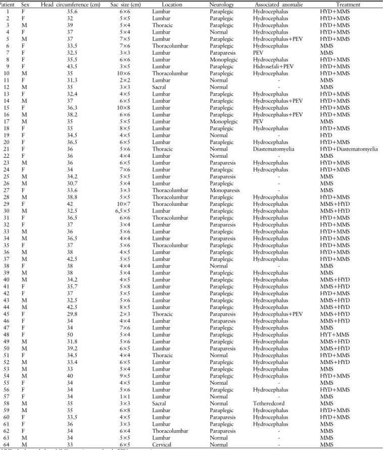
Table 1. Patient characteristics
Patient Sex Head circumference (cm) Sac size (cm) Location Neurology Associated anomalie Treatment
1 F 35.6 6×6 Lumbar Paraplegic Hydrocephalus HYD+MMS
2 F 32 5×5 Lumbar Paraplegic Hydrocephalus HYD+MMS
3 M 39 5×4 Thoracic Paraplegic Hydrocephalus HYD+MMS
4 F 37 5×4 Lumbar Normal Hydrocephalus HYD+MMS
5 M 37 7×5 Lumbar Paraplegic Hydrocephalus+PEV HYD+MMS
6 F 33.5 7×6 Thoracolumbar Paraplegic Hydrocephalus MMS
7 F 32.5 3×3 Lumbar Paraparesis PEV MMS
8 F 35.5 6×6 Lumbar Monoplegic Hydrocephalus HYD+MMS
9 F 43.5 3×5 Lumbar Paraplegic Hidrosefali+PEV HYD+MMS
10 M 35 10×6 Thoracolumbar Paraplegic Hydrocephalus HYD+MMS
11 F 31.3 2×2 Lumbar Normal - MMS
12 M 35 3×3 Sacral Normal - MMS
13 F 32.4 4×5 Lumbar Paraplegic Hydrocephalus HYD+MMS
14 M 37 6×5 Lumbar Paraplegic Hydrocephalus+PEV HYD+MMS
15 F 36.3 10×8 Lumbar Paraplegic Hydrocephalus HYD+MMS
16 M 38.2 6×6 Lumbar Paraplegic Hydrocephalus+PEV HYD+MMS
17 M 35 5×5 Lumbar Monoplegic PEV MMS
18 F 35 8×5 Lumbar Paraplegic Hydrocephalus HYD+MMS
19 F 34.5 4×5 Lumbar Normal - HYD
20 F 36.5 6×5 Lumbar Paraplegic Hydrocephalus HYD+MMS
21 F 36 5×6 Thoracic Normal Diastematomyelia HYD+Diastematomyelia
22 F 36 4×4 Lumbar Normal - MMS
23 M 36 6×5 Lumbar Paraparesis Hydrocephalus HYD+MMS
24 F 34 7×6 Lumbar Paraplegic Hydrocephalus HYD+MMS
25 M 34.2 5×5 Lumbar Paraparesis - MMS
26 M 30.7 5×4 Lumbar Paraplegic - MMS
27 F 33.6 3×3 Thoracolumbar Monoparesis - MMS
28 M 38.8 5×5 Thoracolumbar Paraplegic Hydrocephalus HYD+MMS
29 F 42 10×7 Thoracolumbar Paraplegic Hydrocephalus MMS+HYD
30 M 32.5 6,5×5 Lumbar Paraplegic Hydrocephalus MMS+HYD
31 F 36.5 6×6 Thoracolumbar Paraplegic Hydrocephalus HYD+MMS
32 F 37 3×4 Lumbar Paraparesis Hydrocephalus HYD+MMS
33 M 36 5×6 Lumbar Paraplegic Hydrocephalus HYD+MMS
34 M 36.5 4×4 Lumbar Paraparesis Hydrocephalus HYD+MMS
35 F 37 5×6 Thoracolumbar Paraplegic Hydrocephalus HYD+MMS
36 M 38 4×5 Lumbar Paraplegic Hydrocephalus HYD+MMS
37 M 42.5 5×5 Lumbar Paraplegic Hydrocephalus HYD+MMS
38 F 38 4×4 Lumbar Normal - MMS
39 M 38 5×4 Lumbar Paraplegic Hydrocephalus MMS
40 M 34.2 4×5 Lumbar Paraplegic Hydrocephalus MMS+HYD
41 F 35.7 5×8 Lumbar Paraplegic Hydrocephalus MMS+HYD
42 F 37 5×5 Lumbar Paraplegic Hydrocephalus HYD+MMS
43 M 32.5 5×6 Lumbar Paraplegic Hydrocephalus MMS+HYD
44 M 42.5 8×5 Lumbar Paraplegic Hydrocephalus MMS+HYD
45 F 29.8 2×3 Thoracic Paraparesis Hydrocephalus+PEV MMS+HYD
46 F 34 4×4 Lumbar Paraparesis Hydrocephalus MMS+HYD
47 F 34 7×6 Lumbar Paraplegic Hydrocephalus MMS
48 F 50 5×4 Lumbar Paraplegic Hydrocephalus HYT+MMS
49 M 31.8 5×6 Lumbar Paraplegic Hydrocephalus MMS+HYD
50 M 39.2 6×5 Lumbar Paraparesis Hydrocephalus MMS+HYD
51 F 34.5 4×4 Thoracic Normal Hydrocephalus HYD+MMS
52 M 33.4 6×5 Lumbar Paraplegic Hydrocephalus MMS+HYD
53 M 33 5×4 Lumbar Paraplegic Hydrocephalus MMS
54 M 40 9×5 Lumbar Paraplegic Hydrocephalus HYD+MMS
55 F 34 4×5 Lumbar Normal - MMS
56 F 34 5×6 Lumbar Paraplegic Hydrocephalus HYD+MMS
57 F 34 1×1 Lumbar Normal - MMS
58 M 35 3×3 Sacral Normal Tetheredcord MMS
59 M 35 6×8 Lumbar Paraplegic Hydrocephalus HYD+MMS
60 F 33.5 4×5 Lumbar Paraparesis Hydrocephalus HYD+MMS
61 F 36 3×3 Lumbar Paraplegic Hydrocephalus MMS
62 F 34 6×4 Thoracolumbar Paraparesis - MMS
63 M 34 5×5 Lumbar Normal - MMS
64 M 33 6×5 Cervical Normal - MMS
Determination of the Effect of Diameter of the Sac on Meningomyelocele
Korean J Spine Volume 14 | Number 1 | March 2017 | 9
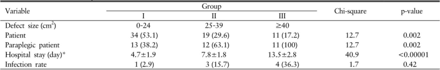
Table 2. Characteristics of patients based on the size of the defect
Variable I GroupII III Chi-square p-value
Defect size (cm2) 0–24 25–39 ≥40
Patient 34 (53.1) 19 (29.6) 11 (17.2) 12.7 0.002
Paraplegic patient 13 (38.2) 12 (63.1) 11 (100) 12.7 0.002
Hospital stay (day)* 4.7±1.9 7.8±1.8 13.5±2.8 40.9 <0.00001
Infection rate 1 (2.9) 3 (15.7) 4 (36.3) 1.7 0.42
Values are presented as number (%) or mean±standard deviation.
*z Group I → Group II=-4.5; **z Group I → Group III=-4.9; ***z Group II → Group III=-4.3; p<0.05; Mann whitney U-Test.
Fig. 1. Preoperative (A) and postoperative (B) images of the patient who were operated on for the cervical meningomyelocele.
Fig. 2. Preoperative (A) and postoperative (B) images of the patient who were operated on for the thoracolumbar meningomyelocele.
area: Ωr2 (Ω: pi number, r: radius). Serum urea and creatinine levels were measured during the first 72 hours in order to assess renal functions in all babies. Neurological examination was per-formed in every patient in order to determine motor deficit. Patients were evaluated for infection, and we performed surgery for those patients without any sign of infection, and the patients were followed up at the postoperative period. We evaluated the effect of sac diameter on hospital stay length, duration of anti-biotherapy, and early morbidity. Babies were followed up throu- ghout the newborn period, and prognosis and complications at the early period were evaluated.
The study data was analyzed with demographic statistics me- thod. Data was expressed as mean±standard deviation. Using SPSS ver. 16.0 (SPSS Inc., Chicago, IL, USA), nonparametric data were analyzed with chi-square test, Kruskal-Wallis H-test and Mann-Whitney U-Test. Determination of risk factors and rela-tion between variables were analyzed with correlarela-tion analysis. A p-value of <0.05 was accepted as statistically significant.
RESULTS
Table 1 shows detailed properties of our cases along with the surgical intervention performed and postoperative conditions. None of the mothers used folic acid supplement, or were expo- sed to radiation before or during their pregnancy. None of the families receiving antenatal diagnosis accepted medical termi-nation recommendation. In the study group, it was determined that most of the mothers were undereducated, and 89% of the mothers were either primary school graduate or did not go to school at all. A great proportion of meningomyelocele location, in 50 of 64 patients (78.1%), was lumbar region. Sac size varied between 1 cm×1 cm and 10 cm×8 cm. MRI was performed to aid in the diagnosis. Seven cases did not have neural tissue within the sac. None of these patients had neurological deficit or hydro- cephalus. Three of these cases were in group 1, and 4 were in group 2 according to the sac diameter.
The defect was covered with flap in 12 cases, while preserving dorsal intercostal artery. All of these cases had neural tissue with-in the sac. Of these patients, 11 were with-in group 3, and 1 was with-in group 2.
The surgery was performed with prone positioning. Neural tissue within the sac was preserved. New dura was formed using fascia, and the skin was closed. For cases with large skin defects, skin was closed with flap. Hydrocephalus was diagnosed in 47 cases (73.4%). Thirty-nine out of 47 patients were treated with V/P shunt. Parents of 8 cases did not approve the recommended shunt surgery. No mortality was observed among our cases at postoperative period, and during the follow-up. Comparison of mean sac diameter with neurological condition, hospital stay length and additional anomalies showed significant difference in Mann-Whitney U-test (p<0.05). As the sac size increases, the length of stay increases (r=0,91, p<0,05). Smaller sac diameter was associated with better neurological condition, and sig-nificantly lower hospital stay and infection rates in our study (p<0.05) (Table 2). In our study we observed that localization of sac was not affecting prognosis (p>0.05) (Table 1) and diame-ter and having neural tissue within the sac were more important prognostic factors than the localization.
DISCUSSION
mal-Eseoğlu M et al.
10 | Volume 14 | Number 1 | March 2017 www.e-kjs.org
formations of CNS; it is a midline line closure defect categorized in spina bifida aperta group. Its frequency varies between 1 and 9 in 1,000 live births10). Spinal cord or nerve roots can protrude
through a vertebra defect into a sac containing meninges as well, or they may be exposed without being covered with any meninx or skin. This congenital anomaly can result in severe neurological dysfunction, and it may be accompanied by other anomalies of
CNS4). The most frequent accompanying anomaly is hydroce-
phalus. For live-born cases, an effective reconstruction is essen-tial to close the exposed neural elements and to protect the pa-tient from sepsis5).
Low socioeconomical state is a risk factor for meningomyelo-cele development. Studies from all around the world and Turkey have reported higher frequency of neural tube defect among fam-ilies with lower educational states2,7). Van 100. Yıl University
is located in eastern part of Turkey and educational level of families in this area is not high. 89% of the mothers had low educational level in our study.
Studies report most frequent location of meningomyelocele
as lumbar region (69%)3). This was supported with our
ob-servation in our study that 78.1% of cases had MM in lumbar region. The most common additional anomaly in MM is hydro-cephalus8). In our study group, the most frequent accompanying anomaly was also hydrocephalus, observed in 73.4% of cases.
During our review of the literature, we did not encounter any study directly investigating the effect of diameter of the me-ningomyelocele sac on prognosis. In cases with meningomyelo-cele, increasing sac diameter is associated with greater amount of neural structures within the sac, which worsens the neuro-logical condition9). Diameter of the sac affects prognosis sig-nificantly in cases with meningomyelocele6). Neurological
con-dition in group I patients, who have the smallest sac diameter, was better in comparison to group III patients who have larger sac diameter (p<0.05). While all of the group III patients had paraplegia, this ratio was 38% in group I patients, who have sac diameter below 24 cm2. Müslüman et al.6) stated that they
closed the defects via primary closure without flap requirement in babies who had sac smaller than 25 cm2, and that these babies
had shorter hospital stay length, lower infection risk, and better prognosis. In our study we determined that localization of sac was not affecting prognosis (Table 1), we observed that sac diam-eter and having neural tissue within the sac were more important prognostic factors than the sac localization.
In our series, infection rate and hospital stay length in group I patients (defect size smaller than 24 cm2) were significantly
lower in comparison to groups II and III patients (p<0.05). In our study group, sac diameter at newborn period was a border-line 2×2-cm size. We observed that these babies with a sac size below this borderline had better outcomes than the group with larger defect in terms of hospital stay length, duration of antibio- therapy, as well as neurological conditions. In their series includ-ing 35 cases with meninclud-ingomyelocele, Wilson et al.9) reported
that apart from 1 case, all patients had sac size below 24 cm2,
and only 8 cases (22.9%) had poor neurological function during
follow-up. In our study group, the ratio of paraplegic cases among babies with sac size below 25 cm2 was 38.2%.
CONCLUSION
Babies with smaller meningomyelocele sac have shorter hospi-tal stay length and fewer complications at early period, and more favorable neurological signs. Increasing size of the sac is asso-ciated with greater amount of neural tissue within the sac, requir-ing flap for skin repair and worsenrequir-ing the prognosis.
CONFLICT OF INTEREST
No potential conflict of interest relevant to this article was reported.
REFERENCES
1. Back SA: Congenital Malformations of the Central Nervous System. ın Taeusch HW, Ballard RA, Gleason CA (eds): Avery’s Diseases of the Newborn, ed 8. Philadelphia: Elsevier, pp938-959, 2005 2. Canfield MA, Ramadhani TA, Shaw GM, Carmichael SL, Waller
DK, Mosley BS, et al: Anencephaly and spina bifida among Hispa- nics: maternal, sociodemographic, and acculturation factors in the National Birth Defects Prevention Study. Birth Defects Res A Clin Mol Teratol 85:637-646, 2009
3. Idowu OE, Apemiye RA: Outcome of myelomeningocoele repair in sub-Saharan Africa: the Nigerian experience. Acta Neurochir (Wien) 150:911-913, 2008
4. Koszutski T, Kawalski H, Kudela G, Wróblewska J, Byrka-Owc- zarek K, Bohosiewicz J: Babies with myelomeningocele in Poland: parents’ attitudes on fetal surgery versus termination of pregnancy. Childs Nerv Syst 25:207-210, 2009
5. Kshettry VR, Kelly ML, Rosenbaum BP, Seicean A, Hwang L, Weil RJ: Myelomeningocele: surgical trends and predictors of outcome in the United States, 1988-2010. J Neurosurg Pediatr 13:666-678, 2014
6. Müslüman AM, Karşıdağ S, Sucu DÖ, Akçal A, Yılmaz A, Sirinoğlu D, et al: Clinical outcomes of myelomeningocele defect closure over 10 years. J Clin Neurosci 19:984-990, 2012
7. Onrat ST, Seyman H, Konuk M: Incidence of neural tube defects in Afyonkarahisar, Western Turkey. Genet Mol Res 8:154-161, 2009
8. Tamburrini G, Frassanito P, Iakovaki K, Pignotti F, Rendeli C, Murolo D, et al: Myelomeningocele: the management of the associated hydrocephalus. Childs Nerv Syst 29:1569-1579, 2013 9. Wilson RD, Johnson MP, Bebbington M, Flake AW, Hedrick HL,
Sutton LN, et al: Does a myelomeningocele sac compared to no sac result in decreased postnatal leg function following maternal fetal surgery for spina bifida aperta? Fetal Diagn Ther 22:348-351, 2007
10. Zeyrek D, Soran M, Cakmak A, Kocyigit A, Iscan A: Serum copper and zinc levels in mothers and cord blood of their newborn infants with neural tube defects: a case-control study. Indian Pediatr 46:675-680, 2009