Cellular and Molecular
Biology
Original Research
Irisin immunostaining characteristics of breast and ovarian cancer cells
T. Kuloglu1, O. Celik2, S. Aydin3*, İ. Hanifi Ozercan4, M. Acet5, Y. Aydin6, G. Artas4, A. Turk1, M. Yardim3, G. Ozan7,
M. Hanifi Yalcin8, N. Kocaman1
1 Firat University, School of Medicine, Department of Histology and Embryology, Elazig 23119, Turkey 2 Private Clinic, Obstetrics and Gynecology, Usak, Turkey
3 Firat University, School of Medicine, Department of Medical Biochemistry (Firat Hormones Research Group), 23119-Elazig, Turkey 4 Firat University, School of Medicine, Department of Pathology, 23119-Elazig, Turkey
5 Istanbul Medipol University Hospital, Department of Obstetrics and Gynecology, İstanbul, Turkey 6 Ankara University, School of Veterinary Medicine, 06200-Ankara, Turkey
7 Firat University, School of Veterinary Medicine, Department of Biochemistry, 23119-Elazig, Turkey 8 Firat University, School of Veterinary Medicine, Department of Histology, 23119-Elazig, Turkey
Abstract: To determine expression pattern of irisin in tissues obtained from human ovarian cancer, breast cancer, and cervix cancer. Tissue
samples obtained from subjects with breast cancer, ovarian cancer cervix cancer, simple endometrial hyperplasia, complex atypical endometrial hyperplasia. At least five sections from each subject were immunohistochemically stained with irisin antibody, and H-score method was used to evaluate irisin intensity. Tissues obtained from healthy breast tissues, proliferative phase endometrium adenomyosis and benign ovarian tumors were accepted as control. Irisin activity was not detected in control breast tissues significantly increased irisin staining was detected in invasive lobular, intraductal papillary, invasive ductal, invasive papillary, and mucinous carcinomas compared to control tissues. Also, significantly in-creased irisin immunoreactivity was detected in both ovarian endometriosis and mucinous carcinomas compared to benign tumors. However irisin staining was not observed at the papillary carcinoma of the ovary while sections obtained from simple and complex atypical endometrial hyperplasia, and cervix carcinoma demonstrated irisin immunoreactivity. Increased irisin immunoreactivity in tissues obtained from breast, ovary, cervix carcinomas, and endometrial hyperplasia suggest critical role of this peptide during carcinogenesis.
Key words: Irisin, Breast cancer, Ovarian cancer, Cervix cancer, Endometrial hyperplasia.
Introduction
Breast cancer (BC) and ovarian cancer (OC) are the leading cancer among women worldwide. 5-10 % of breast cancer cases is of hereditary origin, mainly due to BRCA1/2 mutations (1) or may likely be a relevant some energy associated peptides production (increase or decrease) such as ghrelin and obestatin (2-4). Recently, an energy associated-irisin is an 112-amino acid peptide that was identified in 2013 in the rat and human skeletal muscle by Boström and co-workers (5). After this initial report, irisin was also reported a high level in the adi-pocytes (6), kidneys, gastric mucosa (7), pancreas (8,9), gonads and heart tissue, blood Vessels, and so many other tissues (8-10). Interestingly, the levels in the heart were even higher than those detected in the skeletal muscle (9), indicating a predominant expression of irisin in the heart tissues. The function of irisin is to convert white adipose tissues (WAT) into brown adipose tissue (BAT). From a physiological point of view, the conver-sion of WAT into BAT upregulates mitochondria bear-ing the uncouplbear-ing protein 1 (UCP1), mediates adaptive thermogenesis by releasing heat (5), and thereby con-tributes to the maintenance of body temperature, and glucose homeostasis and also helps weight reduction in obese individuals after exercise because it is released into the blood stream (10). It has been also reported that this molecule has an anticarcinogenic properties (11).
Furthermore, we have recently showed the differ-ent expression patterns of irisin in many healthy (9) and pathological tissues (12). However, when reviewing the
literature there is no comprehensive immunohistochem-ical study investigating whether irisin is expressed in ovarian and/or breast cancer tissues or not. Likewise, studies investigating irisin expression in premalignant endometrial lesions are lacking.
The purpose of this study was to characterize the ex-pression pattern of irisin in tissues from different human ovarian cancer. Due to close relation between ovarian cancer and breast cancer cases in terms of BRCA1/2 mutations patients diagnosed with breast cancer are included to study as control group. Moreover, due to estrogen dependent characteristic of breast and endo-metrial lesions patients having endoendo-metrial hyperplasia were obtained as second control group.
Materials and Methods
This study has been approved by the Clinical Re-search Ethics Committee of Firat University Faculty of Medicine (date 02/10/2015, session #3, decision #4), and conducted based on the principles of the Declara-tion of Helsinki. To determine irisin expression status of malignant and premalignant tissues (endometrial
hyper-Received March 27, 2016; Accepted July 21, 2016; Published July 31, 2016 * Corresponding author: Dr. Suleyman Aydin,Firat University, School of Medicine, Department of Medical Biochemistry (Firat Hormones Research Group), 23119-Elazig, Turkey. Email: [email protected]; [email protected]
Copyright: © 2016 by the C.M.B. Association. All rights reserved.
Kuloglu et al. Cell. Mol. Biol.2016, 62 (8): 40-44 ISSN: 1165-158X
plasia), we obtained tissue samples from subjects with ovarian cancer, aged 36–58 years, subjects with breast cancer, aged 40-66 years, subjects with cervix cancer, aged 44-62 years, subjects with simple endometrial hy-perplasia, aged 30-43 years, and subjects with complex atypical endometrial hyperplasia, aged 33-48 years. Tis-sues obtained from healthy breast tisTis-sues, proliferative phase endometrium, adenommyosis and benign ovarian tumors were accepted as control. All breast and ovarian cancer tissues were moderately differentiated accord-ing to World Health Organization classification (WHO) (13). We examined 3-5 sections from each type of can-cer (n: 10 cases for each individual cancan-cer types) and 10 sections from control tissues. Patients whose tissues were used had received no chemotherapy or radiother-apy before operation and had sedentary lifestyle, and samples were taken from both affected and unaffected (internal control) areas of each tumor.
Irisin immunohistochemistry
Tissue samples for irisin immunostaining were ob-tained from women scheduled for ovarian cancer or breast cancer surgery. Likewise, endometrial tissue samples were obtained from women diagnosed with en-dometrial hyperplasia. Irisin intensity was measured in both healthy and malignant cells of precancerous and cancerous tissue sections. Both endometrial samples and post-surgical breast and ovarian cancer blocks were exhaustively sectioned at a thickness of 4-5 µm using a microtome perpendicular to the long axis of the tumor.
At least five slices were selected from the each tumor using systematic random sampling rules. Sections were immunohistochemically stained with irisin as previ-ously described (14). Quantitation of the staining in the sections was done using the light microscopy to give an immunoreactive histoscore (H-score), based on the modification of Kuloglu and Aydin (14), which was deter mined by screening 100 cells in the staining area for each preparation.
Statistical analysis
SPSS 21.00 software package was used for statisti-cal analyses. H-score evaluations are given as the mean ± standard deviation (SD).
Results
Total 180 subjects were enrolled in the investiga-tion. Three-five slices from each case, totally 540-900 sections, were processed for determination of irisin H-score. The mean irisin H-score for different histological groups of breast cancers are illustrated in Table 1. Irisin immunoreactivity was not detected in control breast tis-sues significantly increased irisin staining was detected in invasive lobular, intraductal papillary, invasive duc-tal, invasive papillary, and mucinous carcinomas com-pared to control tissues (p<0.05). Increase in irisin in-tensity in invasive ductal carcinoma is more severe than those in the other histological subtypes (Figure1a-f).
The mean H-score of irisin in human genital tissues
Biological Samples Benign Cell HistoscoreTumour Cell
Breast Control 0.0±0.0
-Invasive Lobular Carcinoma - 0.58±0.21a
Intraductal Papillary Carcinoma - 1.53±0.49 ab
Invasive Ductal Carcinoma - 2.52±0.37 abc
Invasive Papillary Carcinoma - 0.28±0.12 abcd
Mucinous Carcinoma - 0.24±0.14 abcd
Figure 1. Immunoreactivity of irisin in human breast tissues with and without breast cancers. a) Control; b) invasive lobular carcinoma; c)
in-traductal papillary carcinoma; d) invasive ductal carcinoma; e) invasive papillary carcinoma; and mucinous carcinoma. Irisin immunoreactivity increased in all breast cancer tissues compared with control tissue irisin immunoreactivity.
aControl vs all carcinomas (p<0.05); b invasive lobular carcinoma vsintraductal papillary carcinoma; cintraductal papillary
carcinoma vs invasive ductal carcinoma vs invasive papillary carcinoma vs mucinous carcinoma (p<0.05) and invasive ductal carcinoma vs invasive papillary carcinoma vs mucinous carcinoma (p<0.05).
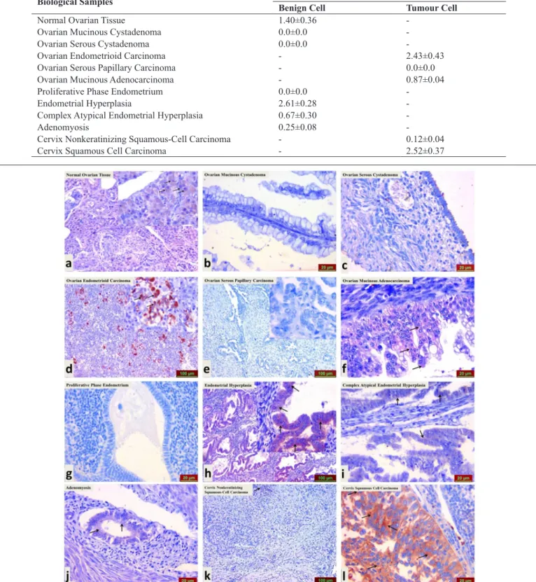
liferative phase endometrial samples did not show irisin staining, sections obtained from simple endometrial hy-perplasia demonstrated intense increase in irisin immu-noreactivity. Interestingly, weak but disseminated irisin staining was detected in complex atypical endometrial hyperplasia. Irisin immunoreactivity was also detected in both adenomyosis and non-keratinized squamous cell carcinoma of the cervix. Increased irisin intensity was found in samples from squamous cell carcinoma of the cervix. All those explanations were given in Figure 2a-l. with and without genital cancer and endometrium
tis-sues was seen in Table 2. Luteal cells of control ovarian tissue displayed irisin expression. However, irisin im-munoreactivity was not detected in both mucinous and serous cystadenoma of the ovary. Although increased irisin immunoreactivity was detected in ovarian endo-metrioid carcinoma, irisin staining was not observed sections obtained from serous papillary carcinoma. Mu-cinous adenocarcinoma of the ovary exhibited weak but widely distributed irisin immunoreactivity. While
pro-Figure 2. Immunoreactivity of irisin in human genital tissues with and without genital cancer and endometrium tissues. a) Control; b) ovarian
mucinous cystadenoma; c) ovarian serous cystadenoma; d) ovarian endometrioid carcinoma; e) ovarian serous papillary carcinoma; f) ovarian mucinous adenocarcinoma; g) proliferative phase endometrium; h) endometrial hyperplasia; I) complex atypical endometrial hyperplasia; j) adenomyosis; k) cervix non-keratinizing squamous-cell carcinoma, and l) cervix squamous-cell carcinoma.
Table 2. Histoscore of irisin immunoreactivity in human genital tissues with and without genital cancer and endometrium
tissues.
Biological Samples HistoscoreBenign Cell Tumour Cell
Normal Ovarian Tissue 1.40±0.36
-Ovarian Mucinous Cystadenoma 0.0±0.0
-Ovarian Serous Cystadenoma 0.0±0.0
-Ovarian Endometrioid Carcinoma - 2.43±0.43
Ovarian Serous Papillary Carcinoma - 0.0±0.0
Ovarian Mucinous Adenocarcinoma - 0.87±0.04
Proliferative Phase Endometrium 0.0±0.0
-Endometrial Hyperplasia 2.61±0.28
-Complex Atypical Endometrial Hyperplasia 0.67±0.30
-Adenomyosis 0.25±0.08
-Cervix Nonkeratinizing Squamous-Cell Carcinoma - 0.12±0.04
Discussion
Our study is the first attempt investigating irisin ex-pression in BC, OC, and endometrial hyperplasia. We have re-addressed the hereditary and estrogen depen-dent nature of BC, OC, and endometrial hyperplasia with entirely new data. Relatively few investigations of irisin have been performed due the difficulty in ob-taining appropriate tumor tissue for evaluation and the laborious nature of tissue preparation from cancerous lesions. Moreover, we have some difficulties related to immunostaining procedures of cancerous tissues with irisin. Fortunately, remarkable animal and human stud-ies from our group have improved our experience re-garding irisin immunostaining in different tissues.
Limitations of our earlier investigations include the use of non-quantitative evaluation techniques for esti-mations of irisin staining intensity in normal and patho-logical tissues. Another limitation is the measurement of irisin staining intensity in either normal or abnormal cells only one section. To overcome these handicaps in present study we have evaluated at least five sections from pathological block’s of one patients. We have also used semi-quantitative H-score method to more accu-rately evaluate tissue irisin levels (12,15).
We believe the immunohistochemical analysis of tissues obtained from breast or ovarian cancer subjects offer considerable advantages over serum-based irisin measurement techniques. Previous study, Provatopou-lou et al measured serum irisin concentration in women with breast cancer. They reported that serum levels of irisin significantly declined in women diagnosed with breast cancer (16). But they did not evaluated the tissue levels of irisin in cancerous tissue. Therefore, without measuring tissue levels of irisin detection of decline in serum irisin in women having breast cancer is not elu-sive. Due to lack of specified irisin receptor in breast tissue we could not make logical comment regarding to transport of serum irisin into the breast tissue. Moreover, since the irisin is an exercise hormone serum levels of it shows fluctuation that leads to under or overestimated irisin measurement in serum samples (10).
Although main production site of irisin is the muscle cells many living tissues express irisin peptide with dif-ferent intensity. Irisin expressions in healthy and non-cancerous cells of tumor tissue support this hypothesis. The possible source of increased irisin expression in breast and ovarian cancer cells is unclear. This might be secondary to increased expression of irisin from muscle cells or increased local production from cancerous cells. Due to age or physiological conditions of subjects with cancer regular exercises in those women are unusual. Hence, it is most likely possible that increased irisin ex-pression in tumor cells is secondary to local production of irisin in cancer cells. Although we did not measure serum levels of irisin, previous study demonstrated the decline in serum levels of irisin in breast cancer women (16). If the increased tissue levels of irisin are related to serum irisin levels they had been detected high serum irisin levels. Together, increase in irisin intensity in BC or OC cells is related to local production rather than sys-temic production and secretion into the circulation.
We exactly do not know how irisin functions in can-cerous cells. It should be noted that our increased irisin
estimates for women at different BC and OC subtypes suggest that irisin accumulation in cancerous tissues is irrespective of histological subtypes of breast or ovarian cancers. Nevertheless, some lack of irisin immunostain-ing some ovarian cancer need further evaluation. Both immunological status and grade and stage of tumors could affect irisin staining of healthy tissue. Therefore, studies evaluating the possible correlation between tu-mor stage, grade, and irisin staining are required. Ac-cumulation of irisin in premalignant endometrial lesions supports the possible inductor or inhibitor role of irisin during cancer development. Since the irisin is a ther-moregulatory peptide it may have a specific role in cell cycle of cancer cells. Concordantly, a previous study conducted by Gannon et al concluded that irisin display cytotoxic impacts against breast and prostate cancer cells (17). Incomplete activation of electron transport system by irisin might lead to thermal injury of tumor cells. It is well known that proliferation of cancer cells is required both high ATP and heating (18). Increased heating activity within the microenvironment of tumor inhibits the apoptosis of cancer cells that lead to occur-rence of immortal cells. Therefore, balanced thermal activity is obligatory to prevent uncontrolled cell prolif-eration. Accordingly, irisin may regulate the thermal ac-tivity inside breast and ovarian tumors and then prohibit the proliferation of cancer cells.
It may be speculative but increased irisin immunore-activity in cancerous tissue might be responsible for the weight loss of women suffering from ovarian and breast cancers. Irisin dependent burning of white adipose tis-sue might lead to cancer cachexia. If our hypothesis is true administration of anti-irisin in patients having can-cer may prevent weight loss. This approach could open new horizons in treatment of cancer cachexia.
In conclusion, we first detected intensity of irisin immunostaining in tissue samples of women diagnosed with ovarian cancer, breast cancer, cervix cancer, or endometrial hyperplasia using immunohistochemistry technique. Second, we measured the irisin intensity control tissues obtained from, normal breast tissue, pro-liferative phase endometrium, adenomyosis, and benign ovarian tumors. In both group H-score method was used to evaluate irisin staining intensity. Accordingly, irisin may regulate the thermal activity inside breast and ovar-ian tumors and then prohibit the proliferation of cancer cells.
References
1. Ferlay J, Steliarova-Foucher E, Lortet-Tieulent J, Rosso S, Coe-bergh JW, Comber H, et al. Cancer incidence and mortality patterns in Europe: estimates for 40 countries in 2012. Eur J Cancer 2013; 49:1374-403.
2. Lien GS, Lin CH, Yang YL, Wu MS, Chen BC. Ghrelin induces colon cancer cell proliferation through the GHS-R, Ras, PI3K, Akt, and mTOR signaling pathways. Eur J Pharmacol 2016; 776:124-31. 3. Dagli AF, Aydin S, Kocdor H, Gurates B, Sahin I, Catak Z, et al. Ghrelin expression of endometrium hyperplasia and endometrioid carcinoma. Gynecol Endocrinol. 2011; 27:199-204.
4. Nurkalem C, Celik H, Dagli F, Gurates B, Kavak B, Dogan Z, et al. Ghrelin and obestatin expression in serous ovarian tumours. Gynecol Endocrinol. 2012; 28:941-4.
A PGC1-α-dependent myokine that drives brown-fat-like develop-ment of white fat and thermogenesis. Nature 2012; 481:463-8. 6. Moreno-Navarrete JM, Ortega F, Serrano M, Guerra E, Pardo G, Tinahones F, et al. Irisin is expressed and produced by human muscle and adipose tissue in association with obesity and insulin resistance. J Clin Endocrinol Metab 2013; 98:E769-78.
7. Lv J, Pan Y, Li X, Cheng D, Ju H, Tian J, et al. Study on the
distribution and elimination of the new hormone irisin in vivo: new discoveries regarding irisin. Horm Metab Res 2015; 47:591-5. 8. Pedersen BK, Febbraio MA. Muscles, exercise and obesity: skeletal muscle as a secretory organ. Nat Rev Endocrinol 2012; 8:457-65.
9. Aydin S, Kuloglu T, Aydin S, Kalayci M, Yilmaz M, Cakmak T, et al. A comprehensive immunohistochemical examination of the distribution of the fat-burning protein irisin in biological tissues. Peptides 2014; 61:130-6.
10. Aydin S. Three new players in energy regulation: preptin, adro-pin and irisin. Peptides 2014; 56:94-110.
11. Tekin S, Erden Y, Sandal S, Yilmaz B. Is Irisin an Anticarcino-genic Peptide? Med-Science 2015; 4:2172-80.
12. Aydin S, Kuloglu T, Ozercan MR, Albayrak S, Aydin S, Bakal U, et al. Irisin immunohistochemistry in gastrointestinal system can-cers. Biotech Histochem 2016; 1-9.
13. Hamilton SR, Aaltonen LA. (Eds.) World Health Organization Classification of Tumours Pathology and Genetics of Tumours of the Digestive System. Lyon IARC Press; 2000.
14. Kuloglu T, Aydin S. Immunohistochemical expressions of adro-pin and ınducible nitric oxide synthase in renal tissues of rats with streptozotocin-ınduced experimental diabetes. Biotech Histochem 2014; 89:104-10.
15. Aydin S, Eren MN, Kuloglu T, Aydin S, Yilmaz M, Gul E, et al. Alteration of serum and cardiac tissue adropin, copeptin, irisin and TRPM2 expressions in DOX treated male rats. Biotech Histochem 2015; 90:197-205.
16. Provatopoulou X, Georgiou GP, Kalogera E, Kalles V, Matia-tou MA, PapapanagioMatia-tou I, et al. Serum irisin levels are lower in patients with breast cancer: association with disease diagnosis and tumor characteristics. BMC Cancer 2015; 15:898.
17. Gannon NP, Vaughan RA, Garcia-Smith R, Bisoffi M, Trujillo KA. Effects of the exercise-inducible myokine irisin on malignant and non-malignant breast epithelial cell behavior in vitro. Int J Can-cer 2015; 136:E197-202.
18. Coffey DS, Getzenberg RH, DeWeese TL. Hyperthermic biol-ogy and cancer therapies a hypothesis for the "Lance Armstrong ef-fect". JAMA 2006; 296:445-8.