Case Reports Anatol J Cardiol 2021; 25: 49-54
50
The patient’s clinical course was favorable, as there was no recurrence of symptoms and there was gradual reduction of cardiac biomarkers. The ECG evolution indicated infero-lateral biphasic T waves. He was discharged 6 days following admis-sion while on dual antiplatelet therapy, statin, and angiotensin-converting enzyme inhibitor. Thirty months after the incident, the patient is still asymptomatic and in good health, on single Anti-platelet therapy and statins.
Discussion
As regards the earlier reported case, we propose that an ana-phylactoid insult was the triggering factor for a type II KS, due to the amino acid histidine found in fresh fish, which is converted in histamine following an infection by gram-negative bacteria, expressing the enzyme histidine decarboxylase (2). Histamine inside the fish is the direct mediator of the syndrome and has various cardiovascular and systemic effects: induces coronary vasoconstriction, activates platelets, induces intimal thickening, and regulates inflammatory cell reactivity (3).
Our results may propose the following pathophysiologic pathway: histamine in spoiled fish induced a coronary vaso-spasm in RCA (possibly responsible for the initial ST segment elevation), leading to the rupture of a silent plaque already found in the artery and the subsequent formation of a non-occluding thrombus. Following spasm release and RCA blood flow restora-tion, a distal embolization probably occurred, causing myocar-dial necrosis.
To the best of our knowledge, this is the first described case of a scombroid syndrome leading to a type II KS, which presented with a distal coronary embolization. The management of this con-dition is challenging for the physician, because there are no es-tablished guidelines and only a few evidences found in literature.
Conclusion
Our case highlights the importance of non-routinely used cardiovascular imaging technologies in the diagnostic workup of particular forms of ACS. A timely CMR can help in the diagnosis of an ACS due to distal embolization, a condition uneasily dis-covered by other imaging techniques; a small necrotic area at CMR induced an OCT that discovered traces of a residual throm-bus in RCA. In this clinical case, the synergy of CMR and OCT had a critical role in the comprehension of the pathophysiology of the disease.
Informed consent: A written informed consent was acquired from the patient.
Video 1. An optical coherence tomography (OCT) was con-ducted, showing a non-occlusive intraluminal thrombus in mid right coronary artery.
References
1. Kounis NG, Zavras GM. Histamine-induced coronary artery spasm: the concept of allergic angina. Br J Clin Pract 1991; 45: 121-8. 2. Kounis NG, Patsouras N, Grapsas N, Hahalis G. Histamine induced
coronary artery spasm, fish consumption and Kounis syndrome. Int J Cardiol 2015; 193: 39-41.
3. Bristow MR, Ginsburg R, Harrison DC. Histamine and the human heart: the other receptor system. Am J Cardiol 1982; 49: 249-51.
Address for Correspondence: Stefano Cornara, MD, Departments of Molecular Medicine,
and Coronary Care Unit,
Fondazione IRCCS Policlinico San Matteo and University of Pavia;
Pavia-Italy
Phone: +390382507027
E-mail: [email protected]
©Copyright 2021 by Turkish Society of Cardiology - Available online at www.anatoljcardiol.com
DOI:10.14744/AnatolJCardiol.2020.28934
Failed transcatheter mitral valve-in-ring
implantation followed by transapical
valve-in-valve within the ring and ad hoc
paravalvular leak closure
Beytullah Çakal, Sinem Çakal, Hacı Murat Güneş, Mehmet Onur Omaygenç, Aydın Yıldırım
Department of Cardiology, Faculty of Medicine, İstanbul Medipol University; İstanbul-Turkey
Introduction
There is a rapid emergence of transcatheter mitral valve-in-valve (TMVIV) and valve-in-valve-in-ring (TMVIR) techniques as an alter-native to the conventional surgical valve replacement in ineli-gible patients requiring repeat surgery (1).
The selection of a new transcatheter heart valve (THV) for de-generated surgical valves is relatively simple; however, the selec-tion of valves for mitral rings is more nuanced because the rigid oval or D-shaped rings will not conform to the round shape of the prosthesis, thus posing a high risk of paravalvular leak (PVL).
Although prior reports have described simultaneous TMVIV and percutaneous PVL closure techniques, our case report pro-vides the first description of simultaneous transapical valve-in-valve implantation and the closure of severe PVL after a failed transseptal valve-in-ring procedure (2, 3).
Case Report
A 64-year-old female with a history of insulin-dependent dia-betes, chronic kidney disease, and hypertension, as well as a
Case Reports
Anatol J Cardiol 2021; 25: 49-54
51
history of failed mitral ring annuloplasty 15 months ago (30-mm Medtronic 3D mitral ring) was deemed ineligible for the surgery and referred to the cardiology clinic for TMVIR and the treatment of severe mitral regurgitation.
After a transseptal puncture under the guidance of 2D transesophageal echocardiography (TEE) and fluoroscopy, we performed balloon dilatation (12 mm
×
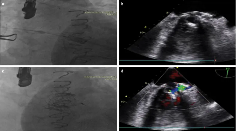
60 mm) of the septum to facilitate the crossing of the septum. We used an Agilis (Abbott Vascular) steerable sheath of 8.5 F to introduce a 0.035-inch an-gled hydrophilic wire across the ring with subsequent exchange for stiff wire. Thereafter, we positioned and slowly mounted the center of the 26-mm Sapien XT stent frame (mounted in the op-posite direction as performed for transfemoral transcatheter aortic valve implantation) within the ring under rapid pacing. Un-fortunately, we were not able to achieve the coaxial deployment of valve in the prior annuloplasty ring (Fig. 1a), thus complicating severe PVL (Fig. 1b) (Video 1). Therefore, we planned paravalvu-lar closure in the same session. However, during this procedure, more dehiscence of the THV from the ring carrying the risk for embolization occurred because of manipulations of catheter (Fig. 1c, 1d) (Video 2), which led to stopping of the procedure without closing the defect.The patient’s hemodynamic status was stable after the first procedure. Transapical TMVIV was scheduled after eight days. We implanted a second 26-mm Sapien XT valve within the previ-ous THV via a coaxial transapical access (Fig. 2a). Nevertheless,
persisting severe PVL was still detected even though kissing bal-loons (20 mm in diameter) were inflated within the valve. There-fore, we deployed 14-mm-long Vascular Plug II (St. Jude Medical, Minneapolis, Minnesota) (Fig. 2b, 2c) (Video 3) within the defect. TEE revealed a competent bioprosthetic valve without residual mitral regurgitation (Fig. 2d) (Video 4). The patient experienced significant symptomatic relief and functional improvement. She remained free from rehospitalization for six months.
Discussion
Technically, the TMVIR procedure is more challenging than the TMVIV implantation because of the larger diameters and geometric variability of the rings. Unlike the frame of the biopros-thesis, the rings have an oval shape to adapt to the configura-tion of the implanted valve, thereby having a higher risk of valve embolization and paravalvular regurgitation (4, 5). The risk of obliquity of the THV in the TMVIR procedures is more important than with bioprostheses because of the absence of frame. In our case, once the position of the THV was deemed adequate, we were not able to provide enough coaxiality under rapid ventricu-lar pacing. The native mitral valve leaflets in the failed ring might have also disrupted the anchoring of THV.
The transseptal route also has several advantages, such as less invasiveness and requirement of local anesthesia; however,
Figure 1. (a) Fluoroscopy of Sapien XT in the mitral bioprosthesis with a suboptimal position. (b) The TEE image showing severe PVL after the deployment of the THV; (c) More dehiscence of the THV after failed PVL closure, (d) Delivery system entrapped in the defect
a
c
b
Case Reports Anatol J Cardiol 2021; 25: 49-54
52
transapical access allows the TMVIV/TMVIR deployment to be performed coaxially while more simply and potentially reducing malpositioning and associated complications as shown in our case.
The current commercially available THVs could be an imper-fect match for the oval saddle-shaped geometry of the mitral an-nulus and rings, thereby leading to inherent PVL. The conversion to a transapical approach might provide a second THV implanta-tion to secure the first one as well as a more feasible closure of the PVL.
Conclusion
Saddle-shaped rings and transseptal approach might pre-clude the optimal placement of TMVIR. The currently available occluder and delivery systems are suboptimal and dedicated ones; therefore, a steerable system is needed for the prevention of malalignment. Transapical implantation enables a better con-trol over the implant position and facilitates defect closure.
Informed consent: Informed consent was signed and given by the patient’s parent.
Video 1. Fluoroscopy of the first procedure indicating cross-ing, malalignment, and displacement of the transcatheter valve during the attempts for paravalvular leak closure.
Video 2. TEE images of the first procedure, wherein central mitral insufficiency turned to severe paravalvular leak after the failure of coaxial placement of the transcatheter valve.
Video 3. Transapical route with persisting paravalvular leak despite kissing balloon inflations after the implantation of the second valve. The defect was closed with 14-mm AVP II.
Video 4. TEE images of the transapical approach that sealed the defect after the deployment of 14-mm AVP II.
References
1. Vohra HA, Whistance RN, Roubelakis A, Burton A, Barlow CW, Tsang GM, et al. Outcome after redo-mitral valve replacement in adult patients: a 10-year single-centre experience. Interact Cardio-vasc Thorac Surg 2012; 14: 575-9. [CrossRef]
2. Asami M, Pilgrim T, Windecker S, Praz F. Case report of simulta-neous transcatheter mitral valve-in-valve implantation and percu-taneous closure of two paravalvular leaks. Eur Heart J Case Rep 2019; 3: ytz123. [CrossRef]
3. Murdoch DJ, Sathananthan J, Cheung A, Webb JG. Combined Transapical Valve-in-Valve/Valve-in-Ring Transcatheter Mitral Valve Implantation and Paravalvular Leak Closure for Failed Mitral Valve Surgery. Can J Cardiol 2018; 34: 1088.e3-6. [CrossRef]
4. Kondo N, Shuto T, McGarvey JR, Koomalsingh KJ, Takebe M, Gor-man RC, et al. Melody valve-in-ring procedure for mitral valve re-placement: feasibility in four annuloplasty types. Ann Thorac Surg 2012; 93: 783-8. [CrossRef]
Figure 2. (a) Transapical route to secure the previous bioprosthetic valve. (b, c) Fluoroscopy and TEE images of 14-mm AVP II device deployed in the defect. (d) TEE confirmed no residual PVL after closure
a
c
b
Case Reports
Anatol J Cardiol 2021; 25: 49-54
53
5. Wunderlich NC, Kische S, Ince H, Bozdağ-Turan I. Transcatheter valve-in-ring implantation after a failed surgical mitral repair using a transseptal approach and a veno-arterial loop for valve place-ment. Catheter Cardiovasc Interv 2014; 84: 1202-8. [CrossRef]
Address for Correspondence: Dr. Beytullah Çakal, İstanbul Medipol Üniversitesi Tıp Fakültesi, Kardiyoloji Anabilim Dalı,
İstanbul-Türkiye Phone: +90 506 284 55 88 E-mail: [email protected]
©Copyright 2021 by Turkish Society of Cardiology - Available online at www.anatoljcardiol.com
DOI:10.14744/AnatolJCardiol.2020.59163
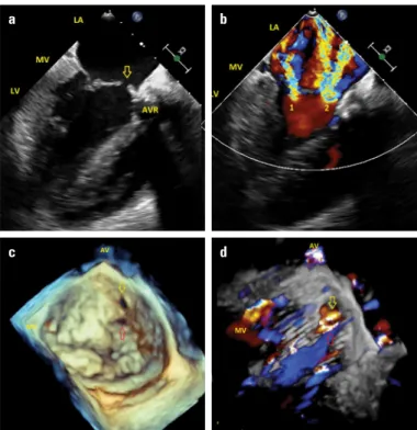
severe MR, together with mild central MR (Fig. 1a, 1b, Videos 1, 2). Three-dimensional (3D) TEE, showing MV from the enface view, revealed two different defects on the A2 scallop of the AML, with sizes of 6
×
3 and 7×
4 mm (Fig. 1c, Video 3), and 3D color Doppler drop-out artifacts were ruled out, and true de-fects were confirmed (Fig. 1d, Video 4). The patient had no his-tory of IE or fever, and all infective markers were within normal limits. Considering that MR developed just after surgery with multiple defects in the AML, iatrogenic perforation of the AML was concluded. Because the patient was asymptomatic with normal EF and ventricular size, conservative follow-up with medical therapy was recommended. However, dyspnea and heart failure symptoms developed after 3 years of follow-up. The patient was referred for MV surgery. Two defects in the AML were seen intraoperatively, as well as in the 3D images. MV replacement was performed because the repair was not successful. Postoperative TTE revealed normal-functioning mechanical prosthetic AV and MV with normal EF. The patient was discharged uneventfully.A rare cause of mitral regurgitation after
aortic valve replacement: Iatrogenic
mitral valve perforation
Begüm Uygur, Ömer Çelik, Anıl Şahin, Ali Rıza Demir, Mehmet Ertürk
Department of Cardiology, University of Health Sciences, İstanbul Mehmet Akif Ersoy Thoracic and Cardiovascular Surgery Training and Research Hospital; İstanbul-Turkey
Introduction
Mitral leaflet perforation is frequently associated with infec-tive endocarditis (IE). Iatrogenic mitral valve (MV) injury after aortic valve (AV) surgery is a well-known but extremely rare re-ported complication. Herein, we report the case of a 19-year-old male patient who underwent AV replacement (AVR) and devel-oped severe mitral regurgitation (MR) immediately postopera-tively.
Case Report
A 19-year-old male patient was admitted because of short-ness of breath and fatigue. He had no past medical history. Electrocardiogram showed sinus rhythm with a heart rate of 98 bpm. On auscultation, grade 5/6 systolic murmur was heard. Transthoracic echocardiography (TTE) revealed that the ejec-tion fracejec-tion (EF) was 60%, 76/42 mm Hg gradient on bicuspid AV with AV area of 0.8 cm2. Other valves were normal in
struc-ture, and mild MR was seen. The patient underwent AVR, but no intraoperative transesophageal echocardiography (TEE) was performed after implantation of the prosthetic valve. Dur-ing intensive care unit follow-up, new-onset severe MR was detected by TTE. TEE was performed to determine the etiology of MR. Two-dimensional (2D) images showed a 4-mm defect on the base of the anterior mitral leaflet (AML), which caused
a
c
b
d
Figure 1. (a) Transesophageal echocardiography, mid-esophageal long-axis view. The yellow arrow indicates the perforation in the anterior mitral leaflet. (b) Transesophageal echocardiography, mid-esophageal long-axis view with color Doppler imaging. 1 shows central mitral regurgitation (MR), 2 shows severe MR from the perforated area in the anterior mitral leaflet. (c) Three-dimensional transesophageal echocardiography image shows MV from surgical view. The yellow and red arrows indicate two different perforations in the anterior mitral leaflet. (d) Three-dimensional transesophageal echocardiography with color Doppler imaging. The yellow and red arrows indicate two different mitral regurgitations from different perforation sites in the anterior mitral leaflet
AVR - aortic valve replacement, LA - left atrium, LV - left ventricle; MV - mitral valve, AV - aortic valve