https://doi.org/10.1177/2515841419835731 https://doi.org/10.1177/2515841419835731 Ther Adv Ophthalmol
2019, Vol. 11 1 –7 DOI: 10.1177/ 2515841419835731 © The Author(s), 2019. Article reuse guidelines: sagepub.com/journals-permissions
Therapeutic Advances in Ophthalmology
journals.sagepub.com/home/oed 1
Introduction
Glaucoma is the most common cause of irreversible blindness worldwide. In 2020, more than 11 million individuals are expected to have bilateral blindness.1
Glaucoma is diagnosed by evaluating all factors such as intraocular pressure (IOP), cupping of the optic nerve head, corneal thickness, thinning of the retinal nerve fiber layer (RNFL), and changes in the visual field.2 Although IOP is not included in the
definition of glaucoma, increased IOP is the most important and the only risk factor that can be changed and slowed or stopped the progress of the disease when it is lowered.3
Therefore, one of the most important steps of the routine examination for early diagnosis is to meas-ure the IOP correctly in order to detect glaucoma patients, most of whom are asymptomatic. Since
Comparison of rebound tonometry and
non-contact airpuff tonometry to
Goldmann applanation tonometry
Goktug Demirci , Sevil Karaman Erdur, Cafer Tanriverdi, Gokhan Gulkilik and Mustafa Ozsutçu
Abstract
Purpose: The aim of this study was to compare the intraocular pressure measurements obtained from healthy subjects with the rebound tonometry, non-contact airpuff tonometry, and Goldmann applanation tonometry in different age groups.
Methods: A total of 180 eyes of 90 healthy subjects were included in the study. According to the subjects’ ages, the eyes were categorized into three groups: group 1 (age: 7–17 years), group 2 (age: 18–40 years), and group 3 (age: 41–75 years). Intraocular pressure was measured on each subject always in the same order: rebound tonometry, non-contact airpuff tonometry, and Goldmann applanation tonometry. Central corneal thickness values were obtained using ultrasonic pachymetry. One-way repeated-measures analysis of variance, Pearson’s correlation coefficient, and Bland–Altman analysis were used for the statistical assessment. Results: The mean corneal thickness was found to be 604 ± 13 µm, 546 ± 15 µm, and 547 ± 15 µm in group 1, group 2, and group 3, respectively. Non-contact airpuff tonometry was significantly higher than both Goldmann applanation tonometry and rebound tonometry measurements in all groups (p < 0.001, for all). No statistical difference between Goldmann applanation tonometry and rebound tonometry measurements was found in group 1 (p = 0.248), group 2 (p = 0.63), and group 3 (p = 0.126). There was a significant positive correlation in the meaning of intraocular pressure measurements between rebound tonometry and non-contact airpuff tonometry; non-contact airpuff tonometry and Goldmann applanation tonometry; and Goldmann applanation tonometry and rebound tonometry in all groups.
Conclusion: As a result, without need for topical anesthesia, fast measurement and ease-of-use rebound tonometry is a reliable alternative to Goldmann applanation tonometry in different age groups.
Keywords: central corneal thickness, glaucoma, Goldmann applanation, I-Care tonometry, rebound tonometry
Received: 29 May 2018; revised manuscript accepted: 8 February 2019.
Correspondence to: Goktug Demirci Department of Ophthalmology, Istanbul Medipol University, Istanbul, 34214, Turkey. [email protected] Goktug Demirci Sevil Karaman Erdur Cafer Tanriverdi Gokhan Gulkilik Mustafa Ozsutçu Department of Ophthalmology, Istanbul Medipol University, Istanbul, Turkey Original Research
the invention of the first tonometry according to the Goldmann Imbert-Fick principles, Goldmann applanation tonometry (GAT) has been accepted as the international gold standard measurement method.4 Non-contact airpuff tonometer (NCT)
is used in the routine patient examination, even though GAT is accepted as the gold standard. NCT was first designed by Zeiss and developed by Grolman in 1972. It measures the IOP without touching the corneal surface and therefore does not increase risk of infection and does not require anesthesia or fluorescein drop.5,6 The rebound
tonometer (RT; I-Care; Tiolat Oy, Helsinki, Finland) is a new player in the field of IOP meas-urement instruments. There is a magnetized probe that moves quickly toward the eye. A solenoid detects the acceleration and deceleration of the probe. Topical anesthesia is not required for measurements with RT, such as NCT, and the risk of spreading infection is considered negligible due to the use of disposable probes.7
The purpose of this study is to compare RT and NCT measurements with the reference method GAT in the eyes of different age groups including healthy subjects.
Methods
This protocol was carried out at Medipol University’s Vatan Health Practice Center follow-ing the 1964 Helsinki Declaration, followfollow-ing approval from the ethics committee of the Istanbul Medipol University (10840098-604.01.01-E.12581) and the approval of research participa-tion from eligible healthy subjects.
A total of 180 eyes of 90 healthy subjects were included in the study. Subjects with any history of ocular disease affecting IOP, astigmatism greater than 2 diopters, contact lens use (within the 2-week period prior to IOP measurement), previ-ous ocular surgery or trauma, ocular inflamma-tion, family history of glaucoma, classical glaucomatous optic disk changes as focal or dif-fuse thinning of the RNFL with consistent glauco-matous visual field defects were excluded from the study. Glaucomatous visual field defects were confirmed if two of the following three conditions were met: presence of a cluster of three points on a pattern deviation probability plot with p < 5%, one of which had p < 1%; a pattern standard devi-ation with p < 5%; or a glaucoma hemifield test result outside normal limits. To avoid the double-organ bias, the mean measurements of two eyes of
one subject were used for statistical analysis. According to the subjects’ ages, the eyes were cat-egorized into three groups: group 1 (age: 7–17 years), group 2 (age: 18–40 years), and group 3 (age: 41–75 years). Subjects below 18 years were accepted as pediatric group. From literature, we know that subjects older than 40 years are at risk of glaucoma that is why those were arranged as a separate group. The subjects aged between 18 and 39 years formed the other group.
Central corneal thickness measurements
Central corneal thickness (CCT) values were obtained by ultrasonic pachymeter (AccuPach VI; Keeler, Malvern, PA, USA) before IOP meas-urements. After one drop of 0.5% proparacaine hydrochloride eye drops (Alcaine®; Alcon
Laboratories Inc., Fort Worth, TX, USA), the pachymeter probe was placed on the center of the cornea, and the mean of three readings was calcu-lated for each eye.
IOP measurements
The IOP values were obtained by RT, NCT, and GAT, respectively. Each of the tonometers was cali-brated according to the manufacturer’s guidelines prior to its use. There was a 15-minute interval between measurements. IOP measurements were made by the same doctor (G.D). The RT (I-Care 100; Tiolat Oy) was positioned near the subject’s eye with the forehead being used as a base support. IOP measurements were taken with the tip of the probe maintained at a distance of approximately 3 to 7 mm from the center of the cornea, according to the manufacturer’s instructions. The mean of six consecutive sets of acceptable final measurements was used for subsequent analyses. The measure-ments with RT were made first in sitting position and after 15 minutes in dorsal decubitus. The NCT (Topcon CT-80; Topcon Corporation, Tokyo, Japan) automatically recorded three IOP readings, with their average per eye being recorded for the study. The GAT (Haag-Streit, Koeniz, Switzerland) values were recorded after three consecutive read-ings and a mean value was calculated for each eye observed. Before acquisition, one drop of 0.5% pro-paracaine hydrochloride eye drops (Alcaine®; Alcon
Laboratories Inc.) was instilled and a fluorescein strip (Fluorescein Sodium Ophthalmic Strip; Optitech Eyecare, Allahabad, India) was applied to the inferior conjunctival fornix. The last IOP measurement was obtained using GAT to avoid a corneal-compression-induced aqueous outflow
increase that would have affected subsequent IOP readings.
Statistical method
Statistical analysis was performed with Statistical Package for the Social Sciences (SPSS Inc., Chicago, IL, USA). The normality of the continu-ous variables was evaluated with the Shapiro–Wilk test. Intraclass correlation coefficient (ICC) test was done before taking average measurements of two eyes. Because ICC was close to 1, the average of measurements from two eyes of a subject was used. The differences between IOP readings were compared with the one-way repeated-measures
analysis of variance (ANOVA). The relationship among three devices in the meanings of IOP read-ings was evaluated by Pearson’s correlation coeffi-cient. Bland–Altman analysis was used to assess the clinical agreement of IOP measurements between the tonometers. Values of p lower than 0.05 were considered as statistically significant. Results
The normality of the continuous variables was evaluated with the Shapiro–Wilk test (Table 1). Demographic data of the all subjects are given in Table 2. When study groups were analyzed, the mean corneal thickness was found to be 604 ± 13 Table 1. Shapiro–Wilk test normality results of the continuous variables.
Statistics Degrees of freedom Significance
Group 1 Age 0.983 16 0.984 Pachymeter 0.935 16 0.295 IOP RT 0.940 16 0.355 NCT 0.954 16 0.552 GAT 0.946 16 0.427 Group 2 Age 0.911 20 0.066 Pachymeter 0.965 20 0.652 IOP RT 0.977 20 0.896 NCT 0.971 20 0.778 GAT 0.976 20 0.872 Group 3 Age 0.970 20 0.765 Pachymeter 0.966 20 0.669 IOP RT 0.962 20 0.579 NCT 0.898 20 0.071 GAT 0.950 20 0.375
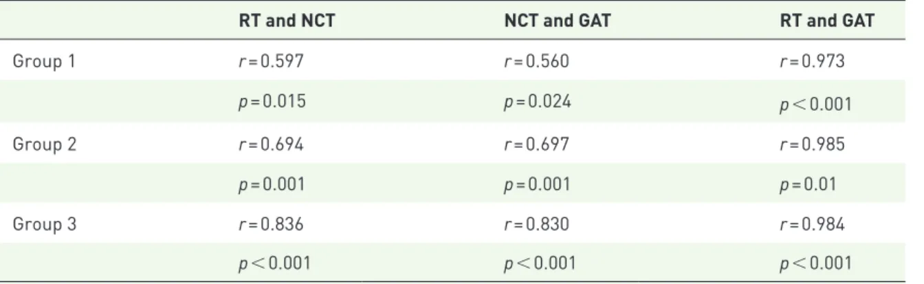
µm, 546 ± 15 µm, and 547 ± 15 µm in group 1, group 2, and group 3, respectively. The mean cor-neal thickness was significantly higher in group 1 compared with groups 2 and 3 (p < 0.001 for both). There was no significant difference in cor-neal thickness between groups 2 and 3 (p = 1.000). The NCT measurements were significantly higher than both GAT and RT measurements in all groups (p < 0.001, for all). No statistical difference between GAT and RT measurements was found in group 1 (p = 0.248), group 2 (p = 0.63), and group 3 (p = 0.126). Comparison of IOP measure-ments obtained by RT, NCT, and GAT among three groups is shown in Table 3. There was a sig-nificant positive correlation in the meaning of IOP measurements between RT and NCT; NCT and GAT; and GAT and RT in all groups (Table 4). The mean ± SD differences and 95% limits of agreement between RT, NCT, and GAT are shown in Table 5.
Discussion
Glaucoma is the most common cause of blindness after cataract in the world.8 Glaucoma treatment
is aimed at slowing or stopping the disease. Today, glaucoma drugs do this by lowering IOP. Therefore, the correct IOP measurement is one of the most important parameters of the eye exami-nation. The most precise measurement is to meas-ure with a manometer by placing a cannula in the anterior chamber, although not in clinical prac-tice.9 For this reason, GAT is accepted as the
international gold standard today.4 However, the
GAT is also affected by the examiner experience, CCT, corneal slope, and axial length.10,11 It is also
important to keep in mind the risk of spreading infection and using smooth and not cracked prism.12,13 In this study, GAT was used as
refer-ence measurement method.
The RT was discovered by Obbink 60 years ago.14
In principle, IOP was found by measuring impact time of the probe that strikes the cornea. The higher the IOP, the shorter the probe’s stroke time. There are various studies showing that they are affected or not affected according to CCT.15,16
The exact advantages are cheapness, easiness, convenience, portability, and fastness. The exact advantages to GAT are that it does not require an Table 2. Demographic data of the subjects.
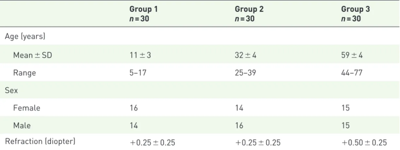
Group 1 n = 30 Group 2n = 30 Group 3n = 30 Age (years) Mean ± SD 11 ± 3 32 ± 4 59 ± 4 Range 5–17 25–39 44–77 Sex Female 16 14 15 Male 14 16 15 Refraction (diopter) +0.25 ± 0.25 +0.25 ± 0.25 +0.50 ± 0.25
Table 3. Comparison of intraocular pressure measurements obtained by RT, NCT, and GAT among three groups.
Devices Group 1
n = 30 (mmHg) Group 2n = 30 (mmHg) Group 3n = 30 (mmHg) p-value
a
RT 17.0 ± 0.4 16.9 ± 2.5 16.7 ± 2.1 0.879
NCT 21.6 ± 0.5 21.8 ± 2.2 21.3 ± 1.9 0.631
GAT 17.2 ± 0.4 17.2 ± 2.5 16.9 ± 2.2 0.878
RT: rebound tonometry; NCT: non-contact tonometry; GAT: Goldmann applanation tonometry.
experienced ophthalmologist, painless, no anes-thesia, and no risk of infection.17 Abraham and
colleagues7 showed that there is no difference
between the experienced and inexperienced tech-nicians of I-Care measurements. No side-effects were observed in any study. Some investigators have shown that the I-Care tonometry results in high CCT cases had high IOP readings.15,18,19
Fernandes and colleagues20 have shown that
I-Care tonometry in the normal population
produces an average of 1.34 mmHg higher results than GAT. I-Care measurements were found to be higher in the studies in which the results of the measurements taken from pediatric cases were compared with those in the GAT.21,22 In another
similar study, I-Care had a significantly lower measurements.23 Brusini and colleagues24 have
shown that the I-Care tonometry is a suitable measurement method for screening healthy indi-viduals. Kim and colleagues25 have shown that
Table 4. Correlation analysis of three devices in all groups.
RT and NCT NCT and GAT RT and GAT
Group 1 r = 0.597 r = 0.560 r = 0.973 p = 0.015 p = 0.024 p < 0.001 Group 2 r = 0.694 r = 0.697 r = 0.985 p = 0.001 p = 0.001 p = 0.01 Group 3 r = 0.836 r = 0.830 r = 0.984 p < 0.001 p < 0.001 p < 0.001
RT: rebound tonometry; NCT: non-contact tonometry; GAT: Goldmann applanation tonometry. Pearson’s correlation coefficient.
Table 5. Results of Bland–Altman analyses of the agreement between RT, NCT, and GAT.
Mean ± SD difference (mmHg) 95% LoA (mmHg)
Group 1 RT and NCT −4.6 ± 1.6 −1.5 to −7.7 NCT and GAT −4.5 ± 1.7 −1.2 to −7.8 GAT and RT 0.1 ± 0.4 0.8 to −0.6 Group 2 RT and NCT −4.9 ± 1.8 −1.4 to −8.4 NCT and GAT −4.7 ± 1.8 −1.2 to −8.2 GAT and RT 0.3 ± 0.4 1 to −0.4 Group 3 RT and NCT −4.6 ± 1.1 −2.5 to −6.7 NCT and GAT −4.4 ± 1.2 −2.1 to −6.7 GAT and RT 0.2 ± 0.4 0.9 to −0.5
I-Care measurements are independent of age, axial length, and CCT parameters. Khanal and colleagues26 found very similar results with I-Care
tonometry in different quadrants of cornea in their study. Thus, they explained that measure-ments of I-Care tonometry were not affected by corneal thickness.
However, Feng and colleagues27 showed that there
were significantly different results taken between NCT, RT, and GAT in a large series of pediatric patients. They observed the highest values with NCT and the lowest values with GAT. They also found that all types of tonometry measurements correlated with CCT. They showed that the CCT affected all types of tonometer measurements. When the whole pediatric group was examined, 93% of cases with NCT, 85% of patients with RT, and 77% of patients with GAT could be meas-ured. This study emphasized the necessity of NCT and RT to measure IOP due to compliance in the pediatric group. We found the highest values with NCT similar to this study but we found similar values in RT and GAT measurements.
Some studies have investigated the effects of topi-cal anesthetic drugs on corneal thickness and IOP measurements.28,29 Rosa and colleagues28 showed
that oxybuprocaine eye drops had no significant effect on corneal volume and CCT measure-ments. Lam and Chen29 also observed that there
was no significant effect on CCT measurements with proparacaine eye drops. For these reasons, we did not take into account the effect of local anesthetic drops on CCT in our study.
NCTs provide air application without touching the eye. After the beginning, air force increases until the cornea is flattened. Then, the tonometer trans-forms the power of applanation to IOP. There are measurement problems compared to similar cor-neal pathologies such as GAT.30 Ogbuehi31 showed
that NCT performed reliable and similar measure-ments with GAT. Tonnu and colleagues30 have
shown that NCT, Tono-Pen XL, and GAT meas-urements were influenced by CCT. Shields32 have
shown that the reliability of NCT in high IOP cases is reduced. A study, comparing RT and GAT, found mean IOP measurements 14.3 ± 3.9 mmHg and 11.7 ± 4.7 mm Hg using GAT and RT, respec-tively.33 Similar to our study, RT measurements
were lower than GAT measurements.
Limitations of this study are small sample size and lack of glaucomatous subjects.
Conclusion
The measurements from the three different age groups with RT were found to be consistent with GAT measurements. The NCT measurements were significantly higher than those obtained by GAT. It can be concluded that RT is a reliable alternative for screening healthy subjects for whom GAT is difficult to implement. However, it is necessary to take into consideration that RT measurements are slightly lower than GAT measurements.
Funding
The authors received no financial support for the research, authorship, and/or publication of this article.
Conflict of interest statement
The authors declared no potential conflicts of interest with respect to the research, authorship, and/or publication of this article.
ORCID iD
Goktug Demirci https://orcid.org/0000-0002 -5079-4713
References
1. Quigley HA. Number of people with glaucoma worldwide. Br J Ophthalmol 1996; 80: 389–393.
2. Kass MA, Heuer DK, Higginbotham EJ, et al. The ocular hypertension treatment study (OTHS). Arch Ophthalmol 2002; 120: 701–713. 3. Davanger M and Holter O. The statistical
distribution of intraocular pressure in the population. Acta Ophthalmol 1965; 43: 314–322. 4. Wessels IF and Oh Y. Tonometer utilization,
accuracy, and calibration under field conditions. Arch Ophthalmol 1990; 108: 1709–1712.
5. Moseley MJ. Non-contact tonometry. Ophthalmic Physiol Opt 1995; 15: 35–37.
6. Grolman B. A new tonometer system. Am J Optom Arch Am Acad Optom 1972; 49: 646–660. 7. Abraham LM, Epasinghe NC, Selva D, et al.
Comparison of the ICare with Goldmann applanation tonometry by experienced and inexperienced tonometrists. Eye 2008; 22: 503–506.
8. Kingman S. Glaucoma is second leading cause of blindness globally. Bull World Health Organ 2004; 82: 887–888.
9. Pahlitzsch M, Brunner J, Gonnermann J, et al. Comparison of ICare and IOPen vs Goldmann applanation tonometry according to international standards 8612 in glaucoma patients. Int J Ophthalmol 2016; 9: 1624–1628.
10. Whitacre MM and Stein R. Sources of error with use of Goldmann-type tonometers. Surv Ophthalmol 1993; 38: 1–30.
11. Kohlhaas M, Boehm AG, Spoerl E, et al. Effect of central corneal thickness, corneal curvature, and axial length on applanation tonometry. Arch Ophthalmol 2006; 124: 471–476.
12. Moses RA. The Goldmann applanation tonometer. Am J Ophthalmol 1958; 46: 865–869. 13. Brandt JD, Beiser JA, Kass MA, et al. Central
corneal thickness in the ocular hypertension treatment study (OHTS). Ophthalmology 2001; 108: 1779–1788.
14. Kniestedt C, Punjabi O, Lin S, et al. Tonometry through the ages. Surv Ophthalmol 2008; 53: 568–591.
15. Martinez-de-la-Casa JM, Garcia-Feijoo J, Saenz-Frances F, et al. Comparison of rebound tonometer and Goldmann handheld applanation tonometer in congenital glaucoma. J Glaucoma 2009; 18: 49–52.
16. Poostchi A, Mitchell R, Nicholas S, et al. The Icare rebound tonometer: comparisons with Goldmann tonometry, and influence of central corneal thickness. Clin Exp Ophthalmol 2009; 37: 687–691.
17. Cervino A. Rebound tonometry: new opportunities and limitations of non-invasive determination of intraocular pressure. Br J Ophthalmol 2006; 90: 1444–1446.
18. Sahin A, Basmak H and Yildirim N. The influence of central corneal thickness and corneal curvature on intraocular pressure measured by tono-pen and rebound tonometer in children. J Glaucoma 2008; 17: 57–61.
19. Tamcelik N, Atalay E, Cicik E, et al.
Comparability of Icare pro rebound tonometer with Goldmann applanation and noncontact tonometer in a wide range of intraocular pressure and central corneal thickness. Ophthalmic Res 2015; 54: 18–25.
20. Fernandes P, Diaz-Rey JA, Queiros A, et al. Comparison of the ICare rebound tonometer with the Goldmann tonometer in a normal population. Ophthalmic Physiol Opt 2005; 25: 436–440.
21. Cook JA, Botello AP, Elders A, et al. Surveillance of ocular hypertension study group. Systematic
review of the agreement of tonometers with Goldmann applanation tonometry. Ophthalmology 2012; 119: 1552–1557.
22. Chan WH, Lloyd IC, Symes RJ, et al. Accuracy of intraocular pressure measurement with the Icare tonometer in children. Asia Pac J Ophthalmol 2015; 4: 357–359.
23. Dabasia PL, Lawrenson JG and Murdoch IE. Evaluation of a new rebound tonometer for self-measurement of intraocular pressure. Br J Ophthalmol 2016; 100: 1139–1143.
24. Brusini P, Salvetat ML, Zeppieri M, et al. Comparison of ICare tonometer with Goldmann applanation tonometer in glaucoma patients. J Glaucoma 2006; 15: 213–217.
25. Kim KN, Jeoung JW, Park KH, et al.
Comparison of the new rebound tonometer with Goldmann applanation tonometer in a clinical setting. Acta Ophthalmol 2013; 91: e392–e396. 26. Khanal S, Walton M and Davey PG. Evaluation
of intraocular pressure estimates obtained using an iCare rebound tonometer. Clin Exp Optom 2017; 100: 179–183.
27. Feng CS, Jin KW, Yi K, et al. Comparison of intraocular pressure measurements obtained by rebound, noncontact, and Goldmann applanation tonometry in children. Am J Ophthalmol 2015; 160: 937–943.
28. Rosa N, De Bernardo M, Borrelli M, et al. of oxybuprocaine eye drops on corneal volume and thickness measurements. Optom Vis Sci 2011; 88: 640–644.
29. Lam AK and Chen D. Effect of proparacaine on central corneal thickness values: an evaluation using noncontact specular microscopy and pentacam. Cornea 2007; 26: 55–58. 30. Tonnu PA, Ho T, Newson T, et al. The
influence of central corneal thickness and age on intraocular pressure measured by pneumotonometry, non-contact tonometry, the Tono-Pen XL, and Goldmann applanation tonometry. Br J Ophthalmol 2005; 89: 851–854. 31. Ogbuehi KC. Assessment of the accuracy and
reliability of the Topcon CT80 non-contact tonometer. Clin Exp Optom 2006; 89: 310–314. 32. Shields MB. The non-contact tonometer. Its
value and limitations. Surv Ophthalmol 1980; 24: 211–219.
33. Brown L, Foulsham W, Pronin S, et al. The influence of corneal biomechanical properties on intraocular pressure measurements using a rebound self-tonometer. J Glaucoma 2018; 27: 511–518.
Visit SAGE journals online journals.sagepub.com/ home/oed