Early second trimester retinol-binding protein-4 values in
cases with or without gestational diabetes mellitus risk
factors: A cross-sectional study
Asli Yarci Gursoy
1, Gulsah Aynaoglu
2, Gamze Sinem Caglar
1and Feride Soylemez
3 1Faculty of Medicine, Department of Obstetrics and Gynecology, Ufuk University,2Numune Education and Research Hospital, and3Faculty of Medicine, Department of Obstetrics and Gynecology, Ankara University, Ankara, TurkeyAbstract
Aim: Retinol-binding protein-4 (RBP-4) has been correlated with different degrees of insulin resistance
includ-ing gestational diabetes mellitus (GDM). Presence of risk factors for GDM is an indication for early screeninclud-ing. We studied RBP-4 values in the early second trimester of pregnancy in pregnant subjects with or without GDM risk factors and compared the results by routine GDM screening methodology.
Methods: Seventy-nine patients with at least one GDM risk factor and 46 patients without any GDM risk
factors were enrolled in the cross-sectional study as risk and control groups, respectively. In the early second trimester, RBP-4 values were measured, in addition to fasting plasma glucose and 50-g glucose challenge test in all subjects.
Results: The RBP-4 values in 16–18th weeks of pregnancy were not significantly different between risk and
control groups (95.3± 20.1 vs 103.1 ± 24.4 μg/mL, respectively; P = 0.055) although fasting plasma glucose levels and 50-g glucose challenge test results were higher in the risk group than the control group (75.3 vs 69.3 mg/dL and 112.4 vs 97.5 mg/dL, respectively; P< 0.05).
Conclusion: Presence of GDM risk factors does not have an impact on early second trimester RBP-4 values in
pregnant subjects.
Key words: early second trimester, gestational diabetes mellitus, retinol-binding protein 4.
Introduction
Gestational diabetes mellitus (GDM) complicates 1.7– 11% of pregnancies1 and is still an important health
problem associated with fetal macrosomia, shoulder dystocia, stillbirth, hypocalcemia, hypoglycemia, hyperbilirubinemia, respiratory distress syndrome, and increased maternal and fetal diabetes mellitus risk. Identifying women with GDM is important to improve the outcomes. Although the criteria for screening and diagnosis of GDM is controversial and an international agreement is lacking, the American Diabetes
Associa-tion and the American College of Obstetrics and Gyne-cologists recommend routine screening for GDM in pregnancy.2–4
Early screening of all pregnant women will help to identify GDM cases that will lead to earlier interven-tions and may decrease associated morbidities. The association between different serum markers mea-sured early in pregnancy, in the first or early second trimester, and GDM were reported previously.5–7
Pla-cental diabetogenic hormones cause insulin resistance (IR) and hyperinsulinemia which predispose women to development of diabetes mellitus in pregnancy. In
Received: August 27 2013. Accepted: May 7 2014.
Reprint request to: Asli Yarci Gürsoy, Faculty of Medicine, Department of Obstetrics and Gynecology, Ufuk University, 86-88 Mevlana Bulvarı (Konya Yolu), Balgat, Ankara 06520, Turkey. Email: [email protected]
Conflict of Interests: The authors declare that no actual or potential conflict of interest in relation to this article exists.
uncomplicated pregnancies8 and pregnancies with
GDM,9Retinol-binding protein-4 (RBP-4) increases
sig-nificantly through gestation parallel to a decrease in insulin sensitivity.
Retinol-binding protein-4 is a 21-kDa polypeptide, an adipokine, secreted from hepatocytes and adipocytes. The main function of RBP-4 is to bind and transport vitamin A (retinol).10 Recent research
indi-cated that RBP-4 levels are correlated with IR.11,12
Although higher RBP-4 values have been reported in women with GDM, comparison of early second trimes-ter RBP-4 values with early second trimestrimes-ter 50-g glucose challenge test (GCT) results, in cases with and without GDM risk factors have not been reported yet. Other than pregnancy and GDM,13 RBP-4 has been
shown to be upregulated in various insulin-resistant states such as obesity, type 2 diabetes mellitus12 and
metabolic syndrome.14However, whether RBP-4 is the
promoter or the result of the IR or a molecule corre-lated with different parameters of metabolic syndrome is not clear yet.15 The elevated RBP-4 levels in type 2
diabetes mellitus was suggested as a secondary and predominantly non-genetic phenomenon. The authors reported that plasma RBP-4 plays a minor role in development of IR in humans.15
This study was designed to evaluate whether there is a difference in RBP-4 levels in the early second trimes-ter between pregnant women with and those without risk factors for GDM.
Methods
Study group
This is a cross-sectional study consisting of data from 125 pregnant subjects admitted to an outpatient clinic of department of obstetrics and gynecology of a uni-versity hospital between January 2007–January 2009 (n= 630). The recruitment of the patients to the study was stopped at the 18th month and the study ended when all the cases delivered. The patients admitted for the routine visit at 16–18 weeks of pregnancy were first evaluated (n= 410) for the study. Second, patients who were informed verbally about the planned study, agreed to participate and provided written consent were included in the study (n= 164). The 164 pregnant patients in their routine 16–18th week visit, were asked about the gestational diabetes risk factors (pre-pregnancy body mass index [BMI],>25; GDM history in previous pregnancy; type 2 diabetes mellitus in first-degree relatives; previously giving birth to a>4000-g infant; poor obstetric history such as missed abortus,
malformed infant, polyhydramnios, stillbirth or preterm birth).3,16Twenty of the patients (n= 10 in the
risk group and n= 10 in the control group; total, n = 20) who did not fulfill the inclusion criteria were excluded. As a result, 144 patients who gave written informed consent and fulfilled the inclusion criteria were eligible and were incorporated either in the risk group (n= 83) in the presence of any risk factors or control group (n= 61) in absence of any. Patients who did not attend their visits and who did not give birth in the same center were excluded (risk group, n= 4 vs control group, n= 15) leading to final data of 125 patients (risk group, n= 79 vs control group, n = 46) who were eli-gible for statistical analysis. Patients with any addi-tional medical conditions (e.g. hypertension, thyroid disorders, pre-existing diabetes mellitus) were excluded from the study. The study was approved by the local ethics committee and informed consent was taken from the participants.
Study design
In the same visit, fasting plasma glucose (FPG), 50-g GCT and RBP-4 values were measured in both groups of patients. In case of FPG of more than 95 mg/dL or 50-g GCT of more than 140 mg/dL, 100-g oral glucose tolerance test (OGTT) was performed with the refer-ence values of Carpenter and Coustan.16 At
24–28 weeks of pregnancy, GDM screening was per-formed in all patients. Similarly, FPG was measured and 50-g GCT was also performed in all of the subjects. In cases with the previously mentioned results above cut-off values, 100-g OGTT was performed. In case of a high 50-g GCT, the patient was put on a diet because abnormal 50-g GCT on its own has been associated with adverse pregnancy outcomes.17–19 Gestational
week at birth, route of delivery (vaginal or cesarean), sex and birthweight of newborn and 1- and 5-min Apgar scores were recorded.
Laboratory
Blood samples were taken into empty separator tubes and let to clot for 30 min at room temperature and centrifuged for 20 min at 1000 g. Serum samples were stored at−80°C until assayed for RBP-4 analysis. RBP-4 measurement was performed by Human RBP-4 Com-petitive ELISA Kit (Invitrogen, San Diego, CA, USA) and enzyme-linked immunosorbent assay (ELISA). The sensitivity was 1 ng/mL and intra- and inter-assay coefficients of variation were 2.6–9.2% and 3.4–10.2%,
respectively. All the blood samples were studied in duplicate according to the manufacturers’ instructions.
Statistical analyses
Data analysis was performed by using SPSS for Windows version 11.5 (SPSS, Chicago, IL, USA). Whether the distributions of continuous variables were normal or not was determined by Shapiro–Wilk test. Levene’s test was used for the evaluation of homoge-neity of variances. Data were shown as mean± stan-dard deviation or median (range), where applicable.
While the mean differences between control and risk groups were compared by using Student’s t-test; oth-erwise, the Mann–Whitney U-test was applied for com-parisons of the median values. Nominal data were analyzed by Pearson’sχ2-test. Degrees of associations
between continuous variables were determined by Spearman’s rank correlation coefficient analysis.
Whether RBP-4 measurements had a statistically sig-nificant effect on GDM risk or not was evaluated by multiple logistic regression analysis after adjustment for both age and BMI. Any variable whose univariate test had a P-value of less than 0.25 was accepted as a candidate for the multivariate model along with all variables of known clinical importance. Odds ratios and 95% confidence intervals for each independent variable were also calculated.
A P-value less than 0.05 was considered statistically significant.
Results
One hundred and twenty-five pregnant patients who completed antenatal follow-up and gave birth in the same hospital were included in the study. The risk group consisted of pregnant subjects with at least one GDM risk factor (n= 79) while subjects without any GDM risk factors constituted the control group
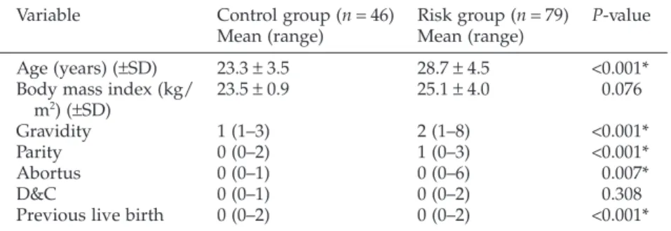
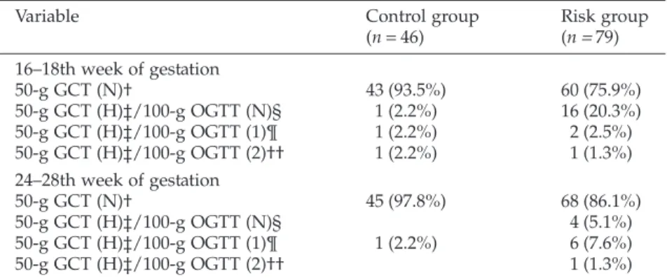
(n= 46). The demographic characteristics of partici-pants are given in Table 1. As expected; the mean age, gravidity, parity, abortus and previous live birth rates were statistically higher in the risk group (P< 0.05). The most common risk factor was maternal age over 25 years (n= 64, 81%) while first-degree relative and history of missed abortus were the following most fre-quent ones (Table 2). Distribution of FPG, 50-g GCT and 100-g OGTT results in risk and control groups are shown in Table 3.
Gestational week at birth was higher in the control group when compared to the risk group (39.4± 1.4 vs 38.6± 1.9 weeks, respectively; P = 0.033). Route of delivery (vaginal or cesarean), sex, birthweight and 1-and 5-min Apgar scores of the newborns did not differ significantly between the two groups (P> 0.05) (Table 4). As expected, 16–18th week FPG and 50-g GCT values were significantly higher in the risk group than the control group (75.3 vs 69.3 mg/dL and 112.4 vs 97.5 mg/dL, respectively; P< 0.05). But RBP-4 values did not differ significantly between the risk and control groups (95.3± 20.1 μg/mL vs
Table 1 Demographic variables of the risk and control groups
Variable Control group (n= 46) Risk group (n = 79) P-value
Mean (range) Mean (range)
Age (years) (±SD) 23.3± 3.5 28.7± 4.5 <0.001*
Body mass index (kg/
m2) (±SD) 23.5± 0.9 25.1± 4.0 0.076
Gravidity 1 (1–3) 2 (1–8) <0.001*
Parity 0 (0–2) 1 (0–3) <0.001*
Abortus 0 (0–1) 0 (0–6) 0.007*
D&C 0 (0–1) 0 (0–2) 0.308
Previous live birth 0 (0–2) 0 (0–2) <0.001*
*P< 0.05. D&C, dilatation and curettage; SD, standard deviation.
Table 2 Distribution of risk factors in risk group
Risk factor Risk group (n= 79)
n (%)
Previous gestational diabetes mellitus
1 (1.2%)
Maternal age (years) 64 (81.0%)
First-degree relative with diabetes mellitus 20 (25.3%) Previous birth of>4000 g 6 (7.6%) Missed abortus 19 (24%) Polyhydramnios history 2 (2.5%) Stillbirth history 2 (2.5%)
Preterm birth history 4 (5.0%)
Child with congenital abnormality 2 (2.5%)
103.1± 24.4 μg/mL respectively; P = 0.055). Also, RBP-4 levels in patients with more than one risk factors were not significantly different than in the control group (96.7± 22.6 μg/mL vs 93.3 ± 16.2 μg/mL;
P= 0.468). Additionally multiple logistic regression analysis revealed that RBP-4 values were not statisti-cally significantly different between the risk and control groups after adjustment for age (P= 0.354). In a similar manner, FPG and 50-g GCT values were statis-tically significantly higher in the risk group than in the control group in routine GDM screening weeks that are at 24–28th weeks of gestation (78.4 vs 68.6 mg/dL and
116.9 vs 97.3 mg/dL, respectively; P< 0.05). Student’s
t-test revealed that the only risk factor associated with
RBP-4 was maternal age. Maternal age over 25 years in the risk group was related with lower RBP-4 values (102± 24.5 vs 93 ± 22.0 μg/mL, respectively; P = 0.047). The double studied RBP-4 values were significantly similar and intraobserver agreement level was found as 0.897 (95% confidence interval, 0.856–0.926).
Spearman’s rank correlation coefficient analysis revealed that maternal age and fetal birthweight were not correlated with RBP-4 values as independent vari-ables (r= −0.11, P = 0.902; r = 0.095, P = 0.811,
respec-Table 3 Distribution of results of 50-g GCT and 100-g OGTT in control and risk groups
Variable Control group
(n= 46) Risk group(n= 79) 16–18th week of gestation 50-g GCT (N)† 43 (93.5%) 60 (75.9%) 50-g GCT (H)‡/100-g OGTT (N)§ 1 (2.2%) 16 (20.3%) 50-g GCT (H)‡/100-g OGTT (1)¶ 1 (2.2%) 2 (2.5%) 50-g GCT (H)‡/100-g OGTT (2)†† 1 (2.2%) 1 (1.3%) 24–28th week of gestation 50-g GCT (N)† 45 (97.8%) 68 (86.1%) 50-g GCT (H)‡/100-g OGTT (N)§ 4 (5.1%) 50-g GCT (H)‡/100-g OGTT (1)¶ 1 (2.2%) 6 (7.6%) 50-g GCT (H)‡/100-g OGTT (2)†† 1 (1.3%)
†50-g GCT result normal (<140 mg/dL). ‡50-g GCT result higher than normal. §100-g OGTT results are within normal range. ¶100-g OGTT with only one abnormal value (glucose intolerance). ††100-g OGTT with two abnormal values (e.g. gestational diabetes mellitus). GCT, glucose challenge test; H, high; N, normal; OGTT, oral glucose tolerance test.
Table 4 Characteristics and laboratory results of the risk and control groups
Variables Control group (n= 46) Risk group (n= 79) P-value
Week of birth 39.4± 1.4 38.6± 1.9 0.033* Route of delivery 0.113 Vaginal delivery 10 (23.8%) 29 (38.2%) Cesarean section 32 (76.2%) 47 (61.8%) Sex (neonate) 0.917 Female 19 (46.3%) 34 (45.3%) Male 22 (53.7%) 41 (54.7%) Birthweight (g) 3240 (2144–4029) 3280 (2000–4305) 0.546 1-min Apgar 8 (6–10) 8 (6–10) 0.963 5-min Apgar 10 (9–10) 10 (8–10) 0.782 16–18th week FPG (mg/dL) 69.3± 8.4 75.3± 14.6 0.006* 50-g GCT(mg/dL) 97.5± 26.6 112.4± 31.2 0.012* RBP-4 (μg/mL) 103.1± 24.4 95.3± 20.1 0.055 24–28th week FPG (mg/dL) 68.6± 8.8 78.4± 36.5 0.005* 50-g GCT (mg/dL) 97.3± 21.8 116.9± 42.9 <0.001*
*P< 0.05. FPG, fasting plasma glucose; GCT, glucose challenge test; RBP-4, retinol-binding protein-4.
tively). RBP-4 values were not significantly correlated with 16–18th week FPG (r= 0.029, P = 0.749) and 50-g GCT (r= −0.074, P = 0.413) levels. RBP-4 values in the 16–18th week were not significantly different in partici-pants with high and normal 50-g GCT (98.2± 17.4 vs 98.2± 23.0 mg/dL, respectively; P = 0.993). IR is expected to increase with progression of pregnancy, so a correlation analysis between the 16–18th week and 24–28th week results have not been performed.
Discussion
The present study revealed that, in the early second trimester of pregnancy, RBP-4 values do not differ between cases with or without GDM risk factors. The significantly higher FPG and 50-g GCT results, but similar RBP-4 values, in cases with GDM risk factors may be due to the impact of many factors that gradually change during pregnancy.
Yang et al. first demonstrated that transgenic overexpression of human RBP-4 or injection of recom-binant RBP-4 in normal mice caused IR.11 RBP-4 was
suggested as the responsible factor increasing the gluconeogenic enzyme phosphoenolpyruvate carboxykinase in hepatocytes. In addition, RBP-4 also inhibits insulin receptor activity by blocking the insulin-stimulated phosphorylation of insulin receptor substrate-1 at serine in position 307. These actions of RBP-4 have been proposed to result from IR.11,20,21
RBP-4 levels have been related with not only IR but also metabolic syndrome, type 2 diabetes mellitus, obesity and dyslipidemia.11,12,22
Pregnancy is a diabetogenic condition and RBP-4 values continue to rise during gestations of non-diabetic women, generally returning to normal range after delivery.8 On the contrary, Inoue et al. reported
that the RBP-4 values were higher in the first trimester and decrease through the second trimester and plateau thereafter in normal pregnancies.23Up to date, various
factors have been shown to affect RBP-4 values such as iron levels,24 renal function,25 lipid profile,22
transthyretin levels,26vitamin A levels27and
inflamma-tory status (RBP-4 is a negative acute phase reactant).28
Physiological changes such as increased iron require-ment and increased glomerular filtration rate in preg-nancy may also interfere with RBP-4 levels. Neither in our study nor in the previous studies has any adjust-ment of RBP-4 been found when the above-adjust-mentioned parameters were performed. This also makes it hard to reach a conclusion about RBP-4 levels in pregnancy.
Presence of previously mentioned risk factors for GDM are proposed to promote IR to some degree. Therefore, early GDM screening is mandatory in cases with risk factors. Considering this, in this study, RBP-4 levels were analyzed in the early second trimester rather than at routine screening weeks (24–28th weeks) in pregnant patients with GDM risk factors. According to the data from previous published work, RBP-4 values were supposed to be in the higher in risk group. However, even if the FPG and 50-g GCT values were higher in the GDM risk group, the results showed that RBP-4 levels did not differ significantly in cases with or without risk factors (95.3± 20.1 vs 103.1 ± 24.4 μg/mL;
P= 0.55). Also no significant difference was found in RBP-4 values that had one or more risk factors (96.7± 22.6 vs 93.3 ± 16.2 μg/mL; P = 0.468).
In the published work, there are many studies in favor of a positive correlation between RBP-4 and IR,11,29,30 while others support the absence of a
correlation31–34 or a negative correlation as well.35–37
Abetew et al. reported that there is modest evidence for a positive association of early pregnancy elevated RBP-4 concentrations with increased GDM risk, par-ticularly among women aged more than 35 years.38In
our study, the number of participants aged more than 35 years were too few (n= 9) to perform statistical analyses. Additionally, although there was not a direct correlation between age and RBP-4 (r= −0.011,
P= 0.902), RBP-4 values were significantly lower in participants aged 25 years or older compared with those of less than 25 years (93.4± 18.8 vs 102.2± 24.5 μg/mL, respectively; P = 0.047). On the other hand, others have shown a positive correlation between age and RBP-4 values.15,29The results in our
study may be due to the effect of the sample size and more accurate conclusions may be reached in further large population-based studies.
Numerous studies reported higher RBP-4 values in pregnant subjects complicated with GDM.13,39 Others
proposed a negative correlation37or denied existence of
any correlation.40RBP-4 values in a pregnant
popula-tion who have different IR states were studied at 24–28 weeks of pregnancy with ELISA and also con-firmed by western blot, resulting in no significant difference in RBP-4 values in GDM, glucose intolerance and high 50-g GCT but normal 100-g OGTT subgroups (36.0± 10.4 vs 35.6 ± 10.9 vs 34.6 ± 6.7 μg/mL, respec-tively; P= 0.69).41 In spite of all technical issues,
Maghbooli et al. suggested that RBP-4 value over 42μg/mL determined by ELISA at 24–28 weeks of pregnancy may predict future GDM.42 Very recently,
Abetew et al. reported that RBP-4 values in the 16th week of pregnancy are associated with increased GDM risk only among women particularly over 35 years of age.38 Before a universally accepted laboratory
tech-nique is available for determining RBP-4 levels in preg-nancy, all the previous data in the published work needs to be validated.
Retinol-binding protein-4 values in the published work are measured by different techniques such as ELISA, competitive enzyme immunoassay and western blot. Graham et al. reported that the most reli-able results were availreli-able with western blot tech-nique.27Howevre, ELISA has been the most frequently
preferred technique to measure RBP-4 levels.43In the
published work, the wide range of reported RBP-4 values measured by ELISA is quite remarkable. In pregnancy, the previously reported RBP-4 values ranged from 0.0169± 0.005 ng/mL to 41.14 ± 21.29 μg/ mL.9,38 Therefore, it is hard to form any conclusions
about RBP-4 levels in pregnant subjects.
The present study was designed as a cross-sectional study but because secondary outcomes such as routine second trimester GDM screening results, route of birth and fetal birthweights were also included, the subjects who did not attend routine visits and did not give birth in the same hospital were excluded in the statistical analysis due to incomplete data. As a result, the sample size of the study was limited. Therefore, this study can be evaluated as a preliminary work for further research. In order to clarify any existence of an association between RBP-4 levels with IR in pregnancy, adjustment for all the possible variables related to pregnancy should be performed. The controversies over the asso-ciation between RBP-4 and IR may be resolved with confirmation of the previous correlations – either posi-tive or negaposi-tive – with the most reliable available labo-ratory technique for RBP-4 measurement currently being western blot.
Disclosure
The authors have no relevant financial or non-financial relationships to disclose.
References
1. Schneider S, Bock C, Wetzel M, Maul H, Loerbroks A. The prevalence of gestational diabetes in advanced economies. J
Perinat Med 2012; 40: 511–520.
2. American Diabetes Association. Diagnosis and classification of diabetes mellitus. Diabetes Care 2006; 29: 43–48.
3. American College of Obstetricians and Gynecologists. Clini-cal management guidelines for obstetrician-gynecologists; Gestational diabetes. ACOG Practice Bulletin 30. Obstet
Gynecol 2001; 98: 525–538.
4. Metzger BE, Gabbe SG, Persson B et al. International Associa-tion of Diabetes and Pregnancy Study Groups Consensus Panel, International association of diabetes and pregnancy study groups recommendations on the diagnosis and classi-fication of hyperglycemia in pregnancy. Diabetes Care 2010;
33: 676–682.
5. Thadhani R, Wolf M, Hsu-Blatman K, Sandler L, Nathan D, Ecker JL. First trimester sex hormone binding globulin and subsequent gestational diabetes mellitus. Am J Obstet Gynecol 2003; 189: 171–176.
6. Wolf M, Sandler L, Hsu K, Vossen-Smirnakis K, Ecker JL, Thadhani R. First trimester C-reactive protein and subsequent gestational diabetes. Diabetes Care 2003; 26: 819– 824.
7. Smirnakis KV, Martinez A, Blatman KH, Wolf M, Ecker JL, Thadhani R. Early pregnancy insulin resistance and subse-quent gestational diabetes mellitus. Diabetes Care 2005; 28: 1207–1208.
8. Ueland T, Dalsoren T, Voldner N, Godang K, Henriksen T, Bollerslev J. Retinol-binding protein-4 is not strongly associ-ated with insulin sensitivity in normal pregnancies. Eur J
Endocrinol 2008; 159: 49–54.
9. Klein K, Bancher-Todesca D, Leipold H et al. Retinol-binding protein 4 in patients with gestational diabetes mellitus.
J Womens Health (Larchmt) 2010; 19: 517–521.
10. Blaner WS. Retinol-binding protein: the serum transport protein for vitamin A. Endocr Rev 1989; 10: 308–316. 11. Yang Q, Graham TE, Mody N et al. Serum retinol binding
protein 4 contributes to insulin resistance in obesity and type 2 diabetes. Nature 2005; 436: 356–362.
12. Graham TE, Yang Q, Blüher M et al. Retinol-binding protein 4 and insulin resistance in lean, obese, and diabetic subjects. N
Engl J Med 2006; 354: 2552–2563.
13. Lewandowski KC, Stojanovic N, Bienkiewicz M et al. Elevated concentrations of retinol-binding protein-4 (RBP-4) in gesta-tional diabetes mellitus: Negative correlation with soluble vascular cell adhesion molecule-1 (sVCAM-1). Gynecol
Endocrinol 2008; 24: 300–305.
14. Qi Q, Yu Z, Ye X et al. Elevated retinol-binding protein 4 levels are associated with metabolic Syndrome in Chinese people. J
Clin Endocrinol Metab 2007; 92: 4827–4834.
15. Ribel-Madsen R, Friedrichsen M, Vaag A, Poulsen P. Retinol-binding protein 4 in twins: Regulatory mechanisms and impact of circulating and tissue expression levels on insulin secretion and action. Diabetes 2009; 58: 54–60.
16. Metzger BE, Buchanan TA, Coustan DR et al. Summary and recommendations of the Fifth International Workshop-Conference on Gestational Diabetes Mellitus. Diabetes Care 2007; 2: 251–260.
17. Melamed N, Hiersch L, Hod M, Chen R, Wiznitzer A, Yogev Y. Is abnormal 50-g glucose-challenge testing an independentpredictor of adverse pregnancy outcome? J
Matern Fetal Neonatal Med 2012; 25: 2583–2587.
18. HAPO Study Cooperative Research Group, Metzger BE, Lowe LP, Dyer AR et al. Hyperglycemia and adverse pregnancy outcomes. N Engl J Med 2008; 358: 1991– 2002.
19. Retnakaran R, Qi Y, Sermer M, Connelly PW, Zinman B, Hanley AJ. Isolated hyperglycemia at 1-hour on oral glucose tolerance test in pregnancy resembles gestational diabetes in predicting postpartum metabolic dysfunction. Diabetes Care 2008; 31: 1275–1281.
20. Polonsky KS. Retinol-binding protein 4, insulin resistance, and type 2 diabetes. N Engl J Med 2006; 354: 2596– 2598.
21. Ost A, Danielsson A, Lide’n M, Eriksson U, Nystrom FH, Strålfors P. Retinol-binding protein-4 attenuates insulin-induced phosphorylation of IRS1 and ERK1/2 in primary human adipocytes. FASEB J 2007; 21: 3696–3704.
22. Mostafaie N, Sebesta C, Zehetmayer S et al. Circulating retinol-binding protein 4 and metabolic syndrome in the elderly. Wien Med Wochenschr 2011; 161: 505–510.
23. Inoue S, Takamoto N, Akahori Y et al. Elevated levelof serum retinol-binding protein 4 in pregnancy-induced hyperten-sion. J Obstet Gynaecol Res 2009; 35: 293–300.
24. Fernández-Real JM, Moreno JM, Ricart W. Circulating retinol-binding protein-4 concentration might reflect insulin resistance-associated iron overload. Diabetes 2008; 57: 1918– 1925.
25. Ziegelmeier M, Bachmann A, Seeger J et al. Serum levels of adipokine retinol-binding protein-4 in relation to renal function. Diabetes Care 2007; 30: 2588–2592.
26. Refai E, Dekki N, Yang SN et al. Transthyretin constitutes a functional component in pancreatic beta-cell stimulus-secretion coupling. Proc Natl Acad Sci USA 2005; 102: 17020– 17025.
27. Graham TE, Wason CJ, Blüher M, Kahn BB. Shortcomings in methodology complicate measurements of serum retinol binding protein (RBP4) in insulin-resistant human subjects.
Diabetologia 2007; 50: 814–823.
28. Fleck A. Clinical and nutritional aspects of changes in acute-phase proteins during inflammation. Proc Nutr Soc 1989; 48: 347–354.
29. Cho YM, Youn BS, Lee H et al. Plasma retinol-binding protein-4 concentrations are elevated in human subjects with impaired glucose tolerance and type 2 diabetes. Diabetes Care 2006; 29: 2457–2461.
30. Jia W, Wu H, Bao Y et al. Association of serum retinol-binding protein 4 and visceral adiposity in Chinese subjects with and without type 2 diabetes. J Clin Endocrinol Metab 2007; 92: 3224–3229.
31. Erikstrup C, Mortensen OH, Nielsen AR et al. RBP-to-retinol ratio, but not total RBP, is elevated in patients with type 2 diabetes. Diabetes Obes Metab 2009; 11: 204–212.
32. Von Eynatten M, Lepper PM, Liu D et al. Retinol-binding protein 4 is associated with components of the metabolic syndrome, but not with insulin resistance, in men with type 2 diabetes or coronary artery disease. Diabetologia 2007; 50: 1930–1937.
33. Lewis JG, Shand BI, Elder PA, Scott RS. Plasma retinol-binding protein is unlikely to be a useful marker of insulin resistance. Diabetes Res Clin Pract 2008; 80: 13–15.
34. Saucedo R, Zarate A, Basurto L et al. Relationship between circulating adipokines and insulin resistance during preg-nancy and postpartum in women with gestational diabetes.
Arch Med Res 2011; 42: 318–323.
35. Janke J, Engeli S, Boschmann M et al. Retinol binding protein 4 in human obesity. Diabetes 2006; 55: 2805–2810.
36. Broch M, Vendrell J, Ricart W, Richart C, Fernandez-Real JM. Circulating retinol-binding protein-4, insulin sensitivity, insulin secretion, and insulin disposition index in obese and nonobese subjects. Diabetes Care 2007; 30: 1802–1806. 37. Krzyzanowska K, Zemany L, Krugluger W et al. Serum
con-centrations of retinol-binding protein 4 in women with and without gestational diabetes. Diabetologia 2008; 51: 1115–1122. 38. Abetew DF, Qiu C, Fida NG et al. Association of retinol binding protein 4 with risk of gestational diabetes. Diabetes
Res Clin Pract 2013; 99: 48–53.
39. Su YX, Hong J, Yan Q et al. Increased serum retinol-binding protein-4 levels in pregnant women with and without gesta-tional diabetes mellitus. Diabetes Metab 2010; 36: 470–475. 40. Tepper BJ, Kim YK, Shete V, Shabrova E, Quadro L. Serum
retinol-binding protein 4 (RBP4) and retinol in a cohort of borderline obese women with and without gestational diabe-tes. Clin Biochem 2010; 43: 320–323.
41. Khovidhunkit W, Pruksakorn P, Plengpanich W, Tharavanij T. Retinol-binding protein 4 is not associated with insulin resistance in pregnancy. Metabolism 2012; 61: 65–69. 42. Maghbooli Z, Hossein-Nezhad A, Mirzaei K et al. Association
between retinol-binding protein 4 concentrations and gesta-tional diabetes mellitus and risk of developing metabolic syn-drome after pregnancy. Reprod Sci 2010; 17: 196–201. 43. Christou GA, Tselepis AD, Kiortsis DN. The metabolic role of
retinol binding protein 4: An update. Horm Metab Res 2012;