S
ystemic lupus erythematosus (SLE) is a clinically diverse and poten-tially life-threatening chronic inflammatory autoimmune disease that can affect almost any organ system, and has high morbidity if un-treated.1Recently, in patients with this condition there has been an increasein life expectancy and quality of life with an estimated 5-year survival rate of 82%-90% of patients due to the development of modern treatments.1The
clinical features of SLE develop through auto-antibody formation, immune
Atypical Multiple Cutaneous Ulcers in
A Patient with Systemic Lupus Erythematosus
Treated with Rituximab: Case Report
AABBSS TTRRAACCTT Intravenous rituximab therapy is an effective treatment alternative for systemic lupus erythematosus patients for whom standard therapies have not been effective. We report on a 20-year-old male with intractable systemic lupus erythematosus presenting with cutaneous ulcers that occurred two weeks after rituximab therapy. There were multiple painless ulcers on the patient’s abdomen, inguinal and gluteal regions. The histopathological examination revealed ulceration com-prising an inflammatory granulation tissue rich in polymorphonuclear leukocytes and eosinophils. The patient was prescribed 16 mg/day methylprednisolone for 3 weeks. One week later, he pre-sented with multiple dyshidrotic vesicular lesions on the palmar surface of his hand and vesicular-ulcerative lesions on the distal pulp of his right thumb. After the discontinuation of the rituximab therapy, the ulcers partially improved using wound dressings. Cutaneous ulcers are very rarely seen in patients with lupus erythematosus and only few cases have been reported. Cutaneous ulceration due to the rituximab treatment has not yet been reported.
KKeeyy WWoorrddss:: Lupus erythematosus, systemic; rituximab; skin ulcer
Ö
ÖZZEETT İntravenöz rituksimab tedavisi standart tedavilerin etkili olmadığı sistemik lupus eritema-tozuslu hastalarda etkili bir tedavi alternatifidir. Rituksimab tedavisinden hemen iki hafta sonra kutanöz ülserler ile prezente olan, tedaviye yanıtsız sistemik lupus eritematozuslu yirmi yaşında erkek hasta sunuldu. Hastanın karın, kasık ve gluteal bölgelerinde çok sayıda ağrısız ülserler vardı. Histopatolojik incelemede polimorfonükleer lökositler ve eozinofillerden zengin inflamatuar gra-nülasyon dokusu ve ülserasyon görüldü. Hastaya 16 mg/gün metilprednizolon tedavisi verildi. Bir hafta sonra, hasta elinin palmar yüzeyinde çok sayıda dishidrotik veziküler lezyonlar ve sağ el baş parmağının distal pulpasında vezikülo-ülseratif lezyonlar ile başvurdu. Rituksimab tedavisinin ke-silmesinin ardından ülserler yara örtüleri ile kısmen iyileşti. Lupus eritematozuslu hastalarda ku-tanöz ülserler oldukça nadir görülmektedir ve sadece birkaç olgu rapor edilmiştir. Rituksimab tedavisine bağlı kutanöz ülserasyon şimdiye kadar bildirilmemiştir.
AAnnaahh ttaarr KKee llii mmee lleerr:: Lupus eritematozus, sistemik; rituksimab; deri ülseri
TTuurrkkiiyyee KKlliinniikklleerrii JJ DDeerrmmaattooll 22001166;;2266((11))::3322--77 Pelin ÜSTÜNER,a
Mustafa ÖZDEMİR,a
Ali BALEVİa
aDepartment of Dermatology,
İstanbul Medipol University Faculty of Medicine, İstanbul Ge liş Ta ri hi/Re ce i ved: 09.02.2015 Ka bul Ta ri hi/Ac cep ted: 11.06.2015 Ya zış ma Ad re si/Cor res pon den ce: Pelin ÜSTÜNER
İstanbul Medipol University Faculty of Medicine,
Department of Dermatology, İstanbul, TÜRKİYE/TURKEY
doi: 10.5336/dermato.2015-44114
complex deposition and cytokine activation.2
Al-though much is still unknown regarding the patho-genesis of SLE, B-cell abnormalities are considered to be central.3High relapse rates and toxicity
asso-ciated with conventional treatments point to the need for more focused approaches towards this very heterogeneous disease. In previous clinical studies, it was suggested that the B-cell depleting agent rituximab (RTX) could be a potent SLE treat-ment.3However, two randomized controlled trials
failed to meet the efficacy endpoints.4,5In addition,
cardiac, gastrointestinal, and heterogeneous neu-rological adverse effects have been reported as a re-sult of the RTX treatment.6Nevertheless, RTX has
continued to be used as an off-label alternative mainly in patients refractory to conventional im-munosuppressive treatments.3
CASE REPORT
A 20-year-old male was referred to our clinic pre-senting with multiple painless ulcers that had de-veloped on his abdomen and on his inguinal and gluteal regions two weeks earlier. The SLE diagno-sis was made based on the clinical and laboratory criteria developed by the American College of Rheumatology (ACR) in 2010. Despite receiving the recommended doses according to the protocols, the patient had refractory to the conventional SLE treatments; such as hydroxychloroquine, high dose steroids, cyclophosphamide, azathioprine, and my-cophenolate mofetil. His current medication con-sisted of 4 mg/day oral methylprednisolone. He reported no previous trauma, friction, sexual con-tact or concon-tact with chemical or erosive materials. He had suffered from the symptoms of Raynaud’s disease for two years, with bilateral symmetric pol-yarteritis and arthralgia on his fingers otherwise his medical history was otherwise unremarkable. He had received intravenous RTX (MabThera®) 375 mg/m2/week in another clinic. The treatment
scheme was based on the protocols used in patients with rheumatoid arthritis (RA) consisting of an ini-tial dose of 1 g and 1 g after two weeks. Our patient had received two doses of intravenous RTX with no subsequent dose. His CD20+ cell counts were not known.
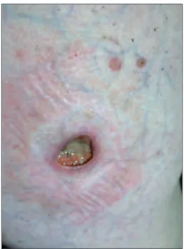
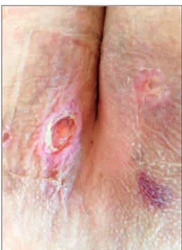
The dermatological examination revealed a deep ulcer of 2x2 cm covered by a serous-purulent exudate on the right lower quadrant of his abdomen (Figure 1a), a punched-out, painless deep ulcer of 0.5-1 cm on his right inguinal area and multiple ul-cers on his medial bilateral gluteal areas (Figure 1b). There were also small superficial erythematosus patches surrounding these deep ulcers. The contrac-tures of his finger joints were noteworthy.
FIGURE 1a: A deep ulcer of 2x2 cm covered with a serous-purulent exudate
on the right lower quadrant of the abdomen.
(See color figure at http://www.turkiyeklinikleri.com/journal/dermatoloji-dergisi/1300-0330/)
FIGURE 1b: Multiple ulcers on the medial bilateral gluteal areas.
The overall result of the laboratory examina-tion was unremarkable except for the leukopenia, chronic anaemia and ANA positivity in 1/32 titres. Anti-phospholipids (aPL) antibodies, cytoplas-mic anti-neutrophil cytoplascytoplas-mic antibodies (c-ANCA), perinuclear anti-neutrophil cytoplasmic antibodies (p-ANCA) and anti-ds DNA were neg-ative. The VDRL test was negneg-ative. Serum Im-munoglobulin (Ig) M and G levels were normal. One week after the first visit, the patient returned to the clinic due to the occurrence of multiple dyshidrotic vesicular lesions on the palmar surface of his hands (Figure 1c) and vesicular--ulcerative le-sions on the distal pulp of his right thumb (Figure 1d). We also noted a recent millimetric pinpoint ulcer with an erythematosus sharp border on the dorsum of his right hand. The differential diagno-sis included possible cutaneous tuberculodiagno-sis, ec-thyma gangrenosum, cutaneous vasculitis, aPL antibody syndrome, pyoderma gangrenosum and drug induced ulcers. The patient’s written in-formed consent was obtained for publication. The histopathological examination of the punch biopsy material taken from his abdominal ulcer revealed
ulceration, consisting of an inflammatory granu-lation tissue rich in polymorphic eosinophilic leukocytes (H-E x200) (Figure 2). The direct im-munofluorescence test was negative. The poly-merase chain reaction (PCR) examinations of the wound scrubs for cytomegalovirus (CMV), DNA and herpes simplex virus (HSV) Type I and Type II DNA were all negative. CMV Ig M, Ig G and HSV Type I and II Ig M and G were also negative.
After the discontinuation of the RTX therapy, the methylprednisolone dose was increased to 16 mg/day. Prontosan Wound Gel X®, a sterile, highly
viscous hydrogel, was prescribed to treat the deep ulcer on the patient’s abdomen and Ag Granuflex®
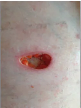
hydrocolloid wound dressings were also prescribed for the remaining ulcers. One month later, the ul-cers had partially healed (Figure 3); therefore, the methylprednisolone dose was gradually reduced to 4 mg/day. After six months of follow-up, he had no further cutaneous ulcers. The patient was called for a follow-up after a year; unfortunately we were in-formed that he had died in another hospital fol-lowing gastrointestinal bleeding.
FIGURE 1c: Multiple dyshidrotic vesicular lesions on the palmar surface of the
hands.
(See color figure at http://www.turkiyeklinikleri.com/journal/dermatoloji-dergisi/1300-0330/)
FIGURE 1d: Vesicular-ulcerative lesions on the distal pulp of the right thumb.
DISCUSSION
The causes of cutaneous ulcers seen in SLE are vari-able. Leg ulceration is a rare skin manifestation af-fecting approximately 5.6% of SLE patients and it is mostly associated with aPL antibodies or leukocy-toclastic vasculitis.6 Therefore, it has been
sug-gested that aPL antibodies can play a pathogenetic
role in ulcerated skin manifestations of aPL-posi-tive SLE patients. However, in the current case the aPL antibodies were negative. SLE has also been found to be associated with pyoderma gangreno-sum (PG) in more than 16 reported cases. It has also been suggested that a predisposition to vascular thrombosis or inflammation in SLE can have a role in PG pathogenesis.7In another study, the
preva-lence of avascular necrosis was found to be 6% in an SLE study group.8 Intractable ulceration has
been reported in a patient with lupus erythemato-sus profundus (LEP) that had been successfully treated with cyclosporine.9 Fat necrosis was
re-ported in 20 of 29 LEP cases, 3 thrombosis in cases with histologically severe lupus panniculitis, and vasculitis in some cases of LEP.9,10
Rituximab, also known as a TNF-α inhibitor, is an anti-neoplastic, immunomodulatory and bio-logic drug that has been approved and is most commonly used in intractable adult RA, psoriatic arthritis, systemic vasculitis anti-neutrophil cyto-plasmic antibodies (ANCA), and positive and
non-Hodgkin lymphoma.2,7 RTX is an anti-CD20
human-murine monoclonal chimeric antibody that causes the selective short-term depletion of ma-tured B-cells through three mechanisms; induction of apoptosis (programmed cell death), comple-ment-dependent cytotoxicity and anti-body de-pendent cytotoxicity.2,11,12
To date, over 540,000 patients worldwide, have received RTX; with only a small minority re-porting serious adverse reactions, and for the great majority of patients, RTX has been reported to be safe and well-tolerated.13However, it has also been
suggested that RTX can only be recommended for organ-specific manifestations of SLE, such as arthritis and autoimmune thrombocytopenic pur-pura and thrombocytopenia.14-16The incidence of
serious infections has been estimated to be 6.6/100-12.6/100 patient-years. The most common side ef-fects associated with RTX include; mild infusion reactions (35%), neutropenia and infections (10%).2,17Certain cutaneous adverse effects of RTX,
such as easy bruising, or bleeding, petechial lesions, painful oral ulcers, redness, blistering, scaly, itchy, or peeling skin have been reported 24 hours after
FIGURE 2: Ulceration, inflammatory granulation tissue rich in
polymor-phonuclear leukocytes and eosinophils (H-E x200).
(See color figure at http://www.turkiyeklinikleri.com/journal/dermatoloji-dergisi/1300-0330/)
FIGURE 3: The partially healing ulcer one month after therapy..
the infusion.4,18,19These adverse effects can persist
for a few weeks or months even if RTX is discon-tinued. So far cutaneous deep ulcers due to RTX treatment have not been reported. Even though RTX has been successfully used in the treatment of cutaneous ulcerations in diseases, such as refractory cases with LEP, polyarteritis nodosa, polymyosi-tis/dermatomyositis and ANCA-associated vasculi-tis, the etiological association of rituximab with these ulcers is still unclear and controversial.20-22
Therefore, we suggest that these atypical ulcers could be a rare mysterious clinical sign of SLE. We think that the cutaneous ulcers reported in the pa-tient in the current paper may also have occurred as a result of necrobiotic changes in the subcuta-neous tissue caused by vascular changes.
In this case, the diagnosis was made according to the clinical setting, the absence of an alternative diagnosis and the partial clinical improvement of the lesion after the initiation of the specific treat-ment. An ulcer biopsy was also performed to elim-inate other conditions presenting with cutaneous ulcers, as follows; malignancy, infections such as sporotrichosis, cutaneous tuberculosis, ecthyma gangrenosum, syphilitic ulceration, amebiasis, HSV Type 2 virus, CMV infection, cutaneous vas-culitis, aPL antibody syndrome, avascular necrosis or PG.7Vasculitic diseases such as granulomatosis
with polyangiitis, polyarteritis nodosa, mixed cryo-globulinemia, Takayasu’s arteritis, leukocytoclas-tic vasculitis, livedoid vasculopathy. Since the histopathological examination revealed no nuclear atypia or abnormal mitosis, cutaneous malignan-cies such as squamous cell carcinoma were elimi-nated. “Lupus profundus is a form of lobular panniculitis characterized by subcutaneous nodules and plaques particularly appearing on the face, trunk and upper proximal extremities.23Due to the
absence of typical histopathological findings of lupus profundus such as hyaline necrosis in the lob-ules of subcutaneous fat tissue, lobular panniculitis accompanied by lymphoplasmocytic infiltration the diagnosis of lupus profundus was quickly elim-inated in this case.23Besides the negative results
ob-tained from the direct immunofluorescence and lupus band tests also excluded lupus profundus.” Furthermore, in our patient the viral markers were all negative, which made it possible to distinguish between the ulcers and herpes according to their clinical features and laboratory results. The ulcers seen in this case also had no symptoms of infection, such as pain, erythema or a local increase in tem-perature. Moreover, tissue cultures were all nega-tive. The negativity of VDRL, aPL antibody, c-ANCA and p-ANCA tests excluded syphilitic ul-cerations and other possible vasculitic diseases from the differential diagnosis. The gastrointestinal bleeding that led to the death of the patient in this case might have been due to either vasculitis that is commonly seen in SLE pathophysiology or the thrombosis of the mesenteric arteries.
In conclusion, RTX has been reported to be an effective treatment alternative for the induction and maintenance of remission in patients with SLE refractory to conventional treatments, particularly in terms of the disease activity, immunologic pa-rameters and steroid-sparing effect. However, we suggest that more high-quality and long-term stud-ies are needed to determine the effect of re-treat-ment and long-term efficacy and safety of the RTX treatment and reveal the exact cause of atypical ul-cerations. We chose to present this case since it is rarely seen. For further research, RTX treatment or other biologic agents should be investigated in SLE patients presenting with cutaneous atypical ulcers without any known etiology.
1. Bonilla-Abadía F, Coronel Restrepo N, Tobón GJ, Echeverri AF, Muñoz-Buitrón E, Castro AM, et al. Rituximab for remission induction and maintenance in refractory systemic lupus erythematosus. Autoimmune Dis 2014;2014: 731806.
2. Kamal A, Khamashta M. The efficacy of novel B cell biologics as the future of SLE treatment: a review. Autoimmun Rev 2014;13(11):1094-101.
3. Ekö SL, van Vollenhoven RF. Rituximab and lupus--a promising pair? Curr Rheumatol Rep 2014;16(9):444.
4. Merrill JT, Neuwelt CM, Wallace DJ, Shana-han JC, Latinis KM, Oates JC, et al. Efficacy and safety of rituximab in moderately-to-severely active systemic lupus erythematosus: the randomized, double-blind, phase II/III sys-temic lupus erythematosus evaluation of rit-uximab trial. Arthritis Rheum 2010;62(1): 222-33.
5. Rovin BH, Furie R, Latinis K, Looney RJ, Fer-venza FC, Sanchez-Guerrero J, et al; LUNAR Investigator Group. Efficacy and safety of rit-uximab in patients with active proliferative lupus nephritis: the Lupus Nephritis Assess-ment with Rituximab Study. Arthritis Rheum 2012;64(4):1215-26.
6. Díaz-Lagares C, Pérez-Alvarez R, García-Hernández FJ, Ayala-Gutiérrez MM, Callejas JL, Martínez-Berriotxoa A, et al; BIOGEAS Study Group. Rates of, and risk factors for, severe infections in patients with systemic autoimmune diseases receiving biological agents off-label. Arthritis Res Ther 2011; 13(4):R112.
7. González-Moreno J, Ruíz-Ruigomez M, Calle-jas Rubio J, Ríos Fernández R, Ortego Cen-teno N. Pyoderma gangrenosum and systemic
lupus erythematosus: a report of five cases and review of the literature. Lupus 2015; 24(2):130-7.
8. Sayarlioglu M, Yuzbasioglu N, Inanc M, Ka-mali S, Cefle A, Karaman O, et al. Risk fac-tors for avascular bone necrosis in patients with systemic lupus erythematosus. Rheuma-tol Int 2012;32(1):177-82.
9. Ishiguro N, Iwasaki T, Kawashima M, Kawakami M. Intractable ulceration in a pa-tient with lupus erythematosus profundus suc-cessfully treated with cyclosporine. Int J Dermatol 2012;51(9):1131-3.
10. Park HS, Choi JW, Kim BK, Cho KH. Lupus erythematosus panniculitis: clinicopathologi-cal, immunophenotypic, and molecular stud-ies. Am J Dermatopathol 2010;32(1):24-30. 11. Bezalel S, Asher I, Elbirt D, Sthoeger ZM.
Novel biological treatments for systemic lupus erythematosus: current and future modalities. Isr Med Assoc J 2012;14(8):508-14. 12. Chan VS, Tsang HH, Tam RC, Lu L, Lau CS.
B-cell-targeted therapies in systemic lupus erythematosus. Cell Mol Immunol 2013;10(2): 133-42.
13. Rajadhyaksha AG, Mehra S, Nadkar MY. Bi-ologics in SLE: the current status. J Assoc Physicians India 2013;61(4):262-7. 14. Kimby E. Tolerability and safety of rituximab
(MabThera). Cancer Treat Rev 2005;31(6): 456-73.
15. Dale RC, Brilot F, Duffy LV, Twilt M, Waldman AT, Narula S, et al. Utility and safety of ritux-imab in pediatric autoimmune and inflamma-tory CNS disease. Neurology 2014;83(2): 142-50.
16. Cobo-Ibáñez T, Loza-Santamaría E, Pego-Reigosa JM, Marqués AO, Rúa-Figueroa I,
Fernández-Nebro A, et al. Efficacy and safety of rituximab in the treatment of non-renal sys-temic lupus erythematosus: a systematic re-view. Semin Arthritis Rheum 2014;44(2): 175-85.
17. Murdaca G, Colombo BM, Puppo F. Emerg-ing biological drugs: a new therapeutic ap-proach for Systemic Lupus Erythematosus. An update upon efficacy and adverse events. Au-toimmun Rev 2011;11(1):56-60.
18. Chen H, Zheng W, Su J, Xu D, Wang Q, Leng X, et al. Low-dose rituximab therapy for re-fractory thrombocytopenia in patients with sys-temic lupus erythematosus-a prospective pilot study. Rheumatology (Oxford) 2011;50(9): 1640-4.
19. Albert D, Dunham J, Khan S, Stansberry J, Kolasinski S, Tsai D, et al. Variability in the bi-ological response to anti-CD20 B cell deple-tion in systemic lupus erythematosus. Ann Rheum Dis 2008;67(12):1724-31.
20. Espírito Santo J, Gomes MF, Gomes MJ, Peixoto L, C Pereira S, Acabado A, et al. In-travenous immunoglobulin in lupus panniculi-tis. Clin Rev Allergy Immunol 2010;38(2-3): 307-18.
21. McArdle A, Baker JF. A case of “refractory” lupus erythematosus profundus responsive to rituximab [case report]. Clin Rheumatol 2009; 28(6):745-6.
22. Unger L, Kampf S, Lüthke K, Aringer M. Rit-uximab therapy in patients with refractory der-matomyositis or polymyositis: differential effects in a real-life population. Rheumatology (Oxford) 2014;53(9):1630-8.
23. Bolognia JL, Jorizzo JL, Schaffer JV. Der-matology. 3rded. Philadelphia: Elsevier
Saun-ders; 2012. p.1653.