e485 Transcanalicular revision surgery for failed dacryocystorhinostomy
Transcanalicular revision surgery for failed
dacryocystorhino-stomy
M. Ozsutcu1, A. Yenigun2, A. Meric2, B. Ozucer2, R. Dogan3, G. Gulkilik1, M.S. Kocabora1
1Department of Ophthalmology, Medipol University, Faculty of Medicine, Bagcilar/Istanbul, Turkey; 2Department of Otolaryngology,
Bezmialem University, Faculty of Medicine, Fatih/Istanbul, Turkey; 3Department of Otolaryngology, Sisli Etfal Research and
Educa-tion Hospital, Sisli/Instanbul, Turkey
Original article
Clin Ter 2013; 164 (6):e485-488. doi: 10.7417/CT.2013.1641Correspondence: Mehmet Selim Kocabora. Medipol Mega Hospital, Faculty of Medicine, Ophthalmology Department, Bagcilar, Istanbul, Turkey. E-mail: [email protected]
Copyright © Società Editrice Universo (SEU)
ISSN 1972-600
Introduction
Dacryocystorhinostomy (DCR) is a surgical operation performed to treat recurrent dacryostenosis and epiphora in which an anastomosis is created between the lacrimal sac and the nasal cavity (1). External DCR is a frequently performed surgical procedure first described by Toti in 1904 (2). DCR is carried out by external, endonasal (END-DCR) or transcanalicular approach; endonasal approach was first described by Caldwell in 1893, followed by its popularity after 1990’s (3, 4).
Abstract
Aim. To evaluate the effectiveness of transcanalicular multi diode laser in revision dacryocystorhinostomy and to determine the outcomes.
Materials and Methods. Fifty-three consecutive patients (19 males, 44 females) who were referred for recurrent dacryostenosis with epiphora after a failed primary dacryocystorhinostomy (DCR) operation were included in the study. Patients were assigned to two separate groups on the basis of primary DCR operation: either endo-nasal dacryocystorhinostomy (END-DCR) or transcanalicular multi diode laser dacryocystorhinostomy (TC-MDL DCR). TC-MDL DCR technique was used for revision surgeries in both groups. The mean age of the patients was 46.5 ± 13.1 (Range, 15 -71) and the average follow-up duration was 12 months.
Results. The success rate was 62% (18/29) in the primarily TC-MDL DCR operated group, and 85.2% (29/34) in the primarily END-DCR operated group respectively. The occlusion of internal ostium with granulation tissue was the leading etiology of unsucces-sful surgical outcome in both group 1 and group 2 (42.8% and 28.5% respectively).
Conclusions. TC-MDL DCR is a minimally invasive surgical method with relatively high success and low complication rates in patients with failed primary DCR requiring revision. Clin Ter 2013; 164(6):e485-488. doi: 10.7417/CT.2013.1641
Key words: epiphora, diode laser, recurrent dacryostenosis,
revi-sion dacryocystorhinostomy
The success rate of the External DCR is over 90%. Re-maining 10% of patients require revision for rhinostomy. These cases can be operated with either external-endonasal or transcanalicular diode-laser (TC-MDL DCR) approaches (5-9). Although rhinostomy formed by diode laser is smaller compared to the rhinostomy formed endonasally (10, 11), it is laborious to locate the obstructed segment with the endonasal approach; furthermore, there is always the risk of inflicting damage to orbital fatty tissue (12, 13).
The aim of the present study was to compare the results of revision TC-MDL DCR cases that underwent DCR with either transcanalicular MDL or endonasal approach.
Materials and Methods
The present study was conducted in accordance to Helsinki Declaration (WMA 2010). All procedures were carried out under the informed consent with the approval of University Ethics Committee and in accordance with the guidelines of the National Health and Medical Research. All volunteers were provided with adequate information and written informed consents were taken in advance.
Study design
Sixty-three consecutive patients (19 males, 44 females) who underwent primary DCR operation and referred with epiphora and recurrent dacryocystitis during postoperative follow-up visits were included in the study. Patients were assigned to two separate groups on the basis of primarily DCR operation: either endonasal or TC-MDL DCR group. TC-MDL DCR technique was used for revision surgeries for both groups. Patients required other nasal procedures such as septoplasty, other ancillary sinus procedures (anterior ethmoidectomy and turbinectomy), and decompartmenta-lized synechia were excluded from the study. Postoperative evaluation was counted into the examination of the lids and adnexa, lacrimal probing, saline irrigation, and dacryo-cystography: nasolacrimal duct patency was documented for each patient.
e486 M. Ozsutcu et al.
Subjects were grouped considering the primary surgical approach followed in a prospective fashion; group 1 (n=29) (mean age=50.8 ± 17.1) comprised of patients who previou-sly underwent DCR through TC-MDL approach and group 2 (n=34) (mean age=44.0 ± 9.7) comprised of patients who previously underwent DCR with endonasal approach. In both groups nasal endoscopy was performed on all patients to evaluate intranasal pathologies; recurrence of epiphora, mucoid efflux, and unsuccessful lavage of nasolacrimal duct was evaluated as unsuccessful surgical outcome. A standar-dized surgical technique was used in all cases:
Operative technique
The patients (n=29) underwent TC-MDL DCR under local anesthesia. Topical anesthetic eye drop containing 0.4% oxybuprocain hydrochloride was applied. Anterior ethmoi-dal nerve was infiltrated with 2% lidocaine anesthesia and nasal mucosa was infiltrated with 1:100.000 epinephrine so-lution containing 2% lidocaine. Nasal packing encompassing 4% lidocaine and 1:100.000 epinephrine was intranasally invoked. Multidiode DCR laser (Intermedic, Barcelona,
Spain) instrument was configured 600-630 nm wavelength; pulse and pause were both configured to 500 ms, power was adjusted to 3 Watts. Lacrimal probe with 0.64 mm diameter was inserted from the superior punctum directed medially for the localization of the obstructed segment. Quartz cable of the laser instrument was inserted following adequate di-lation with the probe and advanced to the nasal cavity. The operational site was observed for a pearl like the tissue in the mucosa. Laser was observed and localized endoscopically and applied until probe recognition with the nasal endoscope. Furthermore, laser probe was inserted from the inferior pun-ctum and advanced to the inferior canaliculus, the passage of the probe from the newly formed rhinostomy was observed. Failure of the passage was an indication for laser application inserted from the inferior canaliculus. Passage of the probe from both of the canaliculi was ascertained. Diameter of the rhinostomy was extended to 15-20 millimeters endonasally. Lacrimal system was profusely irrigated with saline solution and a silicone lacrimal stent was placed to secure patency. All patients were discharged from the hospital on the same day of the surgery.
Post-operative follow up and evaluation
Topical antibiotics and corticosteroid eye drop was pre-scribed four times a day for the first post-operative week. Follow-up periods were as follows: during the first week, in the first month, in the third, sixth and twelfth months after surgery. Silicone stent was removed in the third post-operative month. On the follow up visits, endoscopical examination of the nasal cavity and other evaluations were performed. Mucosal healing was assessed and lacrimal pa-tency was evaluated with nasolacrimal irrigation under nasal endoscopy. The evaluation criteria for successful surgical outcome were as follows: patent nasolacrimal duct system and total absence of initial symptoms for 3 months following postoperative removal of the silicone tube.
Statistical analysis
Data analysis was carried out using SPSS version 17.0 software system (SPSS, Inc., Chicago, IL, USA). Results
were calculated as mean ± standard deviation (SD); Student’s t test was used for comparison of quantitative data and Fi-sher’s exact test was used for comparison of qualitative data. A p value ≤0.05 was considered as statistically significant.
Results
Sixty-three patients underwent revision DCR surgery either with TC-MDL or endonasal surgical approach. The mean age of the patients was 46.5 ± 13.1 (Range: 15-71). No major intra-and post-operative complications such as orbital hematoma, exposure of orbital fatty tissue and diplopia oc-cured. There was no significant difference between the two groups in terms of demographic Characteristics such as age (p=0.068) and gender (p=0.133).
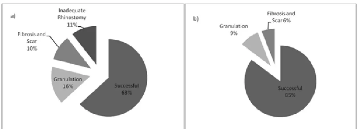
Duration of follow-up was 12 months. Saline irrigation during twelfth postoperative month follow-up visit of group 1 revealed patent nasolacrimal duct in 62% (18/29) patients; whereas 85.2% of patients (29/34) in group 2 had saline drainage. This difference in success rates was not statistically significant (p=0.09). Unsuccessful surgical outcome was further analyzed; occlusion of internal ostium with granu-lation tissue was the leading etiology in both group 1 and group 2 (42.8% and 28.5% respectively) followed by fibrotic scar formation. Two cases primarily treated with TC-MDL DCR in group 1 had unsuccessful surgical outcomes due to inadequate removal of bony tissue; surgical outcome results are schematized Figure 1.
Discussion
Laser DCR is a minimally invasive surgical procedure completed in 20 minutes. It does not leave surgical marks and is well tolerated with local anesthesia. These advantages make laser DCR a preferential approach especially in old patients unable to tolerate general anesthesia (1, 14, 15). Success rates of laser DCR varies from 59% to 100% in different studies (6).
One of the most important reasons of failure in Endo-nasal DCR is Endo-nasal synechia which results from formation of granulation tissue and fibrosis in the surgical site. Septal deviation, concha bullosa, inferior turbinate hyperplasia and hyperpneumatized Agger nasi cells increase this possibility (13). New bone formation following DCR is either non-existent or very limited at the site of rhinostomy; therefore, there is no need for removal of bony structure (16). The main reason of failure in primary DCR surgery is mainly due to aberrant soft tissue regeneration at the surgical site; therefore, revision DCR with endonasal or transcanalicular approaches are applicable methods.
Short-term failure of Laser DCR is associated with dif-ficulty of forming a rhinostomy in lateral nasal bony wall, formation of fibrosis in laser applied region and inflammatory fibrosis causing obstruction in the region of the rhinostomy (17). Wrong localization of the lacrimal sac, granulation,
e487 Transcanalicular revision surgery for failed dacryocystorhinostomy
regrowth of bony tissue and bony spicules in the ostium are other reasons of failure (18). Rhinostomy can be widened and advanced to the appropriate location with endonasal approach. Diode laser enables an effective tissue dissection with minimal tissue damage. Mucosal incisions performed with diode laser also reduces intraoperative bleeding; thereby, providing perfect intraoperative endoscopic view (19).
The success rate of END-DCR has been reported to be between 75% and 94% (20-25). In a study conducted by Zilelioglu et al., 34 primary and 30 revision END-DCRs were performed, and the success rates were reported as 79.4% and 80%, respectively (20). Leong et al demonstra-ted that END-DCR performed as a revision procedure was associated with a better success rate (89%) compared to the primary treatment (85%) (21). Kominek et al. found a total success rate of 84.0 % in revision END-DCR cases (26). Our success rates with revision diode laser DCR following primary END-DCR are similar to these success rates. In the current study, success rate of primarily TC-MDL DCR operated group was 63% and primarily END-DCR operated group was 85% (Fig. 1).
Although the surgical technique is the same in revision surgeries, the different rates of failure is presumably due to previously formed inadequate bony window in TC-MDL DCR group. Higher rates of failure due to granulation, fibrosis and scar formation in the primarily TC-MDL DCR operated group can also be attributed to higher tendency for obstruction due to inadequately enlarged bony windows. This presumption is concomitant with a currently published study, in which patients operated with diode laser DCR were compared to patients operated with enlargement of the neo-ostium via drilling in addition to diode laser DCR; success rates of two groups were 71% and 93% respecti-vely. These dramatic differences in success rates show the importance of widening the bony window in diode laser DCR technique (27).
In conclusion, in our study, subjects with failed primary DCR operation either with endonasal or TC-MDL approach underwent a revision DCR with TC-MDL which is a mini-mally invasive surgical method. Our results showed that it is a suitable method and can be preferred as the treatment of choice in revision DCR patients, especially in patients previously operated with endonasal approach.
References
1. Sadiq SA, Ohrlich S, Jones NS, et al. Endonasal laser dac-ryocystorhinostomy medium term results. Br J Ophthalmol 1997; 81:1089-92 (Grade C)
2. Toti A. Nuovo metodo conservatore di cura radicale delle suppurazioni croniche del sacco lacrimale (dacriocistorino-stomia). Clin Mod 1904; 10:385-7
3. Caldwell GW. Two new operations for obstruction of the nasal duct, with preservation of the canaliculi. Am J Ophthalmol 1893; 10:189-92
4. Gonnering RS, Lyon DB, Fisher JC. Endoscopic laser-assisted lacrimal surgery. Am J Ophthalmol 1991; 111:152-7 5. Tarbet KJ, Custer PL. External dacryocystorhinostomy.
Surgical success, patient satisfaction, and economic cost. Ophthalmol 1995; 102:1065-70
6. Woog JJ, Kennedy RH, Custer PL, et al. Endonasal dacr-yocystorhinostomy: a report by the American Academy of Ophthalmology. Ophthalmol 2001; 108:2369-77
7. Hong JE, Hatton MP, Leib ML, et al. Endocanalicular laser dacryocystorhinostomy: analysis of 118 consecutive surge-ries. Ophthalmology 2005; 112:1629-33
8. Metson R. The endoscopic approach for revision dacryo-cystorhinostomy. Laryngoscope 1990; 100:1344-7
9. Orcutt JC, Hillel A, Weymuller EA. Endoscopic repair of failed dacryocystorhinostomy. Ophthal Plast Reconstr Surg 1990; 6:197-202
10. Rosen N, Barak A, Rosner M. Transcanalicular laser-assisted dacryocystorhinostomy. Ophthalmic Surg Lasers 1997; 28: 723-6
11. Pearlman SJ, Michalos P, Leib ML, et al. Translacrimal tran-snasal laser-assisted dacryocystorhinostomy. Laryngoscope 1997; 107:1362-5
12. Yung MW, Hardman-Lea S. Analysis of the results of surgical endoscopic dacryocystorhinostomy: effect of the level of obstruction. Br J Ophthalmol 2002; 86:792-4
13. Korkut AY, Teker AM, Yazici MZ, et al. Surgical outcomes of primary and revision endoscopic dacryocystorhinostomy. J Craniofac Surg 2010; 21(6):1706-8
14. Alanon FFJ, Alanon FMA, Martinez FA, et al. Transcana-licular dacryocystorhinostomy technique using diode laser. Arch Soc Esp Oftalmol 2004; 79:325-30
15. Drnovsek-Olup B, Beltram M.Transcanalicular diode laser-assisted dacryocystorhinostomy. Indian J Ophthalmol 2010; 58(3):213-7
Fig. 1. Success rates and etiology of failure in Group 1 and Group 2: a) Group 1 (patients previously operated with TC-MDL DCR approach); b) Group 2 (patients previously operated with END-DCR approach).
e488 M. Ozsutcu et al.
16. Korkut AY, Teker AM, Ozsutcu M, et al. A comparison of endonasal with external dacryocystorhinostomy in revision cases. Eur Arch Otorhinolaryngol 2011; 268:377-81 17. Woo KI, Moon SH, Kim YD. Transcanalicular laser-assisted
revision of failed dacryocystorhinostomy. Ophthalmic Surg Lasers 1998; 29:451-5
18. Mirza S, Al-Barmani A, Douglas SA, et al. A retrospective comparison of endonasal KTP laser dacryocystorhinostomy versus external dacryocystorhinostomy. Clin Otolaryngol 2002; 27:347-51
19. Onerci M. Dacryocystorhinostomy. Diagnosis and treatment of nasolacimal canal obstructions. Rhinology 2002; 40:49-65
20. Zilelioglu G, Tekeli O, Ugurba SH, et al. Results of endo-scopic endonasal non-laser dacryocystorhinostomy. Doc Ophthalmol 2002; 105:57-62
21. Leong SC, Karkos PD, Burgess P, et al. A comparison of outcomes between nonlaser endoscopic endonasal and ex-ternal dacryocystorhinostomy: single-center experience and a review of British trends. Am J Otolaryngol 2010; 31:32-7
22. El-Guindy A, Dorgham A, Ghoraba M. Endoscopic revi-sion surgery for recurrent epiphora occurring after external dacryocystorhinostomy. Ann Otol Rhinol Laryngol 2000; 109:425-30
23. Tsirbas A, Davis G, Wormald PJ. Revision dacryocystorhino-stomy: a comparison of endoscopic and external techniques. Am J Rhinol 2005; 19:322-5
24. Agarwal S. Endoscopic dacryocystorhinostomy for acquired nasolacrimal duct obstruction. J Laryngol Otol 2009; 123: 1226-8
25. Metson R. Endoscopic surgery for lacrimal obstruction. Otolaryngol Head Neck Surg 1991; 104:473-9
26. Kominek P, Cervenka S, Pniak T, et al. Revison endonasal dacryocystorhinostomies: analysis of 44 procedures. Rhino-logy 2011; 49(3):375-80
27. Basmak H, Caklı H, Sahin A, et al. Comparison of endo-canalicular laser dacryocystorhinostomy with and without endonasal procedures.Graefes Arch Clin Exp Ophthalmol 2011; 249(5):737-43. Epub 2010 Nov 4