The present study aimed to review the general characteristics of 18 cases diagnosed with transient osteoporosis of the hip (TOH) in our hospital within a 3-year period and to present their follow-up results after conservative treatment. A retrospective evalua-tion was made of the treatment and results of follow-up of TOH cases using physical examination and laboratory findings, hip radiographs and magnetic resonance imaging (MRI) and Harris Hip Scores (HHS). The mean duration of complaints of 6 females (meanage,34.3±4.3years)and12males(meanage, 40.7±10.5years)was6.1±2.7weeksbeforethetreat-ment. Three female patients had a history of giving birth by cesarean delivery. None of the patients had any history of trauma. MRI revealed increased inten-sity in T2 sequences and decreased inteninten-sity in T1 sequencesintheproximalaspectofthefemur.None of the patients had subchondral collapse or intra- articular effusion. For 3 female patients who were breastfeeding,nomedicaltherapywasgiven,butonly hyperbaric oxygen (HBO) therapy and forearm crutches. As standard management, the other pa-tients were prevented from weight-bearing with the use of forearm crutches and medical therapy of di-clofenacsodium,acetylsalicylicacid,andrisedronate sodium was administered and additional HBO thera-py. Clinical and radiological improvements were ob-served in all patients. None of the patients had avas-cular necrosis (AVN) of the femoral head. There was no record of therapy-related complications. While HHSwas55.6±7.8beforethetreatment,itincreased to88.8±5.8inthe3rdmonthandto96.0±1.8inthe 6th month after the treatment. This change in score overtimewasfoundtobesignificant.
Keywords : transient osteoporosis ; bisphosphonate ; risedronate ; hyperbaric oxygen.
INTROduCTION
Transient osteoporosis of the hip (TOH) is a self-limiting condition with good prognosis in general and with poorly enlightened etiology and patho-physiology (24). More than 200 cases have been
reportedinliteraturesince1959whenitwasfirst defined by Curtiss and Kincaid (3) in pregnant
Evaluation of results of conservative therapy in patients
with transient osteoporosis of hip
Olcay Guler, Selahattin Ozyurek, Selami Cakmak, Mehmet Isyar, Serhat mutlu, Mahir mahIrOGullarI
From Orthopedics and Traumatology Department, Medipol University, Medical Faculty, Istanbul, Turkey
n Olcay Guler, MD, Asst. Prof. n Mehmet Isyar, MD.
n Mahir Mahirogullari, MD, Prof.
Orthopedics and Traumatology Department, Medipol
Uni-versity, Medical Faculty, Istanbul, Turkey.
n Selahattin Ozyurek, MD.
Orthopedics and Traumatology Department, Aksaz Military
Hospital, Mugla, Turkey.
n SelamiCakmak,MD,Asst.Prof.
Orthopedics and Traumatology Department, Gulhane
Mili-tary Medical Academy Haydarpasa Training Hospital, Is-tanbul, Turkey.
n Serhat Mutlu, MD.
Orthopedics and Traumatology Department, Kanuni Sultan
Suleyman Training Hospital, Istanbul, Turkey.
Correspondence: Olcay Guler, Orthopedics and Traumatol-ogy Department, Medipol University, Medical Faculty, Fatih Caddesi, Okul Sokak, No:1, 34196, Bahcelievler, Istanbul, Turkey. E-mail : [email protected]
women (24). The etiology and pathophysiology of
transient osteoporosis of hip remains unclear. Al-though pregnancy has been reported as the unique risk factor associated with TOH, the disease has also been reported in non-pregnant women and in middle-aged males (24). The main clinical symptom
is unexplained joint pain. Magnetic resonance im- aging(MRI)isthemostbeneficialmethodindiag-nosis. Osteonecrosis or avascular necrosis (AVN) has been reported as the most important diseases in the differential diagnosis of TOH and some re-searchers have propounded that TOH might be an earlyreversiblephaseofAVN (14,15). Since
prog-nosisandtreatmentvary,thedifferentiationofAVN from TOH is important. Surgical interventions have a substantial place in the treatment of AVN (26).
However, TOH is a condition that does not require surgical intervention and is treated by supportive and conservative therapies including analgesics, non-steroid anti-inflammatory drugs (NSAIDs), benzodiazepines, rest, graduated physiotherapy to prevent contractures of the involved hip, and protected weight bearing (possibly with crutch-es) (24).
The aim of the present study was to review the general characteristics of 18 TOH cases and to pres-ent their follow-up results after conservative treat-ment.
MATERIAl ANd METHOdS
The study comprised 18 patients who were admitted to the orthopedics polyclinic with thigh and inguinal pain and were diagnosed with TOH. The hospital records of the patients between January 2010 and December 2012 were retrospectively evaluated. Approval for the study wasgrantedbytheLocalEthicsCommittee.
A record was made of the physical examination and laboratory results of the patients, hip radiographs and MRI results, HHSs, and results of treatment and follow-up. Clinical improvement of the patients was assessed according to the classical information (2). The
disappear-ance of the initial presenting complaints and the range of motion of the hip joint becoming normal when compared to the contralateral hip were considered clinical improve-ment. Radiological improvement was considered to be thedisappearanceoftheinitialfindingsonthecontrol MRI scans.
The Harris Hip Score was calculated by scoring pain, function (type of walking, usage of support, range of walk, sitting, ability to get on a public transport vehicle, ability to wear shoes and socks, ability to climb stairs), anddeformityandrangeofmovement(flexion,exten-sion, abduction, adduction, rotation) (7). The Harris Hip
Score is graded as follows : < 70 : poor, 70-79 : fair, 80-89 : good, and 90-100 : excellent (12).
Onlyhyperbaricoxygen(HBO)therapyandforearm crutches, but no medical therapy, were recommended for 3femaleswhowerebreastfeeding.Asstandardmanage-ment, the other patients avoided weight-bearing with the use of forearm crutches and received diclofenac sodium (100mg/day)asananti-inflammatoryagentuntiltheal-leviation of pain and risedronate sodium (150 mg/month) for6months.Inaddition,HBOtherapywasappliedata doseof2.5ATAat2hoursperdayfor30days(30ses-sion/60 hours) and acetylsalicylic acid was given at a doseof100mg/dayfor3months.
The patients were assessed at 4-week intervals in the first 6months and at 3-month intervals in the second 6 months and thereafter at one-year intervals. The pa- tientsunderwenthipradiographs+MRIat3-monthin-tervalsinthefirst6monthsandMRIofthehipat3-month intervals in the second 6 months. The Harris Hip Score wasassessedbeforethetreatment,inthe3rd month after
the treatment, in the month when complaints disap-peared, and in the 6th month after the treatment.
Dual-energy X-ray absorptiometry was not performed.
Data were analyzed using the Statistical Package for the Social Sciences (SPSS Inc. Chicago, IL, USA) version 15.0 for Windows. Descriptive statistics were expressed as frequency tables and cross tables for categorical variables and as mean ± standard deviation for numerical variables. Paired group comparison of independent numerical variables was performed using the Mann-Whitney U test where distribution was not normal. Multiple repeated measurement analyses of dependent variables were performed using the Friedman test when distribution was not normal. The repeated measures test was used to analyze the changes over time between the groups. Subgroup analyses were performed usingtheWilcoxontestwithBonferronicorrection.The levelofstatisticalsignificancewassetatp<0.05.
RESulTS
Of the total 18 patients, 6 were female (mean age, 34.3±4.3 years) and 12 were male (mean age, 40.7 ± 10.5 years). The mean duration of complaints
of the patients before the treatment was 6.1 ± 2.7 weeks.Noneofthepatientshadanyhistoryoftrau-ma. Three females had a history of giving birth by cesarean delivery without trauma associated with delivery. On physical examination, movements of the hip joint of the patients were observed to be painful and restricted. All patients had osteopenia or demineralization on hip radigraphs and normal joint space. MRI revealed increased intensity in T2 se-quences and low signal intensity in T1 sese-quences in the proximal aspect of the femur. Subchondral col-lapse or intra-articular effusion was not present in anyofthepatients.Completebloodcount,C-reac-tive protein, brucella immunoglobulin G, and puri-fiedproteinderivativetests,whichwereperformed to exclude septic arthritis, inflammatory arthritis, and malignancy in the differential diagnosis of patients,revealednopathologicalfindings.Techne-tium 99 whole body bone scintigraphy demonstrat-ed increasdemonstrat-ed diffuse homogeneous intensity.
Clinicalandradiologicalimprovementswereob-servedinallpatients(Fig.1,2).Noneofthepatients hadAVNofthefemoralhead.Therewasnorecord of therapy-related complications. The demographic and disease characteristics of the patients are sum-marized in Table I.
Nostatisticallysignificantdifferencewasdeter-mined between gender groups in terms of the mean age, duration of clinical improvement, duration of radiological improvement, and duration of follow-up (Table II).
While HHS was 55.6 ± 7.8 before the treatment, it increased to 88.8±5.8 in the 3rd month and to 96.0 ± 1.8 in the 6th month after the treatment. The changeinHHSovertimewasstatisticallysignifi- cant(TableIII).Nostatisticallysignificantdiffer-ence was determined between genders in terms of change in HHS over time (Table IV).
dISCuSSION
Transient osteoporosis of hip is a clinical entity that is not uncommon but is thought to be underdi-agnosed. In literature, it is usually presented in the form of case reports and it has been reported to be more frequent in middle-aged healthy males and thatthemale:femaleratiois3:1 (11,19,21). Of the
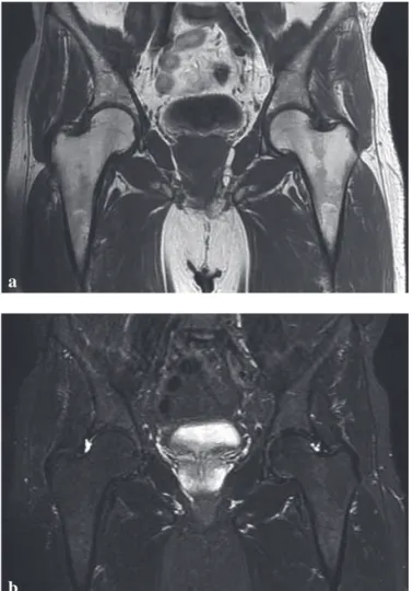
Fig. 1. — (a) Osteopenia in the right femoral head and neck on
hip X-Ray examination. The left hip is normal. (b) The coronal T1-weighted image of the hip joint shows a low signal intensity lesion in the right femoral head (arrow). (c) high signal inten-sity lesion on fat suppressed coronal T2 weighted image (arrow).
a
b
The etiology and pathophysiology of TOH has notbeenclearlydefined.Etiologicalfactorsimpli-cated in TOH include a genetic predisposition, me-chanical compression of obturator nerve, reflex sympathetic dystrophy, bone medullary hyperten-sion and small vessel ischaemia, fatty marrow con-version of the proximal femoral metaphysis, and chemical or hormonal factors related to pregnancy. Angiographical and scintigraphical studies show that nutrient arteries of the femoral head are dilated and that perfusion is higher than in the unaffected contralateralside.ThesefindingssuggestthatTOH may be the result of a vasomotor response to an un-disclosed etiological factor, though ischaemia is most likely. Further risk factors include cigarette smoking, steroid intake, alcoholism, obesity, hemo-globinopathies, and pregnancy. Hypothyroidism may be implicated in the etiology of TOH as well as iatrogenic hyperthyroxinemia. Minor trauma pre-ceding hip pain which may have caused a period of transient ischemia could precipitate to TOH (8,11,
13,34).
present18patients,66.7%weremaleand3of6fe-males had a history of caesarean delivery and pre-sented during the postpartum period. Although it is known that TOH can develop in late pregnancy or in the postpartum period in females, it has also been reported in non-pregnant women without any pre-ceding infection or trauma (8,11).
Table I. — Characteristicsofpatientswithtransient osteoporosis of hip Gender Female 6(33.3) Male 12 (66.7) Age, year 38.6±9.3 Involved hip Left 10 (55.6) Right 8 (44.4)
Pretreatment duration of complaints, week 6.1 ± 2.7 Duration of follow-up, month 17.8±5.3 Duration of clinical improvement, month 3.9±0.9 Duration of radiological improvement, month 8.3±2.2
Table II. — Characteristicsofthepatientsaccordingtogenders Female (n = 6) (Mean ± SD) Male (n = 12) (Mean ± SD) p Age (year) 34.3±4.3 40.7 ± 10.5 0.159
Duration of clinical improvement (month) 4.2 ± 1 3.8±0.8 0.455 Duration of radiological improvement (month) 8.0 ± 2.4 8.5 ± 2.2 0.612 Duration of follow-up (month) 19.7 ± 6 16.9 ± 4.9 0.470
a
b
Fig. 2. — (a-b). Follow-up hip MRI showing no abnormal
tion of spontaneous improvement of symptoms was reportedtobe5.8months(range,2-10months).No focal or subchondral changes were detected on the MRIs of 10 patients.
It has been reported that some TOH cases have been treated by the restriction of weight-bearing only (21). However, there are also reports on the
benefits of various additional medical therapies. Fabbriciani et al (9) reported improvement of
symp-tomsandMRIfindingswithteriparatidetherapyin a 62-year old male case. Seok et al (27) reported a
TOH case improved with a single dose bisphospho-nate (zoledrobisphospho-nate 5 mg) therapy. La Montagna et
al (17) found neridronate, a third-generation amio-bisphosphonate, therapy to be successful in a case with bilateral TOH. Schapira et al (25) reported
suc-cessful treatment of a TOH case with intravenous bisphosphonate.Kibbiet al (16)reported3patients
diagnosed with transient osteoporosis (hip in 2 pa-tients and knee in 1 patient), for whom oral weekly alendronate was effective in shortening the disease duration. Emad et al (6) reported radiological
im-provement demonstrated by MRI in 8 TOH cases treated with alendronate. Varenna et al (33) reported
that effective and rapid results were obtained with pamidronate therapy in 16 TOH cases. In the pres-ent study, acetylsalicylic acid and diclofenac sodi- umwasusedfortheanalgesicandanti-inflammato-ry effect and risedronate, which is a bisphosphonate, was also used. Bisphosphonates modulate bone turnover and reduce remodeling in cases of exces-sive resorption. The bisphosphonate group of drugs have increasingly been used in many bone condi-tions, such as Paget’s disease, osteoporosis, and metastatic cancer (10). Risedronate is an oral
bisphosphonate, which became available after alen-dronate, and is approved by the United States Food Although TOH is usually unilateral, occasional
cases of bilateral involvement have also been re-ported. Xyda et al (34) reported postpartum bilateral
TOHin3cases.Inthepresentstudy,allthepatients had unilateral involvement, with 55.6% left hip and 44.4% right hip.
Inclinicalpractice,therearedifficultiesindif-ferentiatingTOHfromAVN (1,13). While TOH
im-proves spontaneously or with symptomatic treat-ment,AVNmaydisplayaprogressivecourseand may require early surgical intervention(23,31). MRI
is the preferred method for differential diagnosis of painful conditions of the hip (22). MRI
characteris-tics in TOH cases include low signal intensity on T1-weighted images and high signal intensity on T2-weighted images (32). In the present study, the
MRIs of all patients consistently revealed high sig-nal intensity in T2 sequences and low sigsig-nal inten-sity in T1 sequences in the proximal aspect of the femur. Subchondral collapse or intra-articular effu-sion was not present in any of the patients. The mean duration of clinical improvement was 3.9±0.9months and the mean duration of radio-logical improvement was 8.3±2.2months. There was no difference in these durations between the genders. Balakrishnan et al (1) evaluated 10 TOH
cases, all of which were male, and reported the mean duration of radiological improvement as 7.5 months (range, 4-11 months). The mean
dura-Table III. — Harris hip scores before and after the treatment
Mean ± SD p
Beforethetreatment 55.6 ± 7.8
< 0.001 Month3afterthetreatment 88.8 ± 5.8
Month 6 after the treatment 96.0 ± 1.8
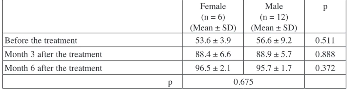
Table IV. — Harris hip scores according to the genders before and after the treatment Female (n = 6) (Mean ± SD) Male (n = 12) (Mean ± SD) p Beforethetreatment 53.6±3.9 56.6 ± 9.2 0.511 Month3afterthetreatment 88.4 ± 6.6 88.9 ± 5.7 0.888 Month 6 after the treatment 96.5 ± 2.1 95.7 ± 1.7 0.372
and were treated conservatively. Uematsu et al (29)
reported 4 TOH cases, who developed TOH during pregnancy and showed clinical and radiological im-provements without recurrence over the course of a mean 70.8 month follow-up period. In the present study, all patients also improved without recurrence overthecourseofamean17.8±5.3-monthfollow-up period.
In conclusion, it is important to make an early and accurate diagnosis by MRI in TOH cases, which are likely to completely recover with conservative therapies without surgical intervention. Early dif- ferentiationofTOHfromAVNwillavoidunneces-sary surgical intervention and ensure appropriate treatment. In the present retrospective study in which 18 patients were evaluated, it was determined that the patients were conservatively treated and followed-up for approximately 2 years. All the pa-tients were observed to have improved without any complications with no significant difference be-tween the genders.
REFERENCES
1. BalakrishnanA,SchemitschEH,PearceD,McKeeMD. Distinguishing transient osteoporosis of the hip from avascular necrosis. Can J Surg2003;46:187-192. 2.Canale ST. Harris Kalça Değerlendirilmesi (Modifiye).
Campbell’s Operative Orthopaedics. Türkçe 10. Basım,
EditorIşıkAKGÜN,Bölüm72007:356.
3. CurtissPHJr,KincaidWE. Transitory demineralization of the hip in pregnancy. A report of three cases. J Bone
Joint Surg Am1959;41-A:1327-1333.
4.Diwanji SR, Cho YJ, Xin ZF, Yoon TR. Conservative treatment for transient osteoporosis of the hip in middle-aged women. Singapore Med J 2008 ; 49 : 17-21.
5.DomachevskyL,KeynanY,MilitianuD,GoldenbergI, Adir Y. Transient osteoporosis associated with hyper-homocystinemia : a possible role for hyperbaric oxygen therapy. Undersea Hyperb Med2004Fall;31:275-279. 6. EmadY,RagabY,El-ShaarawyN,RaskerJJ. Transient
osteoporosis of the hip, complete resolution after treatment with alendronate as observed by MRI description of eight cases and review of the literature. Clin Rheumatol 2012 ; 31:1641-1647.
7.Eren AH.Harriskalçaskoru:Bilimselmakalebasımında bir dizgi hatasının yol açtığı yanlışlık. Acta Orthop
Traumatol Turc1991;31:285-288.
8. EscolàA,PonsM,PasarinA,MajóJ. Idiopathic transient osteoporosis of the pelvis in a non-pregnant young woman : a case study. Hip Int 2009 ; 19 : 71-74.
and Drug Administration (FDA) for the treatment of Paget’s disease of the bones and the prevention and treatment of postmenopausal osteoporosis and glucocorticoid-induced osteoporosis. Risedronate shows its clinical effects by binding to hydroxyapa-tite in bone tissue and inhibiting osteoclast activi-ty (30).
Inthepresentstudy,HBOwasperformedinad-ditiontostandardtherapy.HBOtherapyhasbeen reported to reverse cellular ischemia by enhancing theoxygenconcentrationoftheextracellularfluid and to reduce edema by inducing vasoconstric-tion (23). There have been reported cases of TOH
wherebenefithasbeengainedfromHBOtherapy. Accelerated recovery was observed in the patients withTOHtreatedwithHBO.HBOwouldbeeffec-tive for accelerated recovery time in patients with transient osteoporosis of the hip by suppression of oedema, lowering the intraosseous pressure, restor-ing venous drainage, and rapidly improvrestor-ing the mi-crocirculation (5,20).
Although TOH, which is a self-limiting condition with good prognosis, can be completely improved with conservative therapy, complications may be encountered occasionally. Lamarca et al (18)
report-ed a displacreport-ed subcapital fracture, which was treat-ed by total hip arthroplasty, as a rare complication of TOH. This complication due to weakness of the bone in TOH also might suggests that a period of nonweightbearing or protected weightbearing may be a reasonable component of routine conservative management.
In the present study, no complications were ob-served in any of the patients. According to the HHS, the hip status of the patients was poor before the treatment (mean score, 55.6 ± 7.8 and excellent (mean score, 96.0 ± 1.8) at the 6th month after the treatment. The improvement in HHS over time showed no difference between the genders.
Suresh et al (28) reported a patient who was
suc-cessfully treated for TOH but then developed TOH in the opposite hip 14 months after the initial admis-sion and it was emphasized that TOH cases needed long-term follow-up. Diwanji et al (4) reported no
osteonecrosis over the course of a 2-year follow-up period in 2 middle-aged female cases with TOH, whowerediagnosedonthebasisofMRIfindings
22.RagabY,EmadY,Abou-ZeidA.Bonemarrowedema syndromes of the hip : MRI features in different hip disorders. Clin Rheumatol 2008 ; 27 : 475-482.
23.RajpuraA,WrightAC,BoardTN. Medical management of osteonecrosis of the hip : a review. Hip Int 2011 ; 21 : 385-392.
24.Rocchietti MM, Tovaglia V, Meo A et al. Transient osteoporosis of the hip. Hip Int2010;20:297-300. 25.SchapiraD,BraunMY,GutierrezG,NahirAM. Severe
transient osteoporosis of the hip during pregnancy. Successful treatment with intravenous biphosphonates.
Clin Exp Rheumatol2003;21:107-110.
26.Sen RK. Management of avascular necrosis of femoral head at pre-collapse stage. Indian J Orthop 2009; 43: 6-16.
27.SeokH,KimYT,KimSH,ChaJG. Treatment of transient osteoporosis of the hip with intravenous zoledronate – a case report. Ann Rehabil Med2011;35:432-435.
28.Suresh S, Thomas JK, Raniga S. Migrating transient osteoporosis of the hip in a 30-year-old man. Indian J
Orthop2009;43:301-304.
29.Uematsu N, Nakayama Y, Shirai Y et al. Transient osteoporosis of the hip during pregnancy. J Nippon Med
Sch2000;67:459-463.
30.Umland EM, Boyce EG. Risedronate : a new oral bisphosphonate. Clin Ther2001;23:1409-1421.
31. Van Wagenen K, Pritchard P, Taylor JA. Transient osteoporosis of the hip : A case report. J Can Chiropr Assoc 2013;57:116-122.
32.Vande Berg BE, Malghem JJ et al. MR imaging of avascular necrosis and transient marrow edema of the femoral head. Radiographics1993;13:501-520.
33. Varenna M, Zucchi F, Binelli L et al. Intravenous pamidronate in the treatment of transient osteoporosis of the hip. Bone2002;31:96-101.
34.Xyda A, Mountanos I, Natsika M, Karantanas AH. Postpartum bilateral transient osteoporosis of the hip : MR imagingfindingsinthreecases.Radiol Med2008;113: 689-694. [Article in English, Italian].
9.FabbricianiG,PirroM,ManfredelliMRet al. Transient osteoporosis of the hip : successful treatment with teriparatide. Rheumatol Int2012;32:1367-1370.
10.GanapathyN,GokulnathanS,BalanN,MaheswaranT, Venkatesan.Bisphosphonates:Anupdate.J Pharm Bio
allied Sci2012;4:410-413.
11. Hadidy AM, Al Ryalat NT, Hadidi ST et al. Male transient hip osteoporosis : are physicians at a higher risk ?
Arch Osteoporos 2009 ; 4 : 41-45.
12.Harris Hip Score. http://www.orthopaedicscore.com/
scorepages/harris_hip_score.html
13. HarveyEJ. Osteonecrosis and transient osteoporosis of the hip : diagnostic and treatment dilemmas. Can J Surg2003; 46 : 168-169.
14.HayesCW,ConwayWF,DanielWW. MR imaging of bone marrow edema pattern : transient osteoporosis, transient bone marrow edema syndrome, or osteonecrosis.
Radiographics1993;13:1001-11.
15.Hofmann S, Engel A, Neuhold A et al. Bone-marrow oedema syndrome and transient osteoporosis of the hip. An MRI-controlled study of treatment by core decompression.
J Bone Joint Surg Br1993;75:210-216.
16. KibbiL,ToumaZ,KhouryN,ArayssiT. Oral bisphos-phonates in treatment of transient osteoporosis. Clin
Rheumatol2008;27:529-532.
17.La Montagna G, Malesci D, Tirri R, Valentini G. Successful neridronate therapy in transient osteoporosis of the hip. Clin Rheumatol 2005 ; 24 : 67-69.
18. LamarcaM,HernandezM,CampillosJM,LaprestaM, Tobajas JJ. Subcapital fracture of the hip in transient osteoporosis of pregnancy. Taiwan J Obstet Gynecol 2009 ; 48:423-424.
19.McWalterP,HassanA. Transient osteoporosis of the hip.
Ann Saudi Med 2009 ; 29 : 146-148.
20.Mutluoglu M, Sonmez G, Sivrioglu AK, Ay H. There may be a role for hyperbaric oxygen therapy in transient osteoporosis of the hip. Acta Orthop Belg 2012 ; 78 (5) : 685-7.
21.NiimiR,SudoA,HasegawaM,FukudaA,UchidaA. Changesinbonemineraldensityintransientosteoporosis of the hip. J Bone Joint Surg Br2006;88:1438-1440.