http://dx.doi.org/10.1080/01694243.2016.1240023
Influence of desensitizing procedures on adhesion of resin
cements to dentin
A. Ataya , O. Karaa, H. B. Karab, E. Calc and A. Usumeza
afaculty of dentistry, department of Prosthodontics, Bezmialem Vakıf university, istanbul, Turkey; bfaculty of
dentistry, department of Prosthodontics, istanbul Medipol university, istanbul, Turkey; cfaculty of dentistry,
department of Prosthodontics, ege university, izmir, Turkey
ABSTRACT
This study evaluated the effect of two desensitizer agents with different contents and Nd:YAG laser irradiation on the shear bond strength (SBS) of adhesive resin cements to dentin. New treatment options of Nd:YAG laser irradiation and tetracalcium phosphate-containing agent applications were compared with routinely used glutaraldehyde-containing agents. One hundred and twenty human, caries-free premolars were embedded in acrylic resin blocks 2 mm below the cementoenamel junction. Buccal surfaces of the teeth were ground to expose dentin. The specimens were randomly assigned into three different surface treatments (desensitizing agents, Nd:YAG laser) and the control, then into three different adhesive resin cement applications (n = 10). Resin cements (Panavia SA cement (PA), Panavia SA cement with Clearfil Universal Bond (PACU), and Multilink N (MN)) were applied to the conditioned teeth surfaces using Teflon tubes. The specimens were thermocycled (5000 cycles, 5–55 ± 1 °C, dwell time 30 s). The SBS test was performed in all groups. The results were submitted to two-way ANOVA and Tukey HSD tests (p < .05). Further, SEM analysis was performed on the dentin surfaces. SBS values were significantly difference among the surface treatment groups and also among adhesive resin cement groups (p < .05). The specimen cemented with PA showed lower SBS values than PACU- and MN-applied specimens. The highest SBS value was obtained in the Nd:YAG laser group which was cemented with PACU cement. The lowest SBS value was obtained in the control group which was cemented with PA cement. In addition, SEM evaluation revealed that desensitizing agents and Nd:YAG laser occluded dentin tubules.
Introduction
The short, sharp pain that patients experience when exposed dentin is brought into con-tact with thermal, con-tactile, osmotic, and mechanical stimuli (such as tooth brushing, sweet and sour foods, and hot or cold water) is defined as dentin hypersensitivity (DH).[1] Most DH cases are the result of abrasion, attrition, erosion, abfraction, gingival recession, and improper brushing habits.[2] DH may be seen in patients of any age, and women are affected
KEYWORDS desensitizing agents; calcium–phosphate desensitizer; nd:yag laser; adhesive resin cement; universal bond; shear bond strength
ARTICLE HISTORY received 26 May 2016 revised 5 september 2016 accepted 11 september 2016
© 2016 informa uK limited, trading as Taylor & francis group
more often than men.[1] The prevalence of this condition has been reported to vary from 4 to 73%.[1,3] However, an increase in this prevalence is possible, as more adults retain their teeth into later life.[4]
Among the many theories proposed for the mechanism of DH, Brannstrom’s hydrody-namic mechanism theory notes that in the presence of an external stimulus, fluid inside den-tinal tubules move inwardly or outwardly, causing mechanical deformation of nerve endings at the pulp/dentine interface, which is perceived as a painful sensation.[5] When the dentinal tubules are blocked, fluid shifts and dentin sensitivity can be prevented. Desensitizing agents (DA) may function by desensitizing the nerves (potassium nitrate), precipitating proteins (glutaraldehyde, silver nitrate, zinc chloride), or by plugging dentinal tubules (sodium flu-oride, potassium oxalate). Dentin adhesive sealers, lasers, and homeopathic medication may also be used for this purpose.[6]
Hydroxyethyl methacrylate (HEMA)/glutaraldehyde, the most commonly used desensi-tizer, is a dentin adhesive sealer, which also contains benzalkonium chloride and fluoride. Glutaraldehyde proteins inside the dentinal fluid coagulate and precipitate. Deep resinous tags are formed by HEMA and they occlude the dentinal tubules.[7]
Recently, desensitizers with calcium phosphate have gathered much attention due to their biocompability, and their superior ability to occlude dentinal tubules and reduce dentine permeability.[8,9] Teethmate Desensitizer (TMD; Kuraray Noritake Dental Inc., Tokyo, Japan) is a newly developed calcium–phosphate-containing material; tetracalcium
phosphate (TTCP; Ca4(PO4)2O) and dicalcium phosphate anhydrous (DCPA; CaHPO4),
which can spontaneously transform to hydroxyapatite (HA; Ca10(PO4)6(OH)2).[10] When
TMD powder is mixed with water, the chemical reaction between particles takes place. Within seconds after mixing, calcium and phosphate ions are released, and this layer makes up HA crystals which physically occlude dentin tubules.[11]
Laser applications are another option for DH treatment. Laser desensitization has been suggested as an effective and rapid treatment option for terminating or reducing DH.
Recently, neodymium-doped:yttrium, aluminum and garnet (Nd:YAG), CO2,
galliumealu-minumearsenide (diode), and erbium-doped:yttrium, aluminum and garnet (Er: YAG) have been commonly used lasers for the treatment of DH. The success of the treatment is mainly determined by the laser type and the parameters used.[12,13]
The effect of the Nd:YAG laser is accomplished by the occlusion or narrowing of dentinal tubules causing a form of nerve analgesia. After a short exposure to Nd:YAG laser, dentin fuses, and its surface becomes non-porous and glazed.[14] Followed by the application of the Nd:YAG laser, some physical changes in dentin such as a melted and re-solidified surface as well as craters, cracks, and globules are observed.[15–18]
Long-lasting dentin and luting material bond is a key to the success of the restorations’ retention and avoiding microleakage, secondary caries, and tooth fracture.[19–21] Zinc phosphate, zinc polycarboxylate, conventional glass ionomer cements, and resin-based cements are some of the frequently used luting materials.[22–24] Adhesive resin cements can be used for the cementation of many types of fixed partial dentures.[21] Self-adhesive resin cements have the advantages of having simpler and less technique-sensitive pretreatment steps, eliminating application errors of the cementation process in fixed prosthodontics.[25] The impact of the desensitizer use before cementation with conventional or resin cements is still under debate. Some authors suggest that there is a significant loss in retention in cases using a desensitizer,[21,26–29] while some researchers have concluded that desensitizers
have no negative effect on crown retention.[30,31] However, there is a lack of literature comparing the bond strengths of various desensitizers used before the application of the newly developed self-adhesive resin cements.[32]
The aim of this study was to assess the impact of three different surface treatments and control on the shear bond strength (SBS) of adhesive resin cements to tooth surface. This study also examined dentin surfaces under a field emission scanning electron microscope (SEM) after the application of DAs. The null hypothesis was that DAs and laser irradiation procedures do not affect the SBS of adhesive resin cements to dentin surface.
Materials and methods
Dentin DAs and methods used in the study are shown in Table 1. Dentin surface with no treatment acted as the control group.
One hundred and twenty unrestored, caries-free, extracted human premolars were used in the study. The teeth were cleaned mechanically and stored in distilled water at room temperature until used. They were then embedded in custom made molds using autopol-ymerizing acrylic resin (Vertex Self Curing; Vertex-Dental, Netherlands) with the areas to be treated placed 2 mm above the resin surface. The specimens were then ground with a coarse grit diamond rotary cutting instrument (Mecatome T180, Presi, Grenoble, France) until the dentin surfaces were exposed. To standardize the dentin surface texture, the occlu-sal surface of each tooth was further ground off in a mechanical grinder (Minitech 233, Presi, Grenoble, France) with 600-grit silicon carbide abrasive paper under running water for 30 s.[33] The specimens were randomly assigned into the surface treatments (Gluma, TMD, Nd:YAG laser irradiation, and the control) and then into two different adhesive resin cements and a universal bond which constituted the 12 test groups (n = 10).
Applied resin cement groups were as follows:
PA: Self-adhesive resin cement (Panavia SA cement,) was applied without bonding agent application.
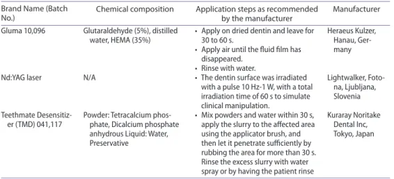
Table 1. The brand names, batch numbers, chemical compositions, application steps, and manufactur-ers of the test groups utilized in the study.
Brand Name (Batch
No.) Chemical composition Application steps as recommended by the manufacturer Manufacturer gluma 10,096 glutaraldehyde (5%), distilled
water, heMa (35%) • apply on dried dentin and leave for 30 to 60 s. • apply air until the fluid film has
disappeared. • rinse with water.
heraeus Kulzer, hanau, ger-many nd:yag laser n/a • The dentin surface was irradiated
with a pulse 10 hz-1 W, with a total irradiation time of 60 s to simulate clinical manipulation.
lightwalker, foto-na, ljubljafoto-na, slovenia Teethmate
desensitiz-er (TMd) 041,117 Powder: Tetracalcium phos-phate, dicalcium phosphate anhydrous liquid: Water, Preservative
• Mix powders and water within 30 s, apply the slurry to the affected area using the applicator brush, and then let it penetrate sufficiently by rubbing the area for more than 30 s. rinse the excess slurry with water spray or by having the patient rinse
Kuraray noritake dental inc, Tokyo, Japan
PACU: Self-adhesive resin cement (Panavia SA cement,) was applied with self-etch bonding agent (Clearfil Universal Bond) application.
MN: Self-etching resin cement (Multilink N) was applied.
The DAs and Nd:YAG laser were applied to the dentin surfaces according to the man-ufacturer’s instructions (Table 1). A steel mold with a hole in the center (3 mm diameter and 2 mm deep) was used for the application of the resin cements (Table 2) to the dentin surfaces according to the manufacturer’s instructions.
The resin cements were then cured by use of an LED-unit (BAOPTIMA 10, B. A.
Internatıonal, Northampton, U.K., 1200 mW/cm2) with a curing time of 20s. After removal
of the mold, the specimens were subjected to thermocycling for 5000 cycles between 5 °C and 55 °C in deionized water. The dwelling time at each temperature was 30 s, and the transfer time from one bath to the other was 2 s according to the International Organization for Standardization (ISO NO:11405).[34] The SBS testing was performed with a universal testing machine (Shimadzu AG-50 kNG, Kyoto, Japan) at a crosshead speed of 0.5 mm/ minute. Results were expressed in megapascal (MPa) values.
Means and standard deviations of bond strengths were calculated, and mean values were compared by two-way analysis (surface treatments and adhesive resin cements) of variance (ANOVA; SPSS 12.0, SPSS Inc., Chicago, IL, U.S.A.) followed by a multiple comparison test performed with a Tukey’s honestly significant difference (HSD) test (p < .05).
To investigate the changes in the dentinal tubules after applying different surface treatments, one specimen from each group was air-dried overnight and gold sputtered with a sputter coater (S150B, Edwards, Crawley, England), and examined by means of a field emission SEM (JSM-6335F, JEOL Ltd., Tokyo, Japan) at 15.0 or 20.0 kV. There was no resin cement applied. For visual assessment, the SEM photomicrographs were developed at 1000× magnification.
Results
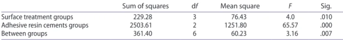
The SBS test results of two-way ANOVA revealed significant differences among the surface treatment groups and also among adhesive resin cement groups (p < .05; Table 3). The mean SBS values and the differences among the groups are shown in Table 4.
There were significant differences among the surface treatment groups. The specimens cemented with PA showed lower SBS values than PACU- and MN-applied specimens (p < .05). However, there were no significant differences between PACU- and MN-applied specimens (p > .05).
The control group’s specimens showed significantly lower SBS values than Gluma and Nd:YAG laser groups (p < .05). However, there was no significant difference between Nd:YAG laser and Gluma group specimen (p > .05). The specimens cemented with TMD did not show any significant differences from other groups (p > .05).
The highest SBS value was obtained in the Nd:YAG laser group which was cemented with PACU cement (20.8 ± 4.52 MPa). The lowest SBS value was obtained in the control group which was cemented with PA cement (5.96 ± 2.9 MPa) (p < .05).
SEM evaluation
The microstructures of the dentin surfaces treated with two different types of DAs and Nd:YAG laser are shown in Figure 1. The specimens treated with Gluma (a) showed
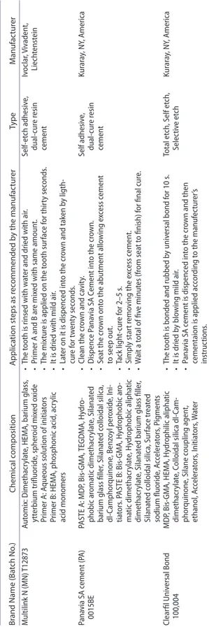
Table 2. The br and names , ba tch numbers , chemical c ompositions , applica tion st eps , and manufac tur ers of the r esin c emen
ts used in the study
. abbr evia tion: Bis-g M a: bisphenol a digly cidylether methacr yla te; M d P: 10-M ethacr ylo ylo xy dec yl dih ydr ogen phospha te; Tegd M a: trieth ylene gly col dimethacr yla te . Br
and Name (Ba
tch No .) Chemical c omposition A pplica tion st eps as r ec ommended b y the manufac tur er Type M anufac tur er Multilink n (M n ) T12873 aut omix: d imethacr yla te , he M a, barium glass , ytt erbium trifluoride , spher oid mix ed o xide Primer a:
aqueous solution of initia
tors Primer B: he M a, phosphonic acid , acr ylic acid monomers • The t
ooth is rinsed with w
at
er and dried with air
.
•
Primer
a and B ar
e mix
ed with same amoun
t. • The mix tur e is applied on the t ooth sur fac e f or thir ty sec onds . •
it is dried with mild air
. • la ter on it is dispenc ed in to the cr
own and tak
en b y ligth- cur e f or t w en ty sec onds . self-et ch adhesiv e, dual-cur e r esin cemen t iv oclar , V iv aden t, liech tenst ein Pana via sa c emen t (P a) 0015B e Pas Te a : M d P, Bis-g M a, Tegd M a, h ydr o-phobic ar oma tic dimethacr yla te , s ilana ted
barium glass filler
, s ilana ted c olloidal silica, dl-camphor quinone , B enz oyl per oxide , i ni -tia tors . P as Te B: Bis-g M a, h ydr ophobic ar o-ma tic dimethacr yla te , h ydr ophobic alipha tic dimethacr yla te , s ilana
ted barium glass filler
, silana ted c olloidal silica, sur fac e tr ea ted sodium fluoride , a cc eler at ors , P ig men ts • clean the cr own and ca vit y. • d ispenc e P ana via sa c emen t in to the cr own. • sea t the cr own on to the abutmen t allo wing e xc ess c emen t to seep out . • Tack ligh t-cur e f or 2–5 s . • simply star t r emo ving the e xc ess c emen t. • W ait a t otal of fiv e minut es (fr om sea t t o finish) f or final cur e. self adhesiv e, dual-cur e r esin cemen t Kur ar ay , n y, america clear fil u niv ersal B ond 100,004 M d P, Bis-g M a, he M a, h ydr ophilic alipha tic dimethacr yla te , c olloidal silica dl-cam -phor quinone , s ilane c oupling agen t, ethanol , a cc eler at ors , i nitia tors , W at er • The t
ooth is bonded and rubbed b
y univ ersal bond f or 10 s . • it is dried b y blo
wing mild air
. • Pana via sa c emen t is dispenc ed in to the cr
own and then
cemen taion is applied ac cor ding t o the manuf ec tur er ’s instruc tions . Total et ch, self et ch, selec tiv e et ch Kur ar ay , n y, america
completely occluded dentinal tubules. In the specimens treated with Nd:YAG laser (b) and TMD (c)-treated group, the dentine tubules were occluded as well. However, TMD com-pletely covered and obliterated the tubules. In the control group specimens (d), exposed dentin tubules were observed.
Discussion
The hypothesis of the present study was that DAs and laser irradiation procedures do not affect the SBS of self-etch, self-adhesive resin cements to dentin surface. However, the study Table 4. Mean and sd values for shear bond strength (MPa) same supercripts in same column and row show not statistically significant differences.
Adhesive resin cements and surface
treatments Gluma (Mean ± SD) Nd: YAG laser (Mean ± SD) Teethmate desensi-tizer (Mean ± SD) (Mean ± SD)Control Pa 14.5 ± 4.71bc 10.84 ± 3.37ab 7.3 ± 4.08a 5.96 ± 2.9a
Pacu 19.06 ± 6.51c 20.8 ± 4.52c 20.17 ± 3.63c 16.6 ± 5.4bc
Mn 17.88 ± 3.33c 20.74 ± 3.56c 20.16 ± 4.07c 19.28 ± 5.01c
Figure 1. scanning electron microscopic views of the dentin surface after treatment with the desensitizing agents: a, gluma; b, nd: yag laser; c, TMd; d, control.
Table 3. Two-way anoVa results.
Sum of squares df Mean square F Sig. surface treatment groups 229.28 3 76.43 4.0 .010 adhesive resin cements groups 2503.61 2 1251.80 65.57 .000
concluded that the SBS was increased after the application of the DAs. Therefore, the null hypothesis was rejected.
Adhesive cementation is a more technique-sensitive procedure compared to conventional cementation, and clinical success may be overridden by the technical challenges involved. Self-adhesive resin cements can be a solution to such difficulties.[32] Clearfil Universal Bond, used in the presented study, is a one-bottle, newly produced universal bond which can be used with self-adhesive resin cement. When used with Panavia SA Cement, no additional primer or activator is needed. In the current study, the combined application of the Panavia SA Cement with Clearfil Universal Bond significantly increased the SBS values.
Because of its chemical composition, TMD is very successful at forming a layer on dentin and at occluding tubules regardless of pretreatment. TMD consists majorly of TTCP and DCPA. When scrubbed on dry dentin surface, thick paste, which is the mixture of the two components, can penetrate into the tubules. This occluding mechanism reduces the dentinal permeability and clinical hypersensitivity.[35] However, in the results of current study, the SBS values of the TMD group were not significantly different from the other groups. In the literature, there is a lack of study on this newly introduced desensitizer on the market.
For a self-etching/self-adhesive system, the monomer infiltration into the dentin and the formation of a resin-infiltrated layer is essential.[19] Self-etching/self-adhesive resin condi-tions and primes the enamel and dentinal surface without any need of rinsing. Therefore, the remnants of DAs cannot be removed from the dentinal surface, and they may cause a decrease in the bond strength.[36] Only a few studies have evaluated the impact of DAs on the self-adhesive resin–dentin interface.[21,28,29,37] Huh et al. [21] evaluated the influence of four different DAs (SuperSeal; Copalite Varnish, Cooley and Cooley, Houston, TX, U.S.A.; MS Coat; and Gluma) on the bond strength of a self-etching adhesive resin cement to dentin. According to their study, the Gluma group showed a lower bond strength value compared to SuperSeal and the control groups. Previously, it had been found that the bond strength of resin cement was mostly influenced by the HEMA concentration, with a maximum of 35% and nearly independent of the glutaraldehyde concentration when greater than 3%.[38] Also, Qin et al. [39] stated that the glutaraldehyde in Gluma is not able to cross-link mineralized dentin. HEMA reduces the surface tension of water and enables monomer diffusion into dentin.[40]
However, Sailer et al. [41] concluded that glutaraldehyde-containing dentin desensitizers and bonding agents (Gluma and Syntac) were likely to increase the SBS of the self-adhesive resin cement (RelyX U200) to dentin. Additionally, they found that the bond strength was increased when glutaraldehyde/HEMA-containing desensitizers were used before self-adhe-sive cement luting.[42] The glutaraldehyde reacts with protein and produces a precipitate on the dentin surface, while HEMA accelerates the diffusion of monomers into dentin regardless of such precipitation.[19,30,43,44] Dündar et al. [46] showed that the application of two brands of adhesive cement in combination with the same DA caused increased SBS. In the researchers’ opinion, HEMA induces rehydration, allowing for the penetration of primer into dentin and causing an increase in the bond strength.[46] In the results of the current study, the HEMA-containing DA (Gluma) group showed a higher bond strength than the control group.
Desensitizing treatment with Nd:YAG laser has also been investigated. This kind of laser irradiation causes a reduction or complete obliteration of the dentinal tubule lumen [47] and closure of exposed dentin tubules,[48] as shown in many of SEM inspections. When self-etch and self-etch-and-rinse systems are used after Nd:YAG laser irradiation, the bond strength usually decreases.[49,50] This might be caused by the tubule obliteration when self-etching
primer is used on the interface of dentin and composite resin. Obliterated tubules may block the penetration of resin monomer and the resin tag formation.[49] However, in a study that evaluated the influence of five different DAs(Gluma, Aqua-Prep F, Bisblock, Cervitec Plus, Smart Protect) and Nd:YAG laser pre-treatment on microtensile bond strength of self-adhesive resin cement to dentin, the results showed higher bond strength values with the Gluma group.[32] In the results of the current study, there were no significant differences between the Nd:YAG laser-irradiated group and the Gluma group.
The present study has several limitations, making it difficult to compare results directly with clinical studies. Resin cement thickness tested in the current study was 2 mm, which is 25–150 μm in the clinical situations. Future investigations should be performed using differ-ent resin cemdiffer-ent thicknesses for greater clinical relevance. Another limitation of the presdiffer-ent study was that it was performed under in vitro conditions, but the results still provide guid-ance for clinicians. The differences among the materials and methods used make the results difficult to compare. In the present study, the effect of two different DAs (of all the types on the market today, with one newly introduced product) and Nd:YAG laser pretreatment on the bonding compatibility of adhesive resin cements were compared. Hence, this protocol is believed to make a more conclusive statement regarding which desensitizing material to use with adhesive resin cements. Also, to the authors’ knowledge, there is no study conducted with all the different kinds of DAs combined with these cements. Future clinical studies are required to evaluate this combined usage of DAs and adhesive resin cements.
Conclusion
Within the limitations of this study, it is concluded that:
(1) The specimen cemented with PA showed lower SBS values than the PACU- and MN-applied specimens.
(2) The highest SBS value was obtained in the Nd:YAG laser group which was cemented with PACU cement. The lowest SBS value was obtained in the control group which was cemented with PA cement.
(3) SEM evaluation revealed that DAs and Nd:YAG laser occluded dentin tubules.
Disclosure statement
No potential conflict of interest was reported by the authors.
ORCiD
A. Atay http://orcid.org/0000-0002-5358-0753 References
[1] Orchardson R, Gillam DG. Managing dentin hypersensitivity. J. Am. Dent. Assoc. 2006;137:990–998.
[2] Walters PA. Dentinal hypersensitivity: a review. J. Contemp. Dent. Pract. 2005;6:107–117.
[3] Rees JS, Addy M. A cross-sectional study of dentine hypersensitivity. J. Clin. Periodontol.
[4] Ipci SD, Cakar G, Kuru B, et al. Clinical evaluation of lasers and sodium fluoride gel in the
treatment of dentine hypersensitivity. Photomed. Laser. Surg. 2009;27:85–91.
[5] Pashley DH, Tay FR, Haywood VB, et al. Consensus-based recommendations for the diagnosis
and management of dentin hypersensitivity. Comp. Cont. Educ. Dent. 2008;29:1–7.
[6] Miglani S, Aggarwal V, Ahuja B. Dentin hypersensitivity: recent trends in management.
J. Conserv. Dent. 2010;13:218–224.
[7] Dondi dall’Orologio G, Lone A, Finger WJ. Clinical evaluation of the role of glutardialdehyde
in a one-bottle adhesive. Am. J. Dent. 2002;15:330–334.
[8] Guentsch A, Seidler K, Nietzsche S, et al. Biomimetic mineralization: long-term observations
in patients with dentin sensitivity. Dent. Mater. 2012;28:457–464.
[9] Shetty S, Kohad R, Yeltiwar R. Hydroxyapatite as an ın-office agent for tooth hypersensitivity:
a clinical and scanning electron microscopic study. J. Periodontol. 2010;81:1781–1789.
[10] Chow LC. Next generation calcium phosphate-based biomaterials. Dent. Mater. J. 2009;28:1–10.
[11] Thanatvarakorn O, Nakashima S, Sadr A, et al. In vitro evaluation of dentinal hydraulic conductance and tubule sealing by a novel calcium-phosphate desensitizer. J. Biomed. Mater.
Res. Part B Appl. Biomater. 2013;101B:303–309.
[12] Kimura Y, Wilder-Smith P, Yonaga K, et al. Treatment of dentine hypersensitivity by lasers: a
review. J. Clin. Periodontol. 2000;27:715–721.
[13] He S, Wang Y, Li X, et al. Effectiveness of laser therapy and topical desensitising agents in treating
dentine hypersensitivity: a systematic review. J. Oral Rehabil. 2011;38:348–358.
[14] Birang R, Poursamimi J, Gutknecht N, et al. Comparative evaluation of the effects of Nd:YAG
and Er:YAG laser in dentin hypersensitivity treatment. Lasers Med. Sci. 2007;22:21–24.
[15] Zapletalová Z, Peřina J Jr, Novotný R, et al. Suitable conditions for sealing of open dentinal
tubules using a pulsed Nd:YAG laser. Photomed. Laser Surg. 2007;25:495–499.
[16] Naylor F, Corrêaaranha AC, Eduardo Cde P, et al. Micromorphological analysis of dentinal structure after ırradiation with Nd:YAG laser and ımmersion in acidic beverages. Photomed.
Laser Surg. 2006;24:745–752.
[17] Corrêa Aranha AC, Domingues FB, Franco VO, et al. Effects of Er:YAG and Nd:YAG lasers on dentin permeability in root surfaces: a preliminary in vitro study. Photomed. Laser Surg.
2005;23:504–508.
[18] Lan WH, Liu HC, Lin CP. The combined occluding effect of sodium fluoride varnish and
Nd:YAG laser irradiation on human dentinal tubules. J. Endod. 1999;25:424–426.
[19] Soeno K, Taira Y, Matsumura H, et al. Effect of desensitizers on bond strength of adhesive luting
agents to dentin. J. Oral Rehabil. 2001;28:1122–1128.
[20] Ivanyi I, Balogh AE, Rosivall L, et al. In vivo examination of the scotchbond multi-purpose
dental adhesive system in rat (vital microscopic study). Oper. Dent. 2000;25:418–423.
[21] Huh JB, Kim JH, Chung MK, et al. The effect of several dentin desensitizers on shear bond
strength of adhesive resin luting cement using self-etching primer. J. Dent. 2008;36:1025–1032.
[22] Owens SE Jr, Miller BH. A comparison of shear bond strengths of three visible light-cured
orthodontic adhesives. Angle Orthod. 2000;70:352–356.
[23] Almuammar MF, Schulman A, Salama FS. Shear bond strength of six restorative materials.
J. Clin. Pediatr. Dent. 2001;25:221–225.
[24] Wassell RW, Barker D, Steele JG. Crowns and other extra-coronal restorations: try-in and
cementation of crowns. Br. Dent. J. 2002;193:17–28.
[25] Behr M, Rosentritt M, Regnet T, et al. Marginal adaptation in dentin of a self-adhesive universal
resin cement compared with well-tried systems. Dent. Mater. 2004;20:191–197.
[26] Awang RAR, Masudi SM, Mohd Nor WZW. Effect of desensitizing agent on shear bond strength
of an adhesive system. Arch. Orofac. Sci. 2007;2:32–35.
[27] Ling TY, Gillam DG, Barber PM, et al. An investigation of potential desensitizing agents in the
dentine disc model: a scanning electron microscopy study. J. Oral Rehabil. 1997;24:191–203.
[28] Yim NH, Rueggeberg FA, Caughman WF, et al. Effect of dentin desensitizers and cementing agents on retention of full crowns using standardized crown preparations. J. Prosthet. Dent.
[29] Sengun A, Koyuturk AE, Sener Y, et al. Effect of desensitizers on the bond strength of a self-etching
adhesive system to caries-affected dentin on the gingival wall. Oper. Dent. 2005;30:430–435.
[30] Swift EJ Jr, Lloyd AH, Felton DA. The effect of resin desensitizing agents on crown retention.
J. Am. Dent. Assoc. 1997;128:195–200.
[31] Wolfart S, Linnemann J, Kern M. Crown retention with use of different sealing systems on
prepared dentine. J. Oral Rehabil. 2003;30:1053–1061.
[32] Acar O, Tuncer D, Yuzugullu B, et al. The effect of dentin desensitizers and Nd:YAG laser pre-treatment on microtensile bond strength of self-adhesive resin cement to dentin. J. Adv.
Prosthodont. 2014;6:88–95.
[33] Hiraishi N, Yiu CK, King NM, et al. Effect of 2% chlorhexidine on dentin microtensile bond
strengths and nanoleakage of luting cements. J. Dent. 2009;37:440–448.
[34] International Organization for Standardiation. ISO TR 11405. Dental material guidence on
testing of adhesion to tooth structure. 1994. p. 1–15.
[35] Thanatvarakorn O, Nakashıma S, Sadr A, et al. Effect of a calcium-phosphate based desensitizer
on dentin surface characteristics. Dent. Mater. J. 2013;32:615–621.
[36] Külünk Ş, Saraç D, Külünk T, et al. The effects of different desensitizing agents on the shear
bond strength of adhesive resin cement to dentin. J. Esthet. Restor. Dent. 2011;23:380–387.
[37] Pashley EL, Tao L, Pashley DH. Effects of oxalate on dentin bonding. Am. J. Dent. 1993;6:116–118.
[38] Munksgaard EC, Asmussen E. Materials science bond strength between dentin and restorative
resins mediated by mixtures of HEMA and glutaraldehyde. J. Dent. Res. 1984;63:1087–1089.
[39] Qin C, Xu J, Zhang Y. Spectroscopic investigation of the function of aqueous
2-hydroxyethylmethacrylate/glutaraldehyde solution as a dentin desensitizer. Eur. J. Oral Sci.
2006;114:354–359.
[40] Nakabayashi N, Watanabe A, Gendusa NJ. Dentin adhesion of ‘modified’ 4-META/MMA-TBB
resin: function of HEMA. Dent. Mater. 1992;8:259–264.
[41] Sailer I, Tettamanti S, Stawarczyk B, et al. In vitro study of the influence of dentin desensitizing and sealing on the shear bond strength of two universal resin cements. J. Adhes. Dent.
2010;12:381–392.
[42] Sailer I, Oendra AE, Stawarczyk B, et al. The effects of desensitizing resin, resin sealing, and provisional cement on the bond strength of dentin luted with self-adhesive and conventional
resincements. J. Prosthet. Dent. 2012;107:252–260.
[43] Perdigão J, Swift EJ Jr, Heymann HO, et al. Effect of a re-wetting agent on the performance of
acetone-based dentin adhesives. Am. J. Dent. 1998;11:207–213.
[44] Perdigão J, Van Meerbeek B, Lopes MM, et al. The effect of a re-wetting agent on dentin bonding.
Dent. Mater. 1999;15:282–295.
[45] Soares CJ, Santos-Filho PC, Barreto BC, et al. Effect of previous desensitizer and rewetting agent application on shear bond strength of bonding systems to dentin. Cienc. Odontol. Bras.
2006;9:6–11.
[46] Dündar M, Çal E, Gökçe B, et al. Influence of fluoride- or triclosan-based desensitizing agents
on adhesion of resin cements to dentin. Clin. Oral Invest. 2010;14:579–586.
[47] Al-Saud LM, Al-Nahedh HN. Occluding effect of Nd:YAG laser and different dentin desensitizing agents on human dentinal tubules ın vitro : a scanning electron microscopy ınvestigation. Oper.
Dent. 2012;37:340–355.
[48] Kumar NG, Mehta DS. Short-term assessment of the Nd:YAG laser with and without sodium fluoride varnish in the treatment of dentin hypersensitivity – a clinical and scanning electron
microscopy study. J. Periodontol. 2005;76:1140–1147.
[49] Akca T, Yazici AR, Çelik Ç, et al. The effect of desensitizing treatments on the bond strength
of resin composite to dentin mediated by a self-etching primer. Oper. Dent. 2007;32:451–456.
[50] de Oliveira MT, de Freitas PM, de Paula Eduardo C, et al. Influence of diamond sono-abrasion, air-abrasion and Er:YAG laser ırradiation on bonding of different adhesive systems to dentin.