Decompressive Hemicraniectomy in
Patients With Transtentorial Herniation
Orhan BARLAS 1,4, Selhan KARADERELER 1, Yakup KRESPİ 2,5, Nilüfer YEŞİLOT 3, Sara BAHAR 3, Rezzan TUNCAY 3,
Reha TOLUN 2,5
Şişli Florence Nightingale Hospital Departments of Neurosurgery1, Neurology2, İstanbul Medical Faculty Departments of Neurology3, Neurosurgery4,
İstanbul Bilim University Medical Faculty Department of Neurology5
✔ Objective: To present the results, technique and indications of decompressive hemicraniectomy in patients
with unilateral transtentorial herniation.
Methods: Ten consecutive patients with intracranial hypertension refractory to medical management and
transtentorial herniation were treated with decompressive hemicraniectomy over a 3-year period with the fol-lowing diagnoses: malignant infarction (3 patients), subarachnoid hemorrhage and vasospasm (2 patients), traumatic brain injury (3 patients), spontanous intracerebral hemorrhage, and venous thrombosis. Neurological status was assessed by the Glasgow Coma Scale (GCS) and by the modified Rankin Scale (mRS) on admissi-on, and by the mRS on discharge, and on follow-up examinations. Computerized tomography (CT) scans performed preoperatively and on the first, 7th postoperative days, and at one month were assessed for trasten-torial herniation and midline shift. A decompressive hemicraniectomy, at least 14 by 11 cm was performed in all patients.
Results: There were three male and 7 female patients, ages 23 to 57 years. Neurological assessment
preope-ratively showed a median GCS score of 5.5, and hemiplegia in all patients, ipsilateral 3rd nerve palsy in eight of ten patients. Median mRS score on admission was 5. CT showed radiological signs of unilateral transtento-rial herniation in every patient. Mean midline shift was 11.2 mm in preoperative CT scans and 3.2 mm on the first postoperative day. Median mRS score at the last follow up examination was 1.5 The overall mortality rate was 10 %. Six patients had favorable outcome (mRS <3).
Conclusion: The results of this series suggest that decompressive hemicraniectomy is a safe and effective
technique that can be a life saving measure in patients with intractable intracranial hypertension and unilateral transtentorial herniation due to various causes.
Key words: Brain edema, craniectomy, decompressive surgery, intracranial pressure, transtentorial herniation
J Nervous Sys Surgery 2008; 1(3):161-168
Transtentoryal Herniasyondaki Hastalarda Dekompresif Hemikranyektomi
✔ Amaç: Unilateral transtentoryal herniasyonu olan hastalarda dekompresif hemikranyektominin sonuçlarını,
tekniğini ve endikasyonlarını ortaya koymak.
Yöntem: Üç yıl içinde, medikal tedaviye dirençli intrakranyal hipertansiyonlu ve transtentoryal herniasyonda,
habis infarct (3 olgu), subaraknoid kanama ve vazospazm (2 olgu), travmatik beyin hasarı (3 olgu), spontan intraserebral kanama ve venöz tromboz tanıları olan 10 ardışık hastaya dekompresif hemikranyektomi yapıldı. Nörolojik durum girişte Glasgow Koma Skalası (GKS) ve modifiye Rankin Skalası (mRS) ile, çıkışta ve kontrollerde mRS ile değerlendirldi. Operasyon öncesinde, 1. ve 7. postoperatif günlerde ve 1. ayda yapılan bilgisayarlı tomografi (BT) incelemeleri değerlendirildi. Hastaların hepsinde en az 14x11 cm’lik dekompresif hemikranyektomi yapıldı.
Bulgular: 23 ila 57 yaşları arasında üç erkek, yedi kadın hasta vardı. Operasyondan hemen önceki nörolojik
değerlendirmeye göre medyan GKS skoru 5.5, mRS skoru 5’ti. Hastaların hepsinde hemipleji, sekiz tanesinde ipsilateral okülomotor felci vardı. BT incelemesi hastaların hepsinde unilateral transtentoryal herniasyon bul-guları gösterdi. Ortalama orta hat itilmesi operasyon öncesi BT’lerde 11.2 mm iken, operasyon sonrası 1. gün BT’lerde 3.2 mm’ydi. Altı hastada iyi sonuç alındı (mRS <3), son kontrol muayenesinde medyan mRS 1.5’ti. Serinin mortalitesi % 10 oldu.
Sonuç: Bu çalışmanın sonuçları dekompresif hemikranyektominin çeşitli nedenlere bağlı dirençli intrakranyal
hipertansiyonu ve unilateral transtentoryal herniasyonu olan hastalarda güvenilir ve hayat kurtaran etkili bir teknik olduğunu göstermektedir.
Anahtar kelimeler: Beyin ödemi, kranyektomi, dekompresif cerrahi, intrakranyal basınç transtentoryal herniasyon
J Nervous Sys Surgery 2008; 1(3):161-168
R
efractory elevated intracranial pressure leading to transtentorial herniation is a life threatening condition with very high mortality. Increased intracranial pressure caused by expanding supratentorial mass lesions push the medial temporal lobe over the tentorial edge, compressing the oculomotor nerve, the brain stem, and the posterior cerebral artery, and if unchecked, lead to the death of the patient. Ipsilateral pupillary dilatation, decreased conci-ousnesss and coma, hemiplegia, and respiratory arrest are clinical signs of unilateral transtentori-al heniation. Refractory edema leading to incre-ased intracranial pressure and transtentorial heniation may result from a number of conditi-ons including trauma, tumor, infarction, suba-rachnoid hemorrhage, intracerebral hemorrhage, and various inflammatory processes. When edema progression occurs despite medical mea-sures and cerebrospinal fluid drainage, decomp-ressive hemicraniectomy may improve survival. In this study we present our clinical experience with decompressive hemicraniectomy in pati-ents with transtentorial herniation in various conditions including malignant middle cerebral artery (MCA) infarction, subarachnoid hemorr-hage (SAH), intracerebral hemorrhemorr-hage (ICH), and traumatic brain injury.PATIENTS and METHODS Patients
Ten consecutive patients with increased intracra-nial refractory to medical management treated with decompressive hemicraniectomy over a 3-year period (2005 to 2007) comprise this seri-es. History was obtained from the patient, relati-ves, or medical personnel. Time of onset of symptoms were recorded. Medical history with particular attention to hypertension, antiplatelet use and anticoagulation was recorded. Neurological status was assessed by the Glasgow Coma Scale (GCS) and by the modified Rankin Scale (mRS) on admission, discharge and during follow-up examinations.
Computerized tomography (CT) in all cases and magnetic resonance (MR) images when possible were performed and the cause and extent of edema as well as signs of herniation were recor-ded. Midline shift was calculated by measuring displacement of the septum pellucidum from a line drawn between the most anterior and most posterior portions of the falx cerebri at the level of maximum displacement. Midline shift was measured and recorded preoperatively, and on the first, 7th postoperative days, and at one
month. Unilateral transtentorial herniation was defined by the presence of the following five findings: i. ipsilateral effacement or obliteration of the suprasellar cistern, ii. brainstem displace-ment, iii. widening of the ipsilateral crural suba-rachnoid space, iv. effacement or obliteration of the contralateral perimesencephalic cisterns, and v. enlargement of the contralateral temporal horn
(10,19).
Medical management included mannitol % 20 in all patients, dexamethasone in selected cases. Patients on anticoagulants and prolonged proth-rombin times received 10 mg vitamin K and 15 ml/kg fresh frozen plasma. All patients received cephazoline intraoperatively.
Outcome at discharge and at follow up examina-tions was assessed by the mRS score.
Surgical Technique
A large question mark skin incision, beginning at the level of the zygoma, extending well behind the ear, then curving anteriorly over the sagittal suture, and crossing the midline to end at the hairline, three cm lateral to the midline, was made. The superficial temporal artery was pro-tected to preserve the blood supply to the flap. The incision was carried through the subcutane-ous layers and the temporalis muscle to the bone, and the muscle was reflected along with the skin flap. Six bur holes were then placed: 1. Inferiorly, posterior to the pterion, 2. Anterolaterally, lateral to the insertion of the
temporalis muscle, 3. Anteriorly 1 cm lateral to the midline, as low on the frontal bone as possib-le without entering the frontal sinus, 4. Medially,
one cm lateral to bregma, 5. Posteriorly, posteri-or to vertex one cm lateral to the midline, and 6. four cm above and behind the external ear canal. A free bone flap that was at least 14 cm by 11 cm in dimensions was created with a power drill or Gigli saws. Laterally, the temporal bone was rongeured down to the floor of the middle crani-al fossa. Medicrani-ally the edge of the craniotomy ran just off the midline assuring that the conve-xity was revealed. Anteriorly the bur hole low on the frontal bone assured that the anterior cra-nial fossa was adequately exposed. Medially and laterally, draining veins were protected by cotto-noids. The dura is opened by multiple radial incisions, to provide maximal relaxation to the brain. Associated hematomas were evacuated when present. In two patients early in the series, the temporal pole and part of the mesial tempo-ral lobe, including the herniated uncus, were removed. The durotomy was left open, and the cortex overlayed with collagen matrix or a dural substitute graft without suturing in all patients. The removed bone flap was kept frozen at -700 Celcius in nine patients, and embedded between the abdominal fat and muscle in one patient (1).
The bone flap was restored during the second postoperative month (Fig 1). Time of recons-truction was indicated by the relaxation of the brain as determined by inspection and palpation. At the time of reconstruction the temporalis muscle needs particular attention. It was dissec-ted carefully from the skin flap and adhesions below, and attached to tension sutures over the bone. Dissection of the skin and muscle could usually be achieved without injuring the arach-noid, obviating the use of dural grafts. When the arachnoid was opened and there was free flo-wing cerebrospinal fluid, dural substitutes and fibrinogen glue were used to reconstruct the dura. The bone flap was fixated by plates and screws.
RESULTS
There were three male and seven female pati-Fig 1. Three dimensional computerized tomography images
showing hemicraniectomy. The lateral aspect (A), and anteri-or aspect (B), after reconstruction of the bone flap (C).
ents, ages 23 to 57 years (mean 45 years). The series included three patients with malignant infarctions (two patients with MCA and one patient with MCA and anterior cerebral artery infarction), two patients with SAH one of which had ICH and the other ischemia in the MCA territory, three patients with traumatic brain injury, one patient with spontaneous ICH, and one pregnant patient with antiphospholipid anti-body syndrome and venous thrombosis.
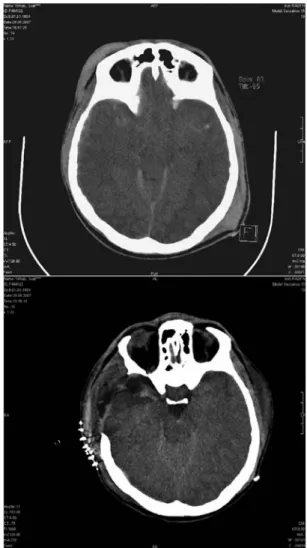
Neurological assessment at the time of surgery showed a median Glasgow Coma Scale (GCS) score of 5.5 (range 4 to 10), and median mRS score of 5. All patients had clinical signs and symptoms of transtentorial herniation (eight of 10 patients had ipsilateral third nerve palsy and all patients had contralateral hemiplegia) (Table 1). Preoperative CT showed signs of unilateral transtentorial herniation in every patient (Fig 2). Mean midline shift was 11.2 mm in the preope-rative CT scans.
CT examination on the first postoperative day showed a mean midline shift of 3.2 mm and resolution of uncal herniation in eight of 10 pati-ents. On the seventh postoperative day midline shift persisted in three patients (mean 3 mm). Mean dimensions of hemicraniectomy as mea-sured at CT was 14 by 10 cm (Fig 3).
Table 1. Summary of clinical findings in 10 patients.
Age, Sex 32, F 54, F 38, F 23, M 36, M 49, F 52, M 57, F 57, F 59, F Mean 47 Etiology Venous thrombosis SAH, vazospasm Traumatic brain injury Traumatic brain injury Traumatic brain injury SAH, ICH, vazospasm Malignant infarction Spontaneous ICH Malignant infarction Malignant MCA infarction
Neurological 3. Nerve palsy -+ + + + -+ + + + Examination Hemiplegia + + + + + + + + + + GCS Initial 8 4 9 4 6 10 5 6 5 5 median 5.5 Initial 4 5 5 5 5 5 5 5 5 5 median 5 mRS Discharge 2 4 3 5 2 4 4 3 6 4 median 4 Last FU 0 3 1 4 1 1 4 0 6 4 median 1.5
Abreviations: FU, follow up examination; GCS, Glasgow coma scale; MCA, middle cerebral artery; mRS, modified Rankin scale; ICH int-racerebral hematoma; SAH, subarachnoid hemorrhage
Figure 2. A. Preoperative computerized tomography shows transtentorial herniation. Note obliteration of the basal cis-terns, medial displacement of the medial temporal lobe, dis-tortion of the brain stem, and dilatation of the contralateral temporal horn. B. Postoperatively the basal cisterns become visible, the mesial temporal lobe and the brainstem have returned to their normal positions.
One patient was treated for postoperative menin-gitis. This patient underwent bone flap resorpti-on and cranioplasty two years postoperatively. Three patients developed subdural effusions requiring taps. One patient with malignant MCA
and anterior cerebral artery infarction developed ventricular enlargement as well as subdural effu-sion and required insertion of a ventriculo-peritoneal shunt. Another patient with malignant MCA infarction developed MCA occlusion in Figure 3. Computerized tomography images showing middle cerebral and anterior cerebral artery infarctions causing midline shift (A), postoperative scans show return of midline structures (B), edematous brain expands outside the skull (C), and cranium is recons-tructed after resolution of edema (D).
the contralateral hemisphere and died. Surviving nine patients were followed for a mean of 14 months.
Neurological examination at discharge showed median mRS score of 4. One patient died, giving an overall mortality rate of 10%. At last follow up examination mean mRS score was 1.5. Six patients had favorable outcomes (mRS <3).
DISCUSSION
The Monro-Kellie doctrine states that the skull is a rigid compartment. It follows that expansion of the contents of the rigid skull, or addition of a mass lesion, leads to elevation of intracranial pressure (ICP). This, in turn impedes cerebral blood flow, leading to secondary ischemia and edema, further elevating the ICP, thus producing a vicious cycle, where increase in edema leads to increase in ICP and increased ICP leads to edema. The process terminates in herniation and death if allowed to propagate. When treatment with osmotic agents and CSF diversion, both directed to lower ICP by reducing intracranial contents, fail to control intracranial hypertensi-on, removal of the rigid skull may break the vicious cycle. The brain can then expand witho-ut the untoward consequences anticipated by the Monro-Kellie doctrine, and without causing her-niation (6,15). Increased ICP may be due to various
causes, such as tumor, trauma, hemorrhage, infarction, etc., but it always leads to the same process of herniation.
Medical management of transtentorial herniati-on attempts to reduce cherniati-ontents of the intracranial compartment mainly by the use of hyperosmolar agents such as mannitol and hypertonic saline, by drainage of cerebrospinal fluid and by hyper-ventilation. Studies evaluating the results of aggressive medical management of transtentori-al herniation in intensive care facilities have shown that although these measures can reverse herniation, long term outcomes are bleak, with
only a small number of patients achieving good functional outcomes (2,9,12).
The results of this series, as well as others, con-firm that refractory edema and unilateral trans-tentorial herniation can be reversed with timely and judicious use of decompressive hemicrani-ectomy, allowing good functional outcomes. This series of patients with life-threatening transtentorial herniation includes patients with various pathologies, including malignant MCA infarction, ICH, SAH, venous infarction, and trauma.
The standart technique of hemicraniectomy employed in this series of patients requires that the bone flap be large, at least 14 by 10 cm, and extend down to the floors of the middle and anterior cranial fossae. The flap must also run close to the midline in order to effectively relie-ve bridging relie-veins draining to the sagittal sinus. The dura was left open in every case in this seri-es, without untoward consequences. Partial tem-poral lobectomy and uncusectomy was perfor-med early in the series. Clinical and radiological observation of reversal of herniation and return of midline structures in patients without uncu-sectomy in later patients suggested that temporal lobectomy may not be necessary in most cases, and this practise was abandoned.
In this series, timing of surgery was determined by the appearance of clinical and radiological signs and symptoms of transtentorial herniation. Every patient in this series had contralateral hemiplegia and CT signs of herniation, and eight of 10 patients had pupillary dilatation. It has been noted that appearance of CT signs of herni-ation usually precede development of full clini-cal signs and symptoms (20). Once signs of
herni-ation became evident, surgery was initiated as soon as possible. Timing of surgery is critical in that delay will lead to irreversible brainstem damage or infarction in the posterior cerebral artery area, as occurred in our patient no.2. On
the other hand, initiation of decompressive sur-gery in patients with MCA infarction or vasos-pasm before the appearance of signs of herniati-on may lead to otherwise avoidable surgical decompression in some patients. Age is another critical consideration in the decision to perform decompressive surgery. There is consensus among authors that results of decompressive surgery in malignant MCA infarction in the elderly are not satisfactory (4,8,18). Most authors
have chosen to offer surgery to patients younger than 60 years of age.
Decompressive surgery was first proposed by Kocher (15) more than a century ago. Since then
traumatic brain injury has been the most frequ-ent indication for decompressive surgery (4,7).
Hemicraniectomy, bifrontal craniectomy and holocraniectomy are performed to decompress posttraumatic intracranial hypertension (3,11,16).
All three of our patients with traumatic brain injury made excellent recoveries. Although good results for decompression after traumatic brain injury are reported by many centers, a prospec-tive randomised study of is lacking (13). On the
other hand, many retrospective series of decomp-ressions performed for MCA occlusion have reported good results, and recent randomised studies have established that in patients with malignant MCA infarction, decompressive sur-gery reduces mortality and increases the number of patients with a favorable functional outcome
(8,16-18). In a pooled analysis of three prospective
randomised studies, the mortality rate of conser-vatively treated malignant MCA infarction in patients younger than 60 years was 71 %, whe-reas decompressive surgery reduced this to 22 %
(18). Several studies have suggested that
decomp-ressive craniectomy is also associated with good outcomes in SAH patients with ICH and vasos-pasm (6,14,15).
In conclusion, our experience suggests that decompressive hemicraniectomy can reverse unilateral transtentorial herniation and lead to
good neurological outcomes when employed without delay in patients with unilateral trans-tentorial herniation.
REFERENCES
1. Acikgoz B, Ozcan OE, Erbengi A, et al.
Histopathological and microdensitometric analysis of craniotomy bone flaps preserved between abdominal fat and muscle. Surg Neurol 1986; 26:557-61.
2. Andrews BT, Pitts LH. Functional recovery after
tra-umatic transtentorial herniation. Neurosurgery 1991; 29:227-31.
3. Bose B. Emergency Decompressive Craniectomy for
Traumatic Malignant Intracranial Hypertension. Neurosurgery Quarterly 2002; 12:171-81.
4. Coplin WM, Cullen NK, Policherla PN, Vinas FC, MD, Wilseck JM, Zafonte, Rengachary SS. Safety
and Feasibility of Craniectomy with Duraplasty as the Initial Surgical Intervention for Severe Traumatic Brain. Injury J Trauma 2001; 50:1050-9.
5. Curry WT, sethi MK, Ogilvy CS, Carter BS. Factors
associated withoutcome after hemicraniectomy for large middle cerebral artery territory infarction. Neurosurgery 2005; 56:681-92.
6. Fisher CM, Ojemann RG. Bilateral decompressive
craniectomy for worsening coma in acute subarachnoid hemorrhage: Observations in support of the procedure. Surg Neurol 1994; 41:65-74.
7. Hutchinson PJ, Kirkpatrick PJ. Decompressive
cra-niectomy in head injury. Current Opinion in Critical Care 2004, 10:101-4.
8. Kilincer C, Asil T, Utku U, Hamamcioglu MK, Turgut N, Hicdonmez T, Simsek O, Ekuklu G, Cobanoglu S. Factors affecting the outcome of
decompressive craniectomy for large hemispheric infarctions: a prospective cohort study. Acta Neurochir (Wien). 2005; 147:587-94.
9. Koenig MA, Bryan M, LewinJL, Mirski MA, Geocadin RG, Stevens RD. Reversal of transtentorial
herniation with hypertonic saline. Neurology 2008; 70:1023-9.
10. Osborn AG. Diagnosis of descending transtentorial
herniation by cranial computed tomography. Radiology 1977; 123:93-6.
11. Polin RS, Shaffrey ME, Bogaev CA, et al.
Decompressive bifrontal craniectomy in the treatment of severe refractory posttraumatic cerebral edema. Neurosurgery 1997; 41:84-94.
12. Qureshi AI, Geocadin RG, Suarez JI, Ulatowski JA.
Long-term outcome after medical reversal of transten-torial herniation in patients with supratentransten-torial mass lesions. Crit Care Med 2000; 28:1556-64.
13. Sahuquillo J, Arikan F. Decompressive craniectomy
for the treatment of refractory high intracranial pressu-re in traumatic brain injury. Cochrane Database Syst Rev. 2006:CD003983. DOI: 10.1002/14651858. CD003983.
14. Schirmer CM, Hoit DA, Malek AM. Decompressive
hemicraniectomy for the treatment of intractable intrac-ranial hypertension after aneurysmal subarachnoid
hemorrhage. Stroke 2007; 38:987-992.
15. Smith ER, Carter BS, Ogilvy CS. Proposed use of
prophylactic craniectomy in poor grade aneurismal subarachnoid hemorrhage patients presenting with associated large sylvian hematomas. Neurosurgery 2002; 51:117-24.
16. Toussaint CP, Origitano TC. Decompressive
Craniectomy Neurosurg Q 2008; 18:45-53.
17. Tuncer N, Elmacı İ, Afşar N, Aktan S, Pamir N. Akut
İskemik İnmede Dekompresif Cerrahi Uygulaması. J Nervous Sys Surgery 2008; 1:20-5.
18. Vahedi Hofmeijer J, Juettler E, et al. Early
decomp-ressive surgery in malignant infarction of the MCA: a pooled analysis of three randomised controlled trials. Lancet Neurol 2007; 6:215-22.
19. Wijdicks FM, Miller GM. MR imaging of progressive
downward herniation of the diencephalon. Neurology 1997; 48:1456-59.
20. Ziai WC, Port JD, Cowan JA, Garonzik IM, Anish Bhardwaj A, Rigamonti D. Decompressive
Craniec-tomy for Intractable Cerebral Edema: Experience of a Single Center. Journal of Neurosurgical Anesthesiology 2003; 15:25-32.