Selection of our books indexed in the Book Citation Index in Web of Science™ Core Collection (BKCI)
Interested in publishing with us?
Contact [email protected]
Numbers displayed above are based on latest data collected. For more information visit www.intechopen.com Open access books available
Countries delivered to Contributors from top 500 universities
International authors and editors
Our authors are among the
most cited scientists
Downloads
We are IntechOpen,
the world’s leading publisher of
Open Access books
Built by scientists, for scientists
12.2%
118,000
130M
TOP 1%
154
Invasive and Noninvasive Approaches in Prenatal
Diagnosis of Thalassemias
Abdullah Tuli and Ebru Dündar Yenilmez
Additional information is available at the end of the chapter http://dx.doi.org/10.5772/intechopen.75570
© 2016 The Author(s). Licensee InTech. This chapter is distributed under the terms of the Creative Commons Attribution License (http://creativecommons.org/licenses/by/3.0), which permits unrestricted use, distribution, and reproduction in any medium, provided the original work is properly cited.
Abdullah Tuli and Ebru Dündar Yenilmez
Additional information is available at the end of the chapter
Abstract
Thalassemia is a significant health problem worldwide. There are two main classifica-tions, α- and β-thalassemias, which are usually caused by the defective synthesis of the α-globin, and which are commonly caused by different mutations of the β-globin chain. Different hemoglobin mutations have been identified to date. Thalassemias can result in profound anemia from early life and, if not treated with regular blood transfusions, can lead to death in the first year. Prenatal diagnosis of thalassemia is the essential part of preventive medicine and is currently dependent on the use of invasive diagnostic tests within the first 2 months of pregnancy. These diagnostic techniques carry a small but sig-nificant risk of fetal loss up to 1%. Molecular diagnostic methods have been developed for genotyping thalassemias based on PCR techniques and high-throughput technologies. Noninvasive tests using cell-free DNA (cfDNA) from a maternal blood sample is also an alternative method, thus eliminating the risk of miscarriage. This chapter summarizes the current invasive approaches and the noninvasive methods using cell-free fetal DNA as new molecular diagnostic methods for genotypic diagnosis of thalassemia in clinical practice. Prevention strategies that encompass carrier screening, genetic counseling, and prenatal diagnosis are discussed.
Keywords: thalassemia, prenatal diagnosis, noninvasive test, cell-free DNA, molecular
method
1. Introduction
Hemoglobin (Hb) is the oxygen carrier molecule in red blood cells. Each adult Hb molecule consists of four subunits: two α-globin and two β- (or β-like) globin chains. The α-globin gene
© 2018 The Author(s). Licensee IntechOpen. This chapter is distributed under the terms of the Creative Commons Attribution License (http://creativecommons.org/licenses/by/3.0), which permits unrestricted use, distribution, and reproduction in any medium, provided the original work is properly cited.
cluster maps near the telomere of the short arm of chromosome 16. The human β-globin spans a region of 70 kb on the short arm of chromosome 11 and contains 5 functional genes. Hb A is the predominant form of Hb molecule in an adult human [1–4]. The α-globin gene cluster undergoes one developmental “switch” but the β-gene cluster undergoes two “switches”. Transcription of the ε gene in the embryonic gene stage switches after the sixth week of gesta-tion to the transcripgesta-tion of the two γ genes in the fetal liver and around the prenatal period, to the δ (minor adult) and β (major adult) genes (Figure 1). At 6 months after birth, hemoglobin F (HbF) constitutes less than 5% of the total hemoglobin and continues to fall, reaching the adult level of <1% at 2 years of age [4].
2. Hemoglobinopathies
Inherited hemoglobinopathies are large groups of autosomal recessive disorders [4]. There are more than 700 defects in globin genes that are found to be responsible for hemoglobin-opathies [5]. The defective (+) or absent (0) production of one of the globin chains of the Hb tetramer causes autosomal recessive inherited disorders. The type of globin chain involved distinguishes α-, β-, and δ-thalassaemias [6]. The meaning of thalassemia comes from the Greek words thalassa; sea and aemia; anemia. It was observed that thalassemia is prevalent in areas in which malaria was seen or was endemic [7, 8].
Figure 1. Alpha beta globin gene cluster [4].
Thalassemia and Other Hemolytic Anemias 44
The resulting imbalance in the ratio of α:β chains underlies the pathophysiology [3]. Both α-thalassemia and β-thalassemia, however, have a high frequency in many populations; although, β-thalassemia is more prevalent and more widely distributed. β-Thalassaemia is a member of an inherited hemoglobin disorder family that is characterized by reduction of β-globin chain synthesis [9]. The high frequency of thalassemia is due to the protective advan-tage against malaria that it confers on carriers, analogous to the heterozygote advanadvan-tage of sickle cell hemoglobin carriers. Thalassemias can result in profound anemia from early life and, if not treated with regular blood transfusions, can lead to death in the first year [7, 10].
3. Prevalence and classification of thalassemias
Thalassaemia was originally thought to be a disease limited to the Mediterranean region, in countries such as Greece, Italy, and Cyprus. There is a characteristic distribution of thalas-semias in a band around the Old World—in the Mediterranean, the Middle East, and parts of Africa, India, and Asia [4, 5]. As a result, clinically important interactions may occur among different alleles of the same globin gene or among mutant alleles of different globin genes [6, 8]. The prevalence of β-thalassemia trait varies within the Mediterranean coastal regions in Turkey; Mediterranean (Adana 3.7%); Aegean (İzmir 4.8%); and Marmara (İstanbul 4.5%) [4] (Figure 2). In highly prevalent regions, an ideal and effective strategy to decrease the birth rate of thalassemia patients is to identify high-risk couples, who are both carriers, before pregnancy by screening (or carrier testing) and then perform a prenatal diagnosis during pregnancy [11]. Thalassemias are genetically classified according to the particular globin chains that are inef-fectively synthesized into α, β, δβ, and εδβ thalassemias. α- and β-thalassemias are important in the public health view [12]. Different hemoglobin mutations have been identified to date, the majority being single nucleotide substitutions, deletions, or insertions of nucleotides lead-ing to a frame shift; rarely does β-thalassaemia result from gene deletions. More than 250 gene defects (more than 1150 mutations have been reported) have been described for different thalassemia phenotypes in different populations [11, 13]. β-Thalassemia have been described, and it has been found that the majority thalassemias can be caused by large deletions or by point mutations. α-thalassemia is often caused by large deletions for single (α+-thal) or both
α-genes (α0-thal) (Table 1) [13].
Thalassemias exhibit a wide spectrum of phenotypes. Depending on the clinical severity, thalassemias are generally divided into three groups: (1) Thalassemia minor/trait: they are carriers who are often asymptomatic and do not need any treatment. (2) Thalassemia inter-media (TI): they have moderate anemia and occasionally require red blood cell transfusion; in α-thalassemia, it is known as Hb H disease. (3) Thalassemia major (TM): they have severe ane-mia and require transfusions for survival; in α-thalasseane-mia, this clinical form was named Hb Bart’s hydrops fetalis. The fetus usually dies in utero or shortly after birth [11]. According to the degree of quantitative reduction in the synthesis of normal β-globin, β-thalassemia muta-tions are classified into three groups: (1) β0-thalassemia mutation, which results in the absence
(3) β++-thalassemia mutation, also known as silent β-mutation, which mildly reduces the
out-put of β-globin (Figure 3) [11].
Clinical presentation for thalassemia carriers is varied from almost healthy to severe anemia requiring blood transfusions all life [14]. A practical way to prevent thalassemia is identifica-tion of carrier couples; genetic counseling and offering prenatal diagnostic services for both carrier couples. Two carrier parents have a 25% chance of having an unaffected child, a 25%
Figure 2. The distribution of common β-thalassemia mutations [7].
Table 1. The common deletions and mutations of thalassemias [13].
Thalassemia and Other Hemolytic Anemias 46
chance of having an affected one, and a 50% risk of having a carrier child. Carriers can only be detected using laboratory methods. Laboratories should increase the diagnostic proficiency for prevention of this blood disorder [13, 15, 16].
In this review, we aim to provide an algorithm consisting of biochemical and molecular meth-ods in the screening of thalassemia carriers and evaluate traditional and new prenatal diagno-sis methods for the disorder.
4. Identification of carrier and diseased individuals with
thalassemias
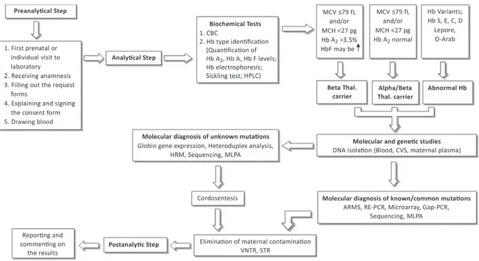
Carrier detection is a necessity in populations in which both α- and β-thalassemia are prev-alent. For laboratory diagnosis of thalassemias, molecular diagnostic algorithms should be produced by screening centers (Figure 4). Combining blood hematology/biochemistry and clinical parameters with laboratory analysis, discussing the clinical results with physicians is important as the beginning step for carrier identification. The hematological information obtained from a complete blood count (CBC) is the first essential test for screening thalas-semias (Figure 1). Specific mutations of hemoglobin will cause hematologic changes.
Hypochromic microcytic parameters and anemia is generally mild in α- or β-thalassemia carri-ers. In cases with rare β-thalassemia, anemia can be intermediate or hemolytic and even severe because it is due to the reduced expression of the thalassemic gene and depends significantly on the degree of β-α imbalance [13].
The first set of carrier detection procedure is the determination of main erythrocyte indices parameters such as the erythrocyte mean corpuscular volume (MCV), erythrocyte mean cor-puscular hemoglobin (MCH) determination and also HbA2 quantitation. The current screening strategies for thalassaemia carriers are to identify individuals with clinically related mutations. Hypochromic microcytic anemia occurs in both iron deficiency anemia and thalassemias. A decrease in red blood cell count (RBC) and an increase in red cell distribution width (RDW) is accompanied by iron deficiency anemia. Increased RBC count and normal RDW values have been seen in hemoglobin disorders [13]. Serum ferritin levels, the golden standard, will usually be normal or elevated in β-thalassemia carriers but might also be borderline or low in α-thalassemia carriers [13]. Decreased hemoglobin (Hb) concentration and MCV levels, altered shape and size of the RBCs (anisopoikilocytosis) and existence of target cells are additional hematologic phe-notypes observed in individuals with hemoglobin abnormalities. When compared to MCV, the MCH is a more stable parameter, and values of <27 pg and <25 pg have been recommended as alternative screening cut-offs for β-thalassemia and α°-thalassemia, respectively (Table 2) [17].
4.1. β-Thalassemia
Heterozygote β-thalassemia (β° or β+) is characterized by high red blood cell count,
micro-cytosis, hypochromia, increased HbA2 levels, and unbalanced α-globin/non-α-globin chain synthesis. Elevation of HbA2 (standard cut-off value is above 3.5%) is the most important characteristic in identifying heterozygote β-thalassemia [7, 19, 20]. A number of heterozy-gotes for β-thalassemia may have normal or borderline HbA2 levels [21]. Some typical car-riers have mild β+ thalassemia mutation, i.e., IVSI-6 (T-C) mutation. The δ- and β-double
heterozygotes exhibit normal HbA2 level, low MCV and MCH values. These double het-erozygote cases should be separated from the α-thalassemia carriers. Carriers of γδβ- and δβ-thalassemias HbA2 levels are also normal. δβ-thalassemias have specific elevated HbF
Figure 4. The algorithim of screening and prenatal diagnosis for thalassemias.
Thalassemia and Other Hemolytic Anemias 48
levels, which distinguish this group from the others [5, 12]. In the presence of a normal MCH, hereditary persistence of fetal hemoglobin (HPFH) should be considered. Another problem-atic group difficult to identify is the silent β-thalassemia and the triple α-gene arrangement. In silent β-thalassemia, the MCV and MCH values are usually normal (i.e., β+-101 C > T
muta-tion). The value of HbA2 and HbF are also normal (Table 3) [21, 22].
4.2. α-Thalassemia
α-Thalassemia mutations are mostly gene deletions. To identify known inherited deletional α-thalassemia mutations, gap-PCR (polymerase chain reaction) is the most common method. A multiplex gap PCR targeting common α gene deletions in a population is most often used as the first step in α-thalassemia genotyping. The sequencing method detects non-deletion α+-thalassemia mutations. The five commonest α°-thalassaemia deletions can be diagnosed
Traits Hb Ferritin MCV MCH RBC RCM
Iron deficiency ↓ ↓↓ ↓ ↓ ↓ APC, HY
β-Thalassemia trait ↓ ↑↑ ↓ ↓ ↑↑↑ APC, HY, TC
α-Thalassemia (-α/αα) ↓or N ↓N↑ ↓ ↓ ↑ Rare TC, HY
α-Thalassemia (--/αα) ↓ N↑ ↓ ↓ ↑↑ APC, HY, TC, rare IB
α-Thalassemia (-α/-α) ↓ N↑ ↓ ↓ ↑↑ APC,HY, TC
α-Thalassemia (--/-α) ↓ ↑↑↑ ↓ ↓ ↓ APC, HY, TC, IB
Hb, hemoglobin; MCV, mean corpuscular volume; MCH, mean corpuscular hemoglobin; RBC, red blood cell; N, normal; APC, anisopoikilocytosis; HY, hypochromia; TC, target cells; IB, inclusion bodies [18].
Table 2. The most significant parameters observed from complete blood count, ferritin, MCV, MCH, RBC, and red cell
morphology tests in the common traits at risk of intermediate or severe conditions for thalassemias.
Phenotype Genotype
Normal red cell indices • α- and β-thalassemia interaction
Normal HbA2 level • Iron deficiency
• Co-inheritance of δ- and β-thalassemmia • Some mild β-thalassemmia mutations • γδβ-thalassemia
Normal red cell indices and HbA2 level
(silent) • Silent β-thalassemmia mutations
• α-globin gene triplication Severe heterozygote β-thalassemmia • Hyper unstable hemoglobin
• Co-inheritance of heterozygote β-thalassemmia and triple α-globin gene
by gap-PCR: the --SEA allele, the --MED and -(α)20.5 alleles; the --FIL allele, and finally the --THAI allele. The two commonest α+-thalassemia deletions are also diagnosed by gap-PCR:
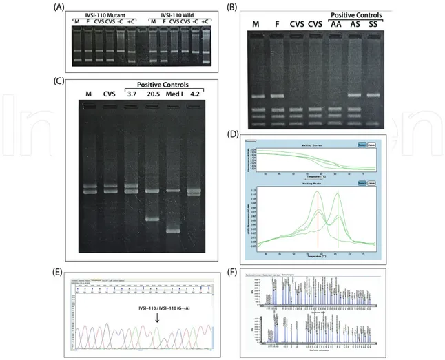
the -α3.7 and -α4.2 alleles. In our laboratory, the common deletions detected with gap-PCR are -α3.7, and -(α)20.5, --MED and -α4.2 (Figure 5).
5. Molecular methods
Blood count is not always consistent with typical β-thalassemia trait. Furthermore, there is no specific screening test for the clear identification of heterozygote α-thalassaemia. Only molecular DNA analysis could give certain results in rare mutations [23, 24].
Molecular methods for DNA analysis of hemoglobinopathies currently in use are based on PCR methods that can be used to detect the globin gene mutations. The PCR-based methods differ in identifying hemoglobin variants. Amplification refractory mutation system (ARMS), denatur-ing gradient gel electrophoresis (DGGE), restriction endonuclease PCR (RE-PCR), real-time PCR, high-resolution melting analysis (HRM), sequencing analysis (Sanger), pyrosequencing,
Figure 5. Applications of (A): ARMS-PCR, (B): RE-PCR for, (C): Gap-PCR, (D): HRMA and (E): sequencing analysis, (F):
Detection unknown mutations using MLPA for deletional alpha, beta gene mutations (M: Mother, F: Father, C: Control, CVS: Chorionic villus sample).
Thalassemia and Other Hemolytic Anemias 50
microarrays can be counted among these PCR-based detecting methods [19, 25]. To detect deletions, Southern blot analysis has been replaced by methods including gap-PCR, mul-tiplex ligation-dependent probe amplification (MLPA), and array comparative genome hybridization (aCGH) [19]. Gene scanning methods such as HRM analysis are also useful for locating possible β-globin gene variants [26]. This technique allows the detection of muta-tions between the primers used in the assay, which is in contrast to more localized tech-niques such as hybridization based technologies or restriction enzyme-based assays [27]. In diagnostic use, for this method, it is mandatory to characterize any nucleotide variation by automated sequencing because they do not determine nucleotide changes. Small deletions can be detected by polyacrylamide gel electrophoresis of an amplified beta gene product. Polyacrylamide gel electrophoresis can be used to detect small deletions in an amplified β gene product. Gap-PCR and recently MLPA identifies larger deletions from the β-globin gene [10]. MLPA detects all common, rare, and novel forms of deletional α-thalassemia (in contrast to gap-PCR) and provides a reliable alternative screening method for the prenatal diagnosis of α°-thalassemia [28, 29, 30]. This technique can also identify triple and qua-druple α-gene re-arrangements [31]. β°-Thalassemia, which is caused by small nucleotide deletions and some larger deletions which the whole β-globin gene removes, can be identi-fied by gap-PCR (Hb Lepore, some δβ-thalassaemia deletions, and the HPFH1/2/3 deletion mutations, etc.).
5.1. Sequencing analysis
The parents’ DNA whose mutations were not found by classic PCR methods were analyzed by ABI 3130 automatic sequencer. The DNA of the cord blood samples were sequenced to confirm the cordosentesis results. BigDye Terminator v3.1 Cycle Sequencing kit (ABI) and primers that cover all the exons, introns,and exon-intron boundaries of the β-globin gene were used for sequencing analysis.
6. Prenatal diagnosis of thalassemias
Prenatal diagnosis created a new option to couples at risk of a major hemoglobinopathy and changed the perspective of screening and counseling for thalassemias [7]. The first step to pre-vent thalassemia is prenatal diagnosis of these hematological disorders. Prenatal diagnosis for thalassemias is still carried out by traditional conventional methods such as amniocente-sis, chorionic villus sampling (CVS), and cordocentesis. These conventional methods have a risk of fetal miscarriage risk around 1% [32].
6.1. Blood sampling from parents
Phenotype of parents should be performed by DNA analysis after whole blood count and electrophoresis. Hematology results should be sent to the molecular diagnostic labo-ratory. Antenatal screening of parents should be performed before the first trimester of pregnancy [17].
6.2. Fetal sampling
Fetal DNA for analysis can be obtained by traditional invasive methods; amniocytes, CVS, and cordosentesis [4, 36].
6.2.1. Chorionic villus sampling
A fetal-derived tissue, genetically reflecting the fetus and easily accessible in the first trimester (up to 11 weeks) can be used for the prenatal diagnosis of hemoglobinopathies. High-quality DNA can be obtained from CVS material to perform DNA analysis. Maternal contamina-tion is low, especially if careful microscopic disseccontamina-tion is performed to remove contaminating maternal tissues prior to DNA extraction and analysis.
6.2.2. Amniocentesis
This is the most commonly used method among invasive prenatal diagnostic methods. Amniotic fluid is the environment of life before the baby is born, and all secretions are in this atmosphere [33]. The prenatal diagnosis result based on an amniocentesis is available later in pregnancy compared to CVS, as amniocentesis is not usually performed earlier than the 15th week [19].
6.2.3. Fetal blood sampling
This sampling method can be used for molecular analysis, globin chain synthesis stud-ies, or high-pressure liquid chromatography (HPLC). It can be useful in women at risk of α-thalassaemia hydrops fetalis. Fetal blood sampling is associated with a higher rate of mis-carriage and results are available much later in pregnancy (after 18–20 weeks) [19].
6.2.4. HPLC analysis of cord blood
Cord blood is taken in ethylenediaminetetraacetic acid (EDTA) by an obstetrician at 18–20 weeks of gestation. The hemograms are measured for all samples by using an automatic blood cell counter (Coulter T-890). HPLC analysis were studied from the prepared hemoly-sate by Agilent 1100 using the thalassemia short program. The levels of HbA, HbF and Hb Barts, and HbS were estimated.
Molecular diagnostic methods that are based on PCR techniques (ARMS, RFLP, GAP-PCR, VNTR, etc.) and high-throughput technologies (Gene expression, HRMA, microarray, MLPA, NGS, etc.) are currently used in prenatal diagnosis. The molecular genetic tests for prenatal diagnosis within the first 2 months of pregnancy is necessary to prevent infants of carrier couples from being thalassemia [19].
6.3. Remove the contamination risk
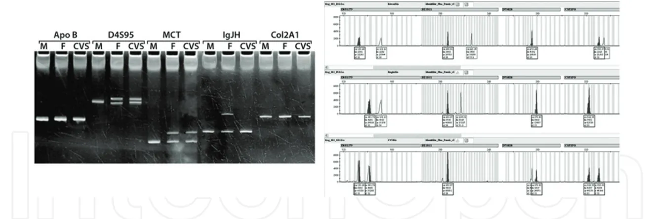
Maternal cell contamination test is recommended in all prenatal samples to remove the risk of contamination of fetal DNA with maternal DNA. It is important to pay attention to all CVS and AF samples (with or without culture) may have maternal contamination. Maternal contamination should be ruled out after careful dissection of CVS from maternal tissue.
Thalassemia and Other Hemolytic Anemias 52
Polymorphic DNA sites in fetal and parental samples can be identified to monitor maternal contamination (Figure 6).
6.4. New diagnostic tests
The discovery of cell-free fetal DNA (cffDNA) in the maternal plasma give the chance to con-duct noninvasive prenatal diagnosis (NIPD) during pregnancy [34]. Some encouraging clini-cal noninvasive approaches to detect paternally inherited mutations such as the detection of fetal sex and RhD status have been improved successfully [35, 36].
Noninvasive tests using cell-free DNA (cfDNA) from a maternal blood sample is also an alternative method, thus eliminating the risk of miscarriage (Figure 7). Cell-free fetal DNA (cffDNA) constitutes approximately 10–15% of the total cfDNA and has been shown to repre-sent the entire fetal genome [35].
6.5. Centrifugation
The noninvasive prenatal detection of paternal mutations in β-thalassemias is achievable using HRM analysis, and next generation sequencing of cell-free fetal DNA [36].
7. Conclusion
β-Thalassemia is an autosomal recessive disorder characterized by hemolytic anemia and micrositosis. It causes decreased synthesis of β-globin chain. This disorder influences 150 mil-lion people in large parts of Asia, North Africa, and in the Mediterranean. A wide range of mutations occurs due to the effects of different cultures living together in Turkey. Genetic heterogenity is more in the southern part of Turkey than in other regions.
Figure 6. VNTR and STR analysis of a family for checking the maternal contamination in CVS. M: Mother, F: Father,
CVS: Choronic Villus Sampling.
Premarital screening and genetic counseling are essential for the prevention and control of thalassemia and hemoglobinopathies [9]. Today, prenatal diagnosis by chorionic villus sam-pling is an accepted method to protect families having children with thalassemia major and to keep the disease under control. The mutation can be identified from samples obtained by chorionic villus sampling (CVS) and amniosentesis methods by classic polymerase chain reac-tion (PCR).
The molecular diagnostic algorithms should be produced by genetic diagnosis centers for screening of the carriers and prenatal diagnosis of the couples both of whom carriage has been detected before pregnancy for protection against thalassemia.
Author details
Abdullah Tuli* and Ebru Dündar Yenilmez
*Address all correspondence to: [email protected]
Faculty of Medicine, Department of Medical Biochemistry, Cukurova University, Adana, Turkey
References
[1] Nussbaum RL, McInnes RR, Willard HF. Thompson & Thompson Genetics in Medicine. Canada: Elsevier Health Sciences; 2015. p. 233
[2] Tuzmen S, Schechter A. Genetic diseases of hemoglobin: Diagnostic methods for eluci-dating β-thalassemia mutations. Blood Reviews. 2001;15(1):19-29
[3] Cappellini M-D, Cohen A, Porter J, Taher A, Viprakasit V. Guidelines for the manage-ment of transfusion dependent thalassaemia (TDT). TIF Publication. 2014;20:236
[4] Cao A, Galanello R, Rosatelli MC. 8 prenatal diagnosis and screening of the haemoglo-binopathies. Baillière's Clinical Haematology. 1998;11(1):215-238
[5] Old J, Traeger-Synodinos J, Galanello R, Petrou M, Angastiniotis M. Prevention of thal-assaemias and other haemoglobin disorders. Thalassaemia International Federation Publications. 2005;2:113-116
[6] Porter J, Taher A, Mufarrij A, Gavalas M. Emergency Management of Thalassaemia. Nicosia, Cyprus: Thalassaemia International Federation; 2012
[7] Acemoglu H, Beyhun NE, Vancelik S, Polat H, Guraksin A. Thalassaemia screening in a non-prevalent region of a prevalent country (Turkey): Is it necessary? Public Health. 2008;122(6):620-624
[8] Ip H-W, So C-C. Diagnosis and prevention of thalassemia. Critical Reviews in Clinical Laboratory Sciences. 2013;50(6):125-141
Thalassemia and Other Hemolytic Anemias 54
[9] Weatherall D, Clegg J. Inherited haemoglobin disorders: An increasing global health problem. Bulletin of the World Health Organization. 2001;79(8):704-712
[10] Shang X, Xu X. Update in the genetics of thalassemia: What clinicians need to know. Best Practice & Research Clinical Obstetrics & Gynaecology. 2017;39:3-15
[11] Giordano P, Smit J, Herruer M, Huisman W, Pouwels J, Verhoef N. Carrier diagnos-tics and prevention of sickle cell disease and thalassemia major; recommendation of the hemoglobinopathies workgroup. Ned Tijdschr Klin Chem Labgeneesk. 2006;31:301-305 [12] Musallam KM, Taher AT, Rachmilewitz EA. Beta-thalassemia intermedia: A clinical
per-spective. Cold Spring Harbor Perspectives in Medicine. 2012;2(7):a013482
[13] Barrett AN, Saminathan R, Choolani M. Thalassaemia screening and confirmation of carriers in parents. Best Practice & Research Clinical Obstetrics & Gynaecology. 2017;
39:27-40
[14] Kaufmann J, Smit J, Huisman W, Idema R, Bakker E, Giordano P. Basic haemoglobinopa-thy diagnostics in Dutch laboratories; providing an informative test result. International Journal of Laboratory Hematology. 2013;35(4):428-435
[15] Ryan K, Bain BJ, Worthington D, James J, Plews D, Mason A, et al. Significant haemo-globinopathies: Guidelines for screening and diagnosis. British Journal of Haematology. 2010;149(1):35-49
[16] Giordano P. Strategies for basic laboratory diagnostics of the hemoglobinopathies in multi-ethnic societies: Interpretation of results and pitfalls. International Journal of Laboratory Hematology. 2013;35(5):465-479
[17] Traeger-Synodinos J, Harteveld CL, Old JM, Petrou M, Galanello R, Giordano P, et al. EMQN best practice guidelines for molecular and haematology methods for carrier identification and prenatal diagnosis of the haemoglobinopathies. European Journal of Human Genetics. 2015;23(4):426-437
[18] Trent R, Higgs D, Clegg J, Weatherall D. A new triplicated α-globin gene arrangement in man. British Journal of Haematology. 1981;49(1):149-152
[19] Galanello R, Barella S, Turco MP, Giagu N, Cao A, Dore F, et al. Serum erythropoi-etin and erythropoiesis in high- and low-fetal hemoglobin beta-thalassemia intermedia patients. Blood. 1994;83(2):561-565
[20] Gonzales-Redondo J, Stoming T, Kutlar F. Hb Monroe or a2 ß230 (B12) Arg→ Thr, a variant associated with ß-thalassemia due to a G→ C sub-stitution adjacent to the donor splice site of the first intron. Hemoglobin. 1989;13:67-74
[21] Sin S, Ghosh A, Tang L, Chan V. Ten years' experience of antenatal mean corpuscular volume screening and prenatal diagnosis for thalassaemias in Hong Kong. Journal of Obstetrics and Gynaecology Research. 2000;26(3):203-208
[22] Leung TN, Lau TK, Chung TK. Thalassaemia screening in pregnancy. Current Opinion in Obstetrics and Gynecology. 2005;17(2):129-134
[23] Yenilmez ED, Tuli A. New perspectives in prenatal diagnosis of sickle cell anemia. In: Sickle Cell Disease-Pain and Common Chronic Complications. Croatia: InTech; 2016. pp. 39-52
[24] Montgomery JL, Sanford LN, Wittwer CT. High-resolution DNA melting analysis in clini-cal research and diagnostics. Expert Review of Molecular Diagnostics. 2010;10(2):219-240 [25] Shih H-C, Er T-K, Chang T-J, Chang Y-S, Liu T-C, Chang J-G. Rapid identification of
HBB gene mutations by high-resolution melting analysis. Clinical Biochemistry. 2009;
42(16):1667-1676
[26] Harteveld CL, Voskamp A, Phylipsen M, Akkermans N, den Dunnen JT, White SJ, et al. Nine unknown rearrangements in 16p13.3 and 11p15.4 causing α-and β-thalassaemia characterised by high resolution multiplex ligation-dependent probe amplification. Journal of Medical Genetics. 2005;42(12):922-931
[27] Liu J-Z, Han H, Schouten JP, Wang L-R, Fan X-P, Duarte HB, et al. Detection of α-thalassemia in China by using multiplex ligation-dependent probe amplification. Hemoglobin. 2008;32(6):561-571
[28] Liu Y, Miles K, Old J, Fisher C, Weatherall D, Clegg J, editors. Rapid Detection of Alpha-thalassaemia Deletions and Alpha-globin Gene Triplications by Multiples PCRs. Ameri-can Journal of Human Genetics. Chicago, IL, USA: Univ. Chicago Press; 1999
[29] Harteveld C, Refaldi C, Cassinerio E, Cappellini M, Giordano P. Segmental duplications involving the α-globin gene cluster are causing β-thalassemia intermedia phenotypes in β-thalassemia heterozygous patients. Blood Cells, Molecules, and Diseases. 2008;
40(3):312-316
[30] Yenilmez ED, Tuli A. A non-invasive prenatal diagnosis method: Free fetal DNA in maternal plasma. Archives Medical Review Journal. 2013;22(3):317-334
[31] Wapner RJ, editor. Invasive Prenatal Diagnostic Techniques. Seminars in Perinatology. Elsevier; 2005
[32] Lo YMD, Corbetta N, Chamberlain PF, Rai V, Sargent IL, Redman CWG, et al. Presence of fetal DNA in maternal plasma and serum. The Lancet. 1997;350(9076):485-487
[33] Moise KJ, Boring NH, O'Shaughnessy R, Simpson LL, Wolfe HM, Baxter JK, et al. Cir-culating cell-free fetal DNA for the detection of RHD status and sex using reflex fetal identifiers. Prenatal Diagnosis. 2013;33(1):95-101
[34] Yenilmez ED, Ozgünen FT, Evrüke IC, Tuli A. Noninvasive fetal RHD genotyping by multiplex real-time PCR in maternal plasma. International Journal of Current Medical Research abbreviation. 2015;4(2):344-347
[35] Dennis Lo YM, Poon LLM. The ins and outs of fetal DNA in maternal plasma. The Lancet. 2003;361(9353):193-194
[36] Yenilmez ED, Tuli A, Evruke IC. Noninvasive prenatal diagnosis experience in the Cukurova region of southern Turkey: Detecting paternal mutations of sickle cell ane-mia and beta-thalasseane-mia in cell-free fetal DNA using high-resolution melting analysis. Prenatal Diagnosis. 2013;33(11):1054-1062
Thalassemia and Other Hemolytic Anemias 56
![Figure 1. Alpha beta globin gene cluster [4].](https://thumb-eu.123doks.com/thumbv2/9libnet/4146934.63494/3.918.100.812.137.597/figure-alpha-beta-globin-gene-cluster.webp)
![Figure 2. The distribution of common β-thalassemia mutations [7].](https://thumb-eu.123doks.com/thumbv2/9libnet/4146934.63494/5.918.107.811.126.603/figure-distribution-common-β-thalassemia-mutations.webp)
![Figure 3. Clinic classification and genotype-phenotype association of thalassemias [11].](https://thumb-eu.123doks.com/thumbv2/9libnet/4146934.63494/6.918.112.813.139.561/figure-clinic-classification-genotype-phenotype-association-thalassemias.webp)
![Table 3. Interpretations to consider when the hematologic is consistent with atypical β-thalassaemia trait [7].](https://thumb-eu.123doks.com/thumbv2/9libnet/4146934.63494/8.918.110.804.803.1127/table-interpretations-consider-hematologic-consistent-atypical-thalassaemia-trait.webp)