[Orthopedic Reviews 2017; 9:6825] [page 19]
A case of schwannoma
of the common peroneal
nerve in the knee
Tayyar Taylan Öz,1Birol Aktaş,1 Korhan Özkan,1Burak Özturan,1 Bulent Kilic,2Murat Demiroğlu1 1Department of Orthopaedics
and Traumatology, İstanbul Medeniyet University, Göztepe Training
and Research Hospital, Istanbul; 2Department of Health Sciences, İstanbul Gelişim University, Istanbul, Turkey
Abstract
Schwannomas are benign neurogenic tumors of peripheral nerves that originate from neural sheath. The aim of this case report is to bring to mind the schwannoma in the patient with knee pain. A 39-year-old woman presented with a complaint of knee pain of three months history. After surgical intervention, the patient’s complaints com-pletely disappeared in the post-operative period. This should come to mind in the dif-ferential diagnosis of knee pain.
Introduction
Neurilemmomas (neurinoma-schwan-noma) are benign, solitary, well-demarcat-ed – usually as a capsule, slowly growing tumors that arise from the proliferation of active peripheral schwann cells.1,2 They
make up 5% of benign soft tissue tumors. They are frequently seen between the ages of 20 and 50. Schwannomas, or neurilem-momas, are the most common benign
peripheral nerve sheath tumours. Cases of common peroneal nerve schwannomas are very rare in the literature (Table 1).3-8They
usually have a clinically silent course, tho-ugh the present case highlights how a com-mon peroneal nerve schwannoma can become symptomatic due to mechanical compression, resulting in pain, swelling or a lump. A thorough examination and prompt investigation helped us make a timely diagnosis and initiate appropriate treatment. Schwannomas should be inclu-ded in the differential diagnosis of lumps in the vicinity of nerves. This case presen-tation highlights the importance of screen-ing for the possibility of a peroneal schwannoma in patients presenting with knee pain despite nonspecific findings of physical examination.
Case Report
A 39-year-old woman presented with a complaint of knee pain of three months his-tory. Physical examination revealed a palpa-ble mass on the peroneal nerve and numb-ness around the deep peroneal nerve.
Routine laboratory diagnostic tests and x-rays showed no specific findings. Therefore, an MRI scan was obtained.
Orthopedic Reviews 2017; volume 9:6825
Correspondence: Murat Demiroğlu, Department of Orthopaedics and Traumatology, İstanbul Medeniyet University, Göztepe Training and Research Hospital, Istanbul, Turkey. Tel: +90.5334847081.
E-mail: [email protected] Key words: Schwannoma; Common peroneal nerve; Knee.
Conflict of interest: the authors declare no potential conflict of interest.
Received for publication: 7 August 2016. Revision received: 15 Januray 2017. Accepted for publication: 29 January 2017. This work is licensed under a Creative Commons Attribution NonCommercial 4.0 License (CC BY-NC 4.0).
©Copyright T.T. Öz et al., 2017 Licensee PAGEPress, Italy Orthopedic Reviews 2017;9:6825 doi:10.4081/or.2017.6825
Figure 1. Intraoperative view of the super-ficial peroneal nerve on the lateral side of the knee, with a mass arising from it.
Figure 2. Microscopic images at various degrees of magnification of different areas of the schwannoma lesion. A) Under the outermost thin fibrous capsule are mostly Antoni A, and to a lesser extent, hypocelluar Antoni B areas, hematoxylin and eosin (H&E), x40. B) The peripheral nerve bundle, located on the left of and contiguous to the schwannoma lesion, contains Antoni A and B areas, from which the roots of the lesion originate, H&E, x100. C) At a larger magnification, microscopic view of the Antoni A areas. Verocay bod-ies, showing a palisade arrangement of the nuclei, form elongated spindle-like cell groups within the network-like fibers at the center of the cytoplasms, H&E, x200. D) At a larger magnification, microscopic view of the Antoni B areas. They are relatively more hypocel-lular and contain enlarged blood vessels that are more hypocelhypocel-lular, edematous, and flac-cid, H&E, x200.
A
B
C
D
Non commercial
[page 20] [Orthopedic Reviews 2017; 9:6825]
Discussion
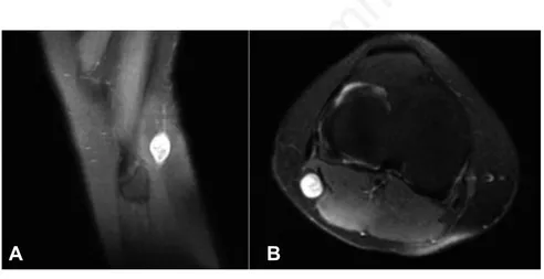
On contrast-enhanced MRI T2 weight-ed imaging, an 11 mm mass was detectweight-ed, with low signal intensity at the center and high signal intensity at the perimeter involving the common peroneal nerve and
demonstrating nerve continuity.
During surgery, a longitudinal incision was made (Figure 1) and the lesion was exposed by longitudinal cutaneous and sub-cutaneous incisions. The peroneal nerve was located and dissected. After longitudi-nal dissection of the perineurioma, the
lesion was excised with its roots in the com-mon peroneal nerve.
The excised material was sent for histopathologic examination which showed a capsule formed in the epineurium, charac-terized by Antoni A and Antoni B areas, compatible with a schwannoma (Figures 2-4). The patient’s complaints completely dis-appeared in the post-operative period.
Conclusions
Schwannomas are benign lesions and their excision is generally curative. Malignant transformation is rare. They can occur in the sacral plexus and sciatic nerve in the pelvis, and in the ulnar and peroneal nerves in the extremities. Lesions often do not interfere with the anatomical or func-tional operations of nerve cells. The tumor can be removed with a careful dissection after a longitudinal incision of the per-ineurium. Nerve continuity must be main-tained during surgery. Nerve dysfunction occurs rarely.
References
1. Akbay A. Periferik sinir tümörleri. Türk Nöroşirürji Der 2005;15:225-6. 2. Houshian S, Freund KG. Gigantic
benign schwannoma in the lateral pero-neal nerve. Am J Knee Surg 1999; 12:41-2.
3. Russell RC, Williams NS, Bulstrode CJ. Bailey and Love’s short practice of sur-gery, 24th ed. London: Arnold; 2004. 4. Stuot AP. Tumours of the peripheral
nervous system. Atlas of tumour patho-logy. Section II, fascicle 6. Washington, DC: Armed Forces Institute of Pathology; 1949.
5. Rafai MA, El Otmani H, Rafai M, et al. Peroneal nerve schwannoma presenting with a peroneal palsy. Rev Neurol 2006,162:866-8.
6. Cato T, Bain LM, Yue JJ, Glick H. Schwannoma of the superficial peroneal nerve presenting as web space pain. J Foot Ankle Surg 1995;34:532-3. 7. Mahitchi E, Van Linthoudt D.
Schwannoma of the deep peroneal nerve. An unusual presentation in rheu-matology. Praxis 2001;96:69-72. 8. Shariq O, Radha S, Konan S. Common
peroneal nerve schwannoma: an unusu-al differentiunusu-al for a symptomatic knee lump. BMJ Case Rep 2012;2012: bcr2012007346.
Case Report
Table 1. Some related case reports.
Authors Location Complaints Outcome
Shariq et al. (2012)8 Knee Knee lump Required intralesional excision
Houshian et al. (1999)2 Knee Pain and numbness Successful
Mahitchi et al. (2001)7 Knee Pain Successful
Cato et al. (1995)6 Knee Web space pain Successful
Figure 3. Strong and diffuse positive cytoplasmic nuclear immunohistochemical staining for S100, x200.
Figure 4. Sagittal (A) and axial (B) T2-weighted magnetic resonance images show a well-defined ovoid mass posterior to the fibula head, arising eccentrically from and which can-not be sepearated from the underlying common peroneal nerve.