SAFETY AND EFFICACY OF TIROFIBAN HYDROCHLORIDE (TH) IN CERULEIN-INDUCED ACUTE PANCREATITIS
AYDIN KAPLAN1, SECKINAKKUCUK2, MUSTAFAUGUR2, AKINAYDOGAN2, TUMAYOZGUR3, YASEMINCALISKAN4, IBRAHIM YETIM2, ILHANPALTACI5, MUHYITTINTEMIZ2
1Siirt State Hospital, General Surgery Clinic, Siirt - 2Medicine School of Mustafa Kemal University, Department of General Surgery, Hatay - 3Medicine School of Mustafa Kemal University, Department of Pathology, Hatay - 4Medicine School of Mustafa Kemal University, Department of, Biochemistry, Hatay - 5Gediz State Hospital, General Surgery Clinic, Kutahya, Turkey
Introduction
Acute pancreatitis (AP) is a life threatening dis-ease and is also an important cause for hospitaliza-tion. Although most of the cases are mild and mortal-ity is under 1%, mortalmortal-ity rate can increase up to %30 due to severity of the disease(1, 2). Alcohol use, gallstones, hypercalcemia, drugs, endoscopic retro-grade cholangiopancreatography and the trauma are the most encountered causes(3).
Despite the mild nature of most cases, intensive care may be indicated, particularly depending on complications. In general, acute pancreatitis is mortal in 2-7 % of cases due to multiple organ failure and
pancreatic necrosis, usually occurs within 1-2 weeks after diagnosis(4).
The tissue perfusion is important during the occurrence and survey of pancreatitis. Microthrombosis and capillary circulation directly affect the tissue perfusion. Therefore, the most important factors in pancreatitis physiopathology are capillary vasodilation, hyperpermeabililty and platelet aggregation(5, 6).
Tirofiban is a non-peptide agent and irreversible antagonist of major thrombotic surface aggregation factor, GP IIb/IIIa and is an antiaggregant agent and is frequently used in coronary artery disease. Many randomized controlled studies reported that tirofiban
Received January 30, 2015; Accepted March 30, 2015 ABSTRACT
Introduction: Tissue perfusion plays an important role in pancreatitis. Free oxygen radicals also have some triggering roles in the severity of acute pancreatitis. Tirofiban hydrochloride (TH) is being used for ischemic disorders for many years and is known as an antiaggregant drug. We aimed to determine the therapeutic effects of tirofiban in cerulein-induced acute pancreatitis in this animal study.
Materials and methods: Twenty-four Wistar-Albino male rats were divided into three equal groups; Group I: Sham-operated group, Group II: cerulein induced pancreatitis group, Group III: cerulein induced pancreatitis + treatment group (intraperitoneal 0.25 mg tirofiban hydrochloride in 1 ml isotonic saline solution). Amylase and lipase levels were studied in blood samples and malondialdehyde, glutathione peroxidase and superoxide dismutase activities were measured in tissue samples.
Results: Amylase and lipase levels were elevated in acute pancreatitis group. The levels of both enzymes were decreased after the rats were medicated with TH. Malondialdehyde (MDA) was higher in pancreatitis group without treatment. There were no significant differences between groups according to glutathione peroxidase (GSH-Px) and superoxide dismutase (SOD) levels.
Conclusion: The present findings suggest that TH has an antiaggregant effect and may reduce the progression and improve the healing of acute pancreatitis.
is a potent inhibitor of thrombocyte aggregation and it prevents arterial thrombosis(7, 8).
In this animal study we aimed to evaluate the therapeutic effects of TH in cerulein induced acute pancreatitis.
Materials and methods
Twenty-four Wistar-Albino male rats, weighing 180-200 g, were housed under constant temperature (21 ± 2 ° C) individually in wire cages with a 12-hour light-dark cycle. No enteral or parenteral antibi-otics were administered during study. The procedures in this experimental study were performed in accor-dance with the National Guidelines for the Use and Care of Laboratory Animals and approved by the Animal Ethics Committee of Mustafa Kemal University.
Rats were divided into three groups, each with 8 rats: Group I: Sham-operated group, Group II: cerulein induced pancreatitis (CIP) group, Group III: Treatment with intraperitoneal 0.25 mg tirofiban hydrochloride in 1 ml isotonic saline solution after CIP group.
On study day, the rats were sedated with 60 mg/kg ketamine hydrochloride and 5 mg/kg xylazine hydrochloride via intramuscular way. Subcutaneous 20 µg/kg of saline solution was administered to the rats in Sham-operated group 4 times an hour apart. Blood and tissue samples were taken after 24 hours of saline infusion. 20 µg/kg of cerulein was injected subcutaneously 4 times an hour apart to Group II and III and then intraperitoneal 0.25 mg tirofiban hydrochloride in 1 ml isotonic saline solution was administered to Group III. All of the animals were sacrificed after 24 hours and blood and then tissue samples were taken. Pancreas tissues were divided into two pieces for biochemical and histopathologi-cal examinations.
Pancreas samples were removed and washed with isotonic saline and kept at -80°C until homoge-nization. The frozen samples resolved under room temperature and then tissues were homogenized in serum physiological solution. Homogenized tissue samples were centrifuged at 4,000 revolutions per minute for 15 min, and upper clear supernatants were used in the assays. The protein level of the clear supernatants was studied. Malondialdehyde (MDA) levels, glutathione peroxidase (GSH-Px) and super-oxide dismutase (SOD) activities were measured.
Blood samples of rats were centrifuged at 4500 rpm for 5 minutes. The spectrophotometric assay of
amylase and lipase were studied in plasma samples with Architect C8000- Abbott auto-analyzer device.
The remained pancreas tissues were fixed in 10% neutral buffered formalin solution for 2 days. Tissues were washed in flowing water and dehydrat-ed with rising concentrations of ethanol for histopathological observation. The tissue sections were taken from embedded paraffin blocs, stained with haemotoxilin eosin, and observed under light microscope. The edema, inflammation, vacuolization and necrosis of pancreatic tissue were evaluated according to Schmidt’s histopathological scoring cri-teria(9).
Statistical analysis
Statistical analyses were performed using the MEDCALC® FOR WINDOWS software (MedCalc Statistical Software version 12.7.4). The variables were investigated using visual (histograms, probabil-ity plots) and analytic (Kolmogorov-Smirnov/Shapiro-Wilk’s test) to determine whether they were normally distributed. Kruskall Wallis test was used to compare data that were not normally dis-tributed. Descriptive analyses were presented using means ± standard deviation and medians (min-max). A p-value less than 0.05 was considered to show a statistically significant result.
Results
All rats were sacrificed 24 hours after the first cerulein administration. There were no signs of intrabdominal abscess, ileus or peritonitis on gross evaluation.
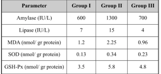
The groups were compared according to amy-lase levels (Table 1). Amyamy-lase levels in treatment group were similar to the levels in Sham group. There was a statistically significant elevation in Group II than Group I and Group III according to amylase levels (p<0.05).
Parameter Group I Group II Group III
Amylase (IU/L) 600 1300 700
Lipase (IU/L) 7 15 4
MDA (nmol/ gr protein) 1.2 2.25 0.96
SOD (nmol/ gr protein) 0.13 0.34 0.23
GSH-Px (nmol/ gr protein) 3.5 5.8 4.8
Table 1: Amylase, Lipase, MDA levels, GSH-Px and
SOD enzyme activities in pancreas tissue and plasma from rats.
* MDA: Malondialdehyde; * SOD: Superoxide dismutase; *GSH-Px: Glutathione peroxidase
Lipase levels were summarized in Table 1. As amylase levels, lipase levels were also higher in group II. The treatment group had lowest lipase lev-els and there was a statistically significant difference between group II and III according to lipase values (p<0.05).
MDA levels were studied within tissue samples (Table 1). The highest MDA values were obtained in group II. Levels of MDA in group III were even lower than Sham group. The difference between Group II and group III was statistically significant (p<0.05).
SOD and GSH-Px analyses were also per-formed (Table 1). Group I and Group III had lower SOD values than group II. In group III, SOD was below than untreated group’s SOD levels, but there were no statistically significant differences between groups according to SOD levels. Glutathione peroxi-dase was also elevated in group II. Although GSH-Px levels were decreased after treatment, there were no statistically significant differences between Group II and group III according to GSH-Px values (p>0.05).
Histopathological evaluation revealed none of the edema, inflammation and necrosis in group I. Group II and III had edema in various degrees (Table 2). Edema in group III was less important than group II and there were no statistically significant differ-ences according to the degree of edema between group II and III.
The inflammation of the pancreatic tissue was summarized in Table 3. Inflammation was detected
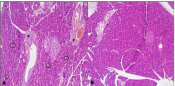
in the pancreatic tissues of rats in groups II and III. The degree of inflammation was slighter in group III (Figure 1 A, B), but there were no statistically signif-icant differences between groups according to inflammation in histopathological evaluation. On the other hand, none of the tissues had necrosis.
Discussion
Acute pancreatitis is a localized inflammation of pancreas at first but can cause severe systemic complications. Ischemia is one of the possible caus-es of severe pancreatitis or may cause the progrcaus-es- progres-sion from edematous pancreatitis to necrotizing pancreatitis(10). There are important data about the roles of oxygen radicals in the pathogenesis of pan-creatitis. In a related experimental study, it was shown that, pancreatic oxidant stress is occurred early after induction(11, 12). In a human study about AP, products of lipid peroxidation were detected in biliary and pancreatic tissues(13). The probable initia-tor of the disease, reactive oxygen species (ROS), is also known as a progressive factor of the disease(14). Different pathogenic mechanisms induce the pro-duction of ROS in various inflammatory diseases as in AP and also ROS have some triggering roles in different inflammatory issues. ROS directly induces the increase in the activation of prostaglandin, thromboxane, and leukotriene(15). ROS are mole-cules with one or more unpaired electrons. Unpaired electrons make ROS remarkably reactive, so free radicals can induce devastating reactions by damaging many structures like proteins, lipids and nucleic acids(16). In our study, we detected remark-able elevation in ROS in the early stages of the pan-creatic inflammation.
Amylase and lipase levels are remarkably increased in AP(17). In our study, we detected remarkably elevated serum levels of amylase and * E -0 *E -1° *E -2° *E-3° *E-4
Group I 8 0 0 0 0
Group II 0 2 5 1 0
Group III 0 5 3 0 0
Table 2: TH leaded to less edema in Group III in the
histopathological evaluation of pancreas tissues.
* E= Edema, scores were determined according to Schmidt’s histopathological scoring criteria
* I -0 * I -1° * I -2° * I -3° *I-4
Group I 8 0 0 0 0
Group II 0 2 5 0 0
Group III 4 3 1 0 0
Table 3: The histopathological evaluation of pancreas
tissues according to inflammation; TH reduced the seve-rity of the inflammation in Group III.
* I= Inflammation, scores were determined according to Schmidt’s histopathological scoring criteria
Figure 1: The histopathological evaluation of pancreatic
tissue. A) edema and inflammation in cerulein-induced pancreatitis B) slighter edema and inflammation after tirofiban therapy.
lipase in cerulein induced pancreatitis group. On the other hand, the applications of tirofiban after pancreatitis induction provided an important improvement in amylase and lipase levels.
MDA is the basic product of lipid peroxida-tion. The measurement of MDA in any tissue helps to detect the lipid peroxidation caused by oxidative stress(16, 18). In an experimental animal study in rats about alcohol-induced pancreatitis, an increase in the ROS and decrease in the buffering mechanisms were detected(19). In our study, MDA levels were increased significantly in CIP without treatment. Additionally, SDO and GSH-Px levels were increased in accordance to literature as well as MDA levels. In this study, the increasing level of serum MDA after induction of pancreatitis was con-sidered as a sign of ongoing oxidative stress and lipid peroxidation.
Glutathione peroxidase and SOD play impor-tant roles against oxidative damage. These enzymes take part in the defense system against oxygen radi-cals. The levels of SOD and GSH-Px are expected to be lower after treatment(20). The levels of SOD and GSH-Px were decreased after tirofiban therapy in our study but there were no statistically signifi-cant differences between groups.
Tribofan is a non-peptide agent and is a reversible antagonist of GP IIb/IIa receptor. Many experimental studies reported that tirofiban inhibits thrombocyte aggregation and prevents arterial thrombosis. Inhibition effect of thrombocyte adhe-sion depends on dose and concentration of the drug. It is being used for the treatment of myocardial ischemia, unstable angina pectoris and acute coro-nary syndromes due to its antithrombotic effect(21-23). According to our knowledge, there is no study about the therapeutic effect of tirofiban in AP in lit-erature. On the other hand, there are some studies settled to point out the useful effects of tirofiban in ischemic disorders. In a previously reported study, TH infusion 36 hours after coronary angioplasty decreased acute ischemic complications(24). There are also a few studies about the therapeutic effects of TH in ischemic brain lesions(25, 26).
Schmidt’s histopathological scoring criteria were used to evaluate the severity of the pancreati-tis(9). Edema, inflammation, vacuolization and the necrosis were evaluated. Despite the lack of statisti-cally significant difference according to edema, inflammation, and necrosis between the treatment and control group, a slight improvement in the pancreatic histopathological parameters was detected with TH.
Although the effects of tirofiban against ischemia-reperfusion injury were studied on vary-ing tissues, there is no study in pancreas up to day in literature. TH is a hopeful and frequently used anti-thrombotic agent in cellular treatment of ischemic injury and inflammation. We suppose that tirofiban which is a highly potent inhibitor of platelet aggregation and a preventer of arterial thrombosis can improve acute pancreatitis and pre-vent the occurrence of pancreas necrosis. Unfortunately there is not enough study about Tirofiban therapy in AP. Hence, advanced and numerous studies are needed to get favorable results.
References
1) Wang YZ, Zhang YC, Cheng JS, Ni Q, Li PW, Han W, Zhang YL. Protective Effects of BML-111 on Cerulein-Induced Acute Pancreatitis-Associated Lung Injury via Activation of Nrf2/ARE Signaling Pathway. Inflammation 2014; 37 (4): 1120-33.
2) De Waele JJ. Acute pancreatitis. Current opinion in critical care 2014, 20:189-195.
3) Hazem ZM. Acute biliary pancreatitis: diagnosis and treatment. Saudi J Gastroenterol 2009; 15: 147-155. 4) Khan AS, Latif SU, Eloubeidi MA. Controversies in
the etiologies of acute pancreatitis. JOP 2010; 11: 545-552.
5) Kinnala PJ, Kuttila KT, Gronroos JM, Havia TV, Nevalainen TJ, Niinikoski JH. Splanchnic and pancre-atic tissue perfusion in experimental acute pancreatitis. Scand J Gastroenterol 2002; 37: 845-849.
6) Bulut NE, Ozkan E, Ekinci O, Dulundu E, Topaloğlu Ü, Şehirli AÖ, Ercan F, Şener G. Beneficial effects of alpha lipoic acid on cerulein-induced experimental acute pancreatitis in rats. Ulusal travma ve acil cerrahi dergisi 2011; 17: 383-389.
7) Ostrowska M, Adamski P, Kozinski M, Navarese EP, Fabiszak T, Grześk G, Paciorek P, Kubica J. Off-target effects of glycoprotein IIb/IIIa receptor inhibitors. Cardiol J 2014 2014; 21(5): 458-64.
8) Tas MH, Simsek Z, Ayan A, Aksu U, Demirelli S, Koza Y, Lazoglu Z, Seven B, Senocak H. Effects of tirofiban maintenance duration on myocardial perfusion defect severity in anterior myocardial infarction. Adv Ther 2013; 30: 834-844.
9) Schmidt J, Rattner DW, Lewandrowski K, Compton CC, Mandavilli U, Knoefel WT, Warshaw AL. A better model of acute pancreatitis for evaluating therapy. Ann Surg1992; 215: 44-56.
10) Gullo L, Cavicchi L, Tomassetti P, Spagnolo C, Freyrie A, D’Addato M. Effects of ischemia on the human pan-creas. Gastroenterology 1996; 111: 1033-1038. 11) Tsai K, Wang SS, Chen TS, Kong CW, Chang FY, Lee
SD, Lu FJ. Oxidative stress: an important phenomenon with pathogenetic significance in the progression of acute pancreatitis. Gut 1998; 42: 850-855.
12) Gough DB, Boyle B, Joyce WP, Delaney CP, McGeeney KF, Gorey TF, Fitzpatrick JM. Free radical inhibition and serial chemiluminescence in evolving experimental pancreatitis. Br J Surg 1990; 77: 1256-1259.
13) Schoenberg MH, Birk D, Beger HG. Oxidative stress in acute and chronic pancreatitis. Am J Clin Nutr 1995; 62: 1306-1314.
14) Closa D. Free radicals and acute pancreatitis: much ado about ... something. Free Radic Res 2013; 47: 934-940.
15) Schoenberg MH, Buchler M, Gaspar M, Stinner A, Younes M, Melzner I, Bültmann B, Beger HG. Oxygen free radicals in acute pancreatitis of the rat. Gut 1990; 31: 1138-43.
16) Halliwell B. Free radicals, antioxidants, and human disease: curiosity, cause, or consequence? Lancet 1994; 344: 721-4.
17) Banks PA, Freeman ML. Practice guidelines in acute pancreatitis. Am J Gastroenterol 2006; 101: 2379-400. 18) Marnett LJ. Lipid peroxidation-DNA damage by
mal-ondialdehyde. Mutat Res 1999; 424: 83-95.
19) Andican G, Gelisgen R, Unal E, Tortum OB, Dervisoglu S, Karahasanoglu T, Burçak G. Oxidative stress and nitric oxide in rats with alcohol-induced acute pancreatitis. World J Gastroenterol 2005; 11: 2340-345.
20) Giordano S, Darley-Usmar V, Zhang J. Autophagy as an essential cellular antioxidant pathway in neurode-generative disease. Redox biology 2014; 2: 82-90. 21) Inhibition of the platelet glycoprotein IIb/IIIa receptor
with tirofiban in unstable angina and non-Q-wave myocardial infarction. Platelet Receptor Inhibition in Ischemic Syndrome Management in Patients Limited by Unstable Signs and Symptoms (PRISM-PLUS) Study Investigators. N Engl J Med1998; 338: 1488-97. 22) Gomma A, Collinson J, Purcell H, Flather M. The role
of tirofiban in acute coronary syndromes. Int J Clin Pract2000; 54: 121-4.
23) Liu Y, Su Q, Li L. Safety and efficacy of early adminis-tration of tirofiban in patients with acute ST-segment elevation myocardial infarction undergoing primary percutaneous coronary intervention: a meta-analysis. Chin Med J (Engl)2014; 127: 1126-32.
24) Spencer B. Effects of platelet glycoprotein IIb/IIIa blockade with tirofiban on adverse cardiac events in patients with unstable angina or acute myocardial infarction undergoing coronary angioplasty. Randomized Efficacy Study of Tirofiban for Outcomes and Restenosis. Circulation 1997; 96: 1445-53. 25) Junghans U, Seitz RJ, Ritzl A, Wittsack HJ, Fink GR,
Freund HJ, Siebler M. Ischemic brain tissue salvaged from infarction by the GP IIb/IIIa platelet antagonist tirofiban. Neurology 2002; 58: 474-6.
26. Seitz RJ, Meisel S, Moll M, Wittsack HJ, Junghans U, Siebler M. The effect of combined thrombolysis with rtPA and tirofiban on ischemic brain lesions. Neurology 2004; 62: 2110-2.
Acknowledgement
This study was granted by Scientific Research Projects Coordination Unit of Mustafa Kemal University.
_______
Corresponding author AYDINKAPLAN Siirt State Hospital General Surgery Clinic 56000 Siirt
(Turkey)
View publication stats View publication stats