ISSN 1308-8084 Online; ISSN 1308-5301 Print 11/3 (2018) 64-70
Research article/Araştırma makalesi
Changes in pigment content of green algae (Desmodesmus sp. and Chodatodesmus mucranulatus) exposed to alumina oxide (Al2O3) nanoparticles
Betül Yılmaz ÖZTÜRK *1, Yeşim DAĞLIOĞLU2, Baran AŞIKKUTLU3
,Cengiz AKKÖZ3 1 Eskişehir Osmangazi University Central Research Laboratory Application and Research Center, Turkey
2 Ordu University Faculty of Arts & Science, Department of Molecular Biology and Genetics, Turkey 3 Selçuk University, Faculty of Science, Department of Biology, Konya, 42250, Turkey
Abstract
Nanoparticles (NPs) have emerged as a new class of environmental pollutants with the emergence of nanotechnology. Al2O3 NPs released from different industries, personal care products and wide range of applications
necessarily end up in aquatic environments. Algal growth inhibition tests are an significant indicator model of monitoring programs designed to predict the effect of NPs on aquatic environments. This study investigated the effects of varying-duration and concentration exposure on the chlorophyll (Chl) contents of Al2O3 NPs to two species of freshwater green
algae (Desmodesmus sp., and Chodatodesmus mucranulatus) recommended for use in standard algal growth inhibition tests. To induce Al2O3 NPs effect, we exposed algae to 3- 96 mg/L for 96 hours. In the test groups treated with Al2O3 NPs
in both algae cells, chlorophyll content decreased in 48 hours exposure compared to the control groups and a clear increase in exposure time to 72 hours was observed. As a result, it was noted that the chlorophyll content of this study varied at the varying duration and concentrations. Variation in chlorophyll (Chla and Chlb) concentrations for Desmodesmus sp. and Chodatodesmus mucranulatus.was recorded at the significance level of p<0.01.
Key words: alumina, chlorophyll, nanotoxicology, nanoparticles, microalgae --- ---
Alümina oksit (Al2O3) nanopartiküllerine maruz kalan yeşil alglerin (Desmodesmus sp. ve Chodatodesmus mucranulatus) pigment içeriğindeki değişiklikler
Özet
Nanopartiküller (NP’ler), nanoteknoloji ile birlikte çevre kirleticilerinin yeni bir sınıfı olarak ortaya çıkmıştır. Al2O3 NP’leri kişisel bakım ürünleri, farklı endüstriler ve geniş uygulama yelpazesinden salınır ve mutlaka sucul çevrelere
ulaşır. Algal büyüme inhibisyon testleri, bu sucul çevrelerde NP’lerin etkisini öngörmek için tasarlanmış izleme programlarının önemli bir göstergesidir. Bu çalışmada, Al2O3 NP’lerinin değişen süresi ve konsantrasyonlarda, iki tatlısu
yeşil alg (Desmodesmus sp. ve Chodatodesmus mucranulatus) türlerinde, standart alg büyüme inhibisyon testi ile klorofil (klf) muhteviyatını üzerine etkisi araştırılmıştır. Al2O3 NP etkisini değerlendirmek için algler 72 saat boyunca 3-96 mg/L
konsantrasyonlarında Al2O3 NP’lerine maruz bırakıldı. Al2O3 NP’leri uygulanan test gruplarında, kontrol grupları ile
karşılaştırıldığında klorofil muhtevasında net bir azalma gözlemlendi. Her iki alg hücresinde Al2O3 NP’leri ile muamele
edilen test gruplarında klorofil muhteviyatı kontrol gruplarına kıyasla 48 saat sonra azaldı, maruz kalma süresi 72 saate uzadığında ise belirgin bir klorofil muhteviyatında artış gözlendi. Sonuç olarak, bu çalışmanın klorofil muhteviyatı Al2O3
NP’lerinin değişen süre ve konsantrasyonlarda değiştiği kaydedilmiştir. Desmodesmus sp. ve Chodatodesmus mucranulatus için klorofil (klfa ve klfb) konsantrasyonlarında değişimi p<0.01 anlamlılık seviyesinde önemli olduğu kaydedilmiştir.
Anahtar kelimeler: alüminyum oksit, klorofil, nanotoksikoloji, nanopartiküller, mikroalgler
*Corresponding author / Haberleşmeden sorumlu yazar: Tel.: +902222393750; Fax.: +902222394106; E-mail: [email protected]
1. Introduction
Nanotechnology is one of the most effective research fields in technology science. Nanotechnology involves the creation and manipulation of materials at the nanometer level. Nanotechnology uses engineering materials or devices with nanometer-scale dimensions, typically ranging from 1 to 100 nm. The application and development of many industrial nanotechnologies increases the potential for harmful effects of nanoparticles (NPs) on the environment. NPs display entirely new, improved and unique properties based on specific properties such as size, distribution, synthesis method and morphology (Sadiq et al., 2011; Özkan et al., 2015). Recently, the widespread use of NPs, especially metal oxide NPs, has drawn great attention. It is now widely known that the ability of nanoparticle-sized materials to respond is potentially dangerous for the environment (Gosteva et al., 2015; Çolak and Nas 2016). It is necessary to assess the NP toxicity of aquatic ecosystems as the surrounding water resources are contaminated by the nanotechnology industry products in various ways. Among these, the most popular NP are aluminum oxide (Al2O3) NPs. Aluminum is one of the
most produced chemicals in nano-sized particles. Aluminum is estimated to account for about 20% of the 2005 world market NPs (Arul Prakash et al., 2011). Al2O3 NPs have been applied in catalysis, reinforcement, polymer modification,
functionalization of textiles, heat transfer fluids, wastewater treatment and structural ceramics. In addition, Al2O3 NPs
have broad biological applications such as biosensors, antigen presentations for biofiltration, drug delivery and immunization purposes (Arul Prakash et al., 2011). Despite the potential benefits of NPs, they cause concern because of their negative effects on human health and the environment impacts. Although Al2O3 NPs are published data on biological
effects in the aquatic environment, their results are inconsistent. These contradictions in the literature are due to a variety of factors such as physicochemical properties of NPs, form of synthesis, experimental conditions, indicator organisms susceptibility, etc., but the real problem is the lack of valid and common analytical evaluations of nanoparticle toxicity.
Alg toxicity tests are widely used to assess the aquatic effects of hazardous substances. Algae play an important role in the balance of aquatic ecosystems that are at the first level of the organic and oxygen producing trophic chain (Sadiq et al., 2011). Microalgae have a very rich carbohydrate content, especially fatty acid content (Çiçek et al., 2017). Algae differs basically in terms of cell structure, pigment composition, storage nutrient and presence, number and structure of flagella (Shelknanloymılan et al., 2012; Coşan et al., 2015). Therefore, the purpose of the current research was to study the differences changes in pigment content response of nanosized alumina particles toward green algae (Desmodesmus sp. and Chodatodesmus mucranulatus) isolated from the aquatic environment.
2. Materials and methods 2.1. Nanoparticles preparation
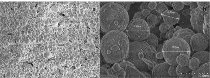
Nanopowder alumina oxide (Al2O3 ) were obtained from Nanografi Co. Ltd. (Purity 99+%, average particle size
20–80 nm, Hydrophilic) (Ankara, Turkey). Nanopowder alumina oxide SEM photographs were taken and dimensions were measured Figure 1. SEM photographs were obtained by using Jeol brand SEM.
Figure 1 a and b) Nanopowder alumina oxide (Al2O3 ) SEM image
2.2. Test organisms and cultivation conditions
Environmental water samples were taken from the following localities Apa Dam Lake (37°22ˈ10ˈˈN 32°29ˈ40ˈˈE), and Eber Lake (38°39ˈ09ˈˈN 31°10ˈ08ˈˈE). The local samples like stone, mud and plants were collected from their natural habitats carried to the laboratory in glass bottles filled with lake water. In order to obtain monocultures, the dilution method was applied Rippka. Specimens taken from various aquatic environments placed in glass bottles and
brought to the laboratory. Inverted microscope was used to obtain single cells from mixed species with pasteur pipette technique. Subsequently, the single cells obtained were transferred to BG 11 medium (Table 1). These cell were then inoculated into test tubes containing medium BG-11 medium to form a pure culture according to Rippka's method (Rippka, 1988). The cultures were incubated at 25 °C for 15-20 days in accordance with photosynthesis, with 12 h light, 12 h darkness under 3000 lux fluorescein light.
2.3. Preparation of Test Solutions
The stock solution of Al2O3 NPs was prepared in deionized water. The solution was then vortexed for 20 seconds
and sonicated for 30 minutes in an ultra sonic water bath (Bandelin, SonoRex) to ensure maximum distribution in water. After all these steps, the test concentrations determined by preliminary studies were prepared by stock solution dilution. The prepared test solutions were added to the BG-11 medium.
2.4. Acute toxicity studies
Cells were counted with thoma slide after they were increased. 90 ml of BG-11 medium and 10 ml of test solution containing 106 cells were added to 200 ml of the erlenmeyer to perform the exposure of the algae species (Desmodesmus
sp., Chodatodesmus mucranulatus) to the Al2O3 NPs. Exposure studies were also carried out in orbital shakers (wiseshake)
to prevent agregations in the stationary environment of Al2O3 NPs, to achieve the desired constant temperature and natural
conditions of algae. The speed of the shaker was set so that the cells would not be damaged, but at the same time would block the aggregate formation of the particles (85 rpm). The test conditions were set at 25 °C, 12:12 (daylight:darkness). After the experiment was established, 2 ml samples were taken at 24, 48, 72 hours after each concentration. The algal chlorophyll content measurements were performed at 72 h as described in the OECD method (Test, 1984).Cell counts and pigment measurements were made on these samples. Exposure studies were performed in 3 replicates independently of each other.
Table 2. BG-11 medium used in the purification of species
BG-11 Medium g/L
NaNO3 15 Trace elements mg/L
K2HPO4 0.4 H3BO3 61
MgSO4. 7H2O 0.75 MnSO4 . H2O 169
CaCl2. 2H2O 0.36 ZnSO4 .7H2O, 287
Citric acid 0.06 CuSO4. 5H2O 2.5
Iron (III) ammonium citrate 0.06 (NH4)6Mo7O24. 4H2O 12.5
Na2-EDTA 0.01
Na2CO3 0.2
2.5. Chlorophyll measurement
Chlorophyll a (Chla), chlorophyll b (Chlb) and carotenoids (Chlc) were identified according to Lichtenthaler and Wellburn (1983). Briefly, 2 ml algae cultures were centrifuged at 2000 rpm, then the supernatant was discarded and 80% (v/v) acetone was added to the samples and placed at 4 °C for 24 hours. Then, the light absorption of these samples were measured with a spectrophotometer (Hange-Lange brand DR 2800) at 663, 646 and 470 nm. Pigment contents were calculated with the formula given below.
Chla =12.21A663-2.81A646
Chlb =20.13A646-5.03A663
Chlc =(1000A470-3.27CChla -104CChlb)/229
2.6. Statistical analyzes
All experiments were repeated independently three times and the data were recorded as mean value with standard deviation. Analyzes were performed using SPSS one-way analysis of variance (ANOVA), tukey multiple comparison analysis.
3. Results 3.1. Algae culture
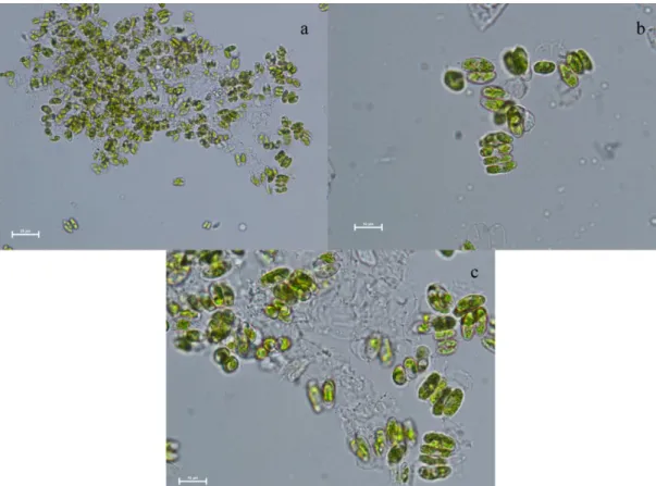
When isolating the algae, an inverted microscope was used and continued until the single cell was lowered. single cells were transferred to the BG-11 medium and the result was determined as Desmodesmus sp. and Chodatodesmus mucranulatus species were obtained from the Apa Dam lake and Eber lake. Light microscope images of species after Al2O3 NPs exposure are given in Fig. 2 and 3.
Figure 2. Light microscope images of Desmodesmus species after Al2O3 NPs exposure: a and b) X40, c) X100
Figure 3. Light microscope images of Chodatodesmus mucranulatus after Al2O3 NPs exposure: a and b) X40, c) X100
3.2. Pigment content
Desmodesmus sp. and Chodatodesmus mucranulatus were exposed to 5-80 nm Al2O3 nanoparticles for 72 h at
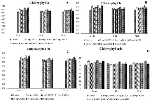
Figure 4. Chlorophyll (a, b, a+b, a/b) values of Chodatodesmus mucranulatus exposed to Al2O3 NPs
In treatment groups, Chla content increased with prolonged exposure duration. Generally, In the treatment groups of Chodatodesmus mucranulatus, the Chla content increased as the duration of exposure prolonged. However, at the 48 mg /L test concentration, the Chla content decreased for 48 hours exposure and increased again when the exposure duration prolonged to 72 h. Generally, when we look at the content of chlb, the content of Chlb is increased as the duration of exposure is prolonged. As is the case in Chla, during 48 h exposure at 48 mg/L, Chlb content decreased by 7% compared to 24 h and increased again when exposure duration prolonged to 72 (Figure 4).
Figure 5. Chl (a, b, a+b, a/b) values of Desmodesmus sp. exposed to Al2O3 NPs
In the Desmodesmus sp. as Chodatodesmus mucranulatus was generally increased Chla content in the treatment groups as the duration of exposure prolonged. In the 96 mg/L only, Chla content reduced at 72 h exposure. Chlb content of Desmodesmus sp. increased as exposure duration prolonged, as was Chla in Chlb content. At 24 h of exposure at 24 mg/L and at 72 h at 96 mg/L, Chlb content reduced by 8% and 44% respectively (Figure 5). Compared the two green algae pigments, the content of Chla and Chlb in the control and treatment groups of Desmodesmus sp. is generally higher.. 4. Conclusions and discussion
NPs have begun to be used more and more in the last 30 years due to their unique properties such as many different consumer products, electronics, catalysis, biomedical applications, drugs and drug delivery, cosmetics, energy, and materials (Nowack and Bucheli, 2007). For this reason, NPs have a potential to pollute the environment nowadays
much more than in the past (Burklew et al., 2012). However, the positive and hazardous effects of NPs on ecosystems are still entirely unknown (Oberdörster et al., 2005). Thus, the environmental risk of NPs, the physicochemical properties of NPs are referable their nano/small size, synthetic, impurity ratio, large surface area, chemical composition, surface reactivity, charge, shape and environment interactions (Oberdorster et al., 2005; Qukarroum et al., 2012).
Metal oxide nanoparticles, such as aluminum oxide (Al2O3) are interesting for a wide variety of applications due
to their unique physical and chemical properties (Huang et al., 2010). It has important applications especially in the ceramic industry and is used as abrasive materials, absorbents and biomaterials and strengthens metal matrix composites. (Sadiq et al., 2009). Since Al2O3 NPs are highly preferred, many in vivo and in vitro studies have been conducted in the
literature on many cell lines (such as human, mouse), as well as a large number of indicator organisms such as yeast, bacteria and nematodes. For example, Zhang et al., 2011 studied the toxic effects of nanoparticles of Al2O3 NPs in human
fetal lung fibroblasts (HFL1) in vitro. The results show that Al2O3 NPs lead to cellular mitochondrial dysfunction,
morphological modifications and apoptosis at a concentration range of 0.25-1.50 mg /mL, and that toxic effects are clearly visible in a dose-dependent manner. Jeng and Swanson 2006, in their study of toxicity of Al2O3 NPs in Neuro-2A (mouse
neuroblastoma) cells, noted that Al2O3 NP reduced mitochondrial function at 100 μg/mL concentration. The effect on the
cellular plasma membrane was demonstrated by measuring LDH leakage and did not cause LDH leakage after 24 hours exposure. In addition, it showed less than 2% apoptosis at 100 μg / mL. Shrivastava et al., 2014 show subacute exposure effects of Al2O3 NPs with oxidative stress and histological changes in mouse brain and liver. As a result, it is confirmed
that the interaction of absorpted Al2O3 NPs with cellular components can produce reactive oxygen species (ROS), and
that the size of ROS production may lead to cellular toxicity if the cell undergoes antioxidant defense. Pakrashi et al., 2011 evaluated the cytotoxicity of Al2O3 NPs at low exposure levels in freshwater bacterial isolates (Bacillus
licheniformis). Exposure to 1 μg / mL Al2O3 NP for 2 hours caused a 17% decrease in cell viability according to the
results of the plate count and MTT assay. Wang et al., 2009, evaluated the toxicity of Al2O3 NPs in Aenorhabditis elegans
(nematode). Al2O3 NPs are toxic to C. elegans, especially their reproductive ability. García-Saucedo et al., 2011 noted
that low toxicity of Al2O3 NPs did not show low or no toxicity in Saccharomyces cerevisiae (yeast) cells.
Algal toxicity tests are widely used to assess effects of hazardous substances in the aquatic environments. Algae plays a significant task in the stabilization of water environments, the first level of the trophic chain producing oxygen and organic matter. In our study, the aqueous media employed was BG-11 (Stanier et al., 1971). Because, these media contain metal ions in trace amounts to maximize the growth of algae. Algae as other plants cells have cell walls that form the primary site for interaction. This cell wall is a barrier to entry of the NPs into the cells. The main cell wall components are carbohydrates which are bound to form a complex network and proteins (Sadiq et al., 2011; Knox 1995). In our study, responses of Desmodesmus sp. and Chodatodesmus mucranulatus to the Al2O3 NPs were dependent on consantration and
duration of exposure. The chla, chlb, chl a+b, chl a/b contents of the six group of treatments are found in Figure 4, 5. chl contents of the Al2O3 NPs outspread an order of magnitude. In the obtained data, the content of the pigment in the
Desmodesmus sp. is calculated more than the Chodatodesmus mucranulatus. This suggests that Al2O3 NPs give more
toxicity to the C. mucranulatus. However, it is interesting that when the exposure time is up to 72 hours, the content of chlorophyll is very low of Desmodesmus sp., and the C. mucranulatus remain almost the same. This is probably due to the increased internalization of Al2O3 NPs by Desmodesmus sp. , which increased the content of chlorophyll in the first
days of the ending exposure and then showed a severe toxic effect. Sadiq et al., 2011, a marked decrease in chl content was observed in cells treated with Al2O3 NPs compared to those not applied and it was noted that cells were more effective.
Namely, during the experiment a concentration-dependent reduce in chl content was recorded, corroborating that growth inhibitory effect was due to increased concentration of the particles. In another study, the toxic effect of (ZnO-TiO2)NCM
on photosynthetic pigment contents was investigated on freshwater Desmodesmus multivariabilis which was exposed for 72 h to 0.1, 0.01 and 0.001 mg l–1 of (ZnO-TiO2)NCM. According to this study, the effect of the photosynthetic pigment
contents of (ZnO-TiO2)NCM in varying concentrations indicated differences depending on the exposure duration and concentration (Dağlıoğlu and Öztürk, 2018). In the Kulacki and Cardinale (2012) study, the most common ten species of freshwater pelagic algae in North America were exposed to n-TiO2. The results indicated that TiO2 NPs may affect some
aspects of the population growth of phytoplankton, but the effects on environmentally relevant concentrations are low. Dağlıoğlu and Türkiş at study, effects on the amount of pigment after exposure to the TiO2 NPs of duckweed (Lemna
minor) Which the indicator organism of the aquatic environment have researched. At the end of the 96-h exposure period, chlorophyll a and b levels were discovered important differences between 50-200 mg-1 concentrations at p<0.001 level.
Dağlıoğlu and Öztürk (2016) have been observed that boron particles (nano and micro) accumulate in different amounts in the Desmodesmus multivariabilis.
Our study; there are studies on the photosynthetic activity and growth of different algae species of aluminum nanoparticles. Aluminum oxide exhibited toxicity, both different time duration, and varying concentrations. In both species of algae (Desmodesmus sp. and Chodatodesmus mucranulatus) the concentration-dependent decrease in chlorophyll content was observed. However, the responses of the algae to the aluminum oxide has similar.
Acknowledgements
References
Arul Prakash, F., Dushendra Babu, G.J., Lavanya, M., Shenbaga Vidhya, K., Devasena, T. (2011). Toxicity studies of aluminium oxide nanoparticles in cell lines. Int J Nanotechnol Appl, 5(2), 99-107.
Burklew, C.E., Ashlock, J., Winfrey, W.B., Zhang, B. (2012). Effects of aluminum oxide nanoparticles on the growth, development, and microRNA expression of tobacco (Nicotiana tabacum). PloS one, 7(5), e34783.
Coşan, E.D., Tezcan, N., Coşan, D.T. (2015). The effects of chemical and biological plant fertilizer types for used to ıncrease agricultural production on Chlorophyll A, Chlorophyll B, Vitamin C, the length of plants, mitosis and chromosomes of Onion (Allium cepa). Biological Diversity and Conservation, 8(2) (2015) 16-22
Çiçek, L.N., Ertan, O.Ö., Erdoğan, Ö., Didinen H., Boyacı, Ö.Y., Kara, D., Zeybek, M., Diken, G. (2017). Distribution of phytoplankton and its relationship with physicochemical parameters in Lake Eğirdir (Isparta/Turkey). Biological Diversity and Conservation, 10(3), 150-162
Çolak, A.D., Nas, B. (2016). NiFe2O4 Nanokompozitinin Olası Toksik Etkisine Karşı Olueropein’in Koruyucu Rolü. Erzincan University Journal of Science and Technology, 9 (Special Issue I), 172-183.
Dağlıoğlu Y, Özturk Y.B. (2016b). Desmodesmus multivariabilis’in bor partikullerine maruz kalmada biyolojik birikiminin değerlendirilmesi. Biological Diversity and Conservation. 9(3), 204–209.
Dağlıoğlu, Y., Çelebi, S.M., Önalan, Ş., (2016a). Determination of Acute Toxic Effects of Poly (Vinylferrocenium) Supported Palladium Nanoparticle (Pd/PVF+ ) on Artemia salina". Pakistan Journal of Zoology vol. 48(1), 187-193.
Dağlıoğlu, Y., Öztürk, Y.B. (2018). Effect of concentration and exposure time of ZnO-TiO2 nanocomposite on photosynthetic pigment contents, ROS production ability, and bioaccumulation of freshwater algae. Caryologia, 71(1), 13-23.
Dağlıoğlu, Y., Türkiş S., (2017). TiO2 nanopartikül Uygulamasının Su Mercimeğinin (Lemna minor L.) Fotosentetik Pigment İçeriği Üzerine Etkisi. Acta Biologica Turcica, 30(4), 108-115.
García-Saucedo, C., Field, J.A., Otero-Gonzalez, L., Sierra-Álvarez, R. (2011). Low toxicity of HfO2, SiO2, Al2O3 and CeO2 nanoparticles to the yeast, Saccharomyces cerevisiae. Journal of hazardous materials, 192(3), 1572-1579.
Gosteva, I., Morgalev, Y., Morgaleva, T., Morgalev, S. (2015). Effect of Al2O3 and TiO2 nanoparticles on aquatic organisms. In IOP Conference Series: Materials Science and Engineering, Vol. 98, No. 1, p. 012007. IOP Publishing.
Huang, Y.W., Wu, C.H., Aronstam, R.S. (2010). Toxicity of transition metal oxide nanoparticles: recent insights from in vitro studies. Materials, 3(10), 4842-4859.
Jeng, H. A., Swanson, J. (2006). Toxicity of metal oxide nanoparticles in mammalian cells. Journal of Environmental Science and Health Part A, 41(12), 2699-2711.
Knox, J.P. (1995). The extracellular matrix in higher plants. 4. Developmentally regulated proteoglycans and glycoproteins of the plant cell surface. The FASEB Journal, 9(11), 1004-1012.
Kulacki, K. J., Cardinale, B. J. (2012). Effects of nano-titanium dioxide on freshwater algal population dynamics. PLoS One, 7(10), 1-7.
Lichtenthaler, H. K., Wellburn, A. R. (1983). Determinations of total carotenoids and chlorophylls a and b of leaf extracts in different solvents, 603rd Meeting, Liverpool, 591-592.
Nowack, B., Bucheli, T.D. (2007). Occurrence, behavior and effects of nanoparticles in the environment. Environmental pollution, 150(1), 5-22.
Oberdorster, G., Oberdorster, E., Oberdorster, J., (2005). Nanotoxicology: an emerging discipline evolving from studies of ultrafine particles. Environmental Health Perspectives, 113, 823–839.
Özkan, Y., İrende, İ., Akdeniz, G., Kabakçı, D., Sökmen, M., (2015). Evaluation of the Comparative Acute Toxic Effects of TiO2, Ag-TiO2 and ZnO-TiO2 Composite Nanoparticles on Apis mellifera (Honey Bee) ". Jornal of International. Environmental Application&Science, 10(1), 26-36.
Pakrashi, S., Dalai, S., Sabat, D., Singh, S., Chandrasekaran, N., Mukherjee, A. (2011). Cytotoxicity of Al2O3 nanoparticles at low exposure levels to a freshwater bacterial isolate. Chemical Research in Toxicology, 24(11), 1899-1904.
Qukarroum, A., Bras, S., Perreault, F., Popovic, R. (2012). Inhibitory effects of silver nanoparticles in two green algae, Chlorella
vulgaris and Dunaliella tertiolecta. Ecotoxicology and Environmental Safety, 78, 80-85.
Rippka, R. (1988). [1] Isolation and purification of cyanobacteria. In Methods in enzymology, Vol. 167, 3-27. Academic Press. Sadiq, I.M., Chowdhury, B., Chandrasekaran, N., Mukherjee, A. (2009). Antimicrobial sensitivity of Escherichia coli to alumina
nanoparticles. Nanomedicine: Nanotechnology, Biology and Medicine, 5(3), 282-286.
Sadiq, I.M., Pakrashi, S., Chandrasekaran, N., Mukherjee, A. (2011). Studies on toxicity of aluminium oxide (Al2O3) nanoparticles to microalgae species: Scenedesmus sp and Chlorella sp. Journal of Nanoparticle Research, 13, 3287-3299
Shelknanloymılan, L., Atıcı, T., Obal, O. (2012). Removal of nitrogen and phosphate by using Choleralla vulgaris on synthetic and organic materials waste water. Biological Diversity and Conservation. 5/2 (2012) 89-94.
Shrivastava, R., Raza, S., Yadav, A., Kushwaha, P., Flora, S.J. (2014). Effects of sub-acute exposure to TiO2, ZnO and Al2O3 nanoparticles on oxidative stress and histological changes in mouse liver and brain. Drug and chemical toxicology, 37(3), 336-347.
Stanier, R. Y., Kunisawa, R., Mandel, M., Cohen-Bazire, G. (1971). Purification and properties of unicellular blue-green algae (Order Chroococcales). Bact. l~ev. 85, 171-205.
Test, O.A.G.I. (1984). OECD guideline for testing of chemicals 201. Paris, France: Organisation of Economic Cooperation and Development.
Wang, H., Wick, R. L., Xing, B. (2009). Toxicity of nanoparticulate and bulk ZnO, Al2O3 and TiO2 to the nematode Caenorhabditis
elegans. Environmental Pollution, 157(4), 1171-1177.
Zhang, X. Q., Yin, L. H., Meng, T.A.N.G., Pu, Y.P. (2011). ZnO, TiO2, SiO2, and Al2O3 nanoparticles-induced toxic effects on human fetal lung fibroblasts. Biomedical and Environmental Sciences, 24(6), 661-669.